Email Alert | RSS    帮助

中国防痨杂志 ›› 2023, Vol. 45 ›› Issue (5): 499-506.doi: 10.19982/j.issn.1000-6621.20220507

• 论著 • 上一篇    下一篇

颅内结核合并急性脑梗死的危险因素分析

李翔1, 杞敏1, 姜建杰2, 魏佳璐1, 付旭文1, 李海雯1, 张乐3()   

  1. 1 昆明市第三人民医院/云南省传染性疾病临床医学中心医学影像科, 昆明 650041
    2 昆明市第三人民医院/云南省传染性疾病临床医学中心互联网医疗部, 昆明 650041
    3 昆明市第三人民医院/云南省传染性疾病临床医学中心重症医学科, 昆明 650041
  • 收稿日期:2022-12-25 出版日期:2023-05-10 发布日期:2023-04-25
  • 通信作者: 张乐 E-mail:9035643@qq.com
  • 基金资助:
    云南省临床医学重点专科建设项目(云卫医发〔2017〕15号);昆明市卫生科研课题项目(2022-09-01-001)

Analysis of risk factors for acute cerebral infarction combined with intracranial tuberculosis

Li Xiang1, Qi Min1, Jiang Jianjie2, Wei Jialu1, Fu Xuwen1, Li Haiwen1, Zhang Le3()   

  1. 1 Department of Radiology, Kuming Third People’s Hospital/Yunnan Clinical Medical Center for Infectious Diseases, Kunming 650041, China
    2 Department of Internet Medicine, Kuming Third People’s Hospital/Yunnan Clinical Medical Center for Infectious Diseases, Kunming 650041, China
    3 Department of Intensive Care Unit, Kuming Third People’s Hospital/Yunnan Clinical Medical Center for Infectious Diseases, Kunming 650041, China
  • Received:2022-12-25 Online:2023-05-10 Published:2023-04-25
  • Contact: Zhang Le E-mail:9035643@qq.com
  • Supported by:
    Yunnan Provincial Clinical Medicine Major Specialist Construction Project (2017-15);Scientific Research Projects of Kunming Science and Technology Bureau(2022-09-01-001)

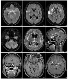
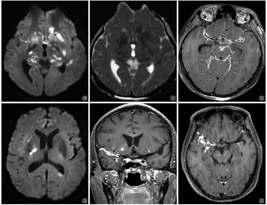
摘要: 目的 分析颅内结核患者的临床特征及合并急性脑梗死的危险因素。方法 采用回顾性研究方法,收集昆明市第三人民医院2019年7月至2021年12月收治的脑脊液结核分枝杆菌培养阳性的84例患者作为研究对象。收集整理研究对象的临床症状、实验室检查结果及影像学检查结果等资料;依据颅脑磁共振成像(magnetic resonance imaging,MRI)表现,将研究对象分为脑梗死组32例(38.1%)和非脑梗死组52例(61.9%)。采用单因素和多因素logistic回归分析颅内结核合并急性脑梗死的危险因素。结果 颅内结核临床症状常见的有头痛(95.2%,80/84)、发热(73.8%,62/84)、呕吐(52.4%,44/84)及意识障碍(48.8%,41/84),抽搐(7.1%,6/84)少见。非脑梗死组患者外周血血镁浓度[中位数(四分位数)]为0.80(0.75,0.85)mmol/L(正常值0.73~1.06mmol/L),明显低于脑梗死组的0.84(0.81,0.89)mmol/L,差异有统计学意义(Z=2.079,P=0.038)。脑梗死组颅脑MRI表现为基底池/环池脑膜增厚者31例(96.9%),外侧裂池脑膜增厚者21例(65.6%),合并动脉血管壁增厚者31例(96.9%);非脑梗死组颅脑MRI表现为基底池/环池脑膜增厚者26例(50.0%),外侧裂池脑膜增厚者18例(34.6%),合并动脉血管壁增厚者28例(53.8%)。脑梗死组基底池/环池脑膜增厚、外侧裂池脑膜增厚及动脉管壁增厚的发生率均明显高于非脑梗死组,差异均有统计学意义(χ2=19.956,P=0.000;χ2=7.659,P=0.006;χ2=17.545,P=0.000)。多因素logisitic回归分析显示,动脉血管壁增厚是颅内结核合并急性脑梗死的独立危险因素[OR(95%CI)=27.128(3.393~216.917)]。结论 颅内结核常见临床症状有头痛、发热、呕吐及意识障碍,MRI提示动脉血管壁增厚是发生急性脑梗死的独立危险因素。

关键词: 结核,脑膜, 脑梗死, 磁共振成像, 危险因素

Abstract:

Objective: To analyze the clinical characteristics and the risk factors of acute cerebral infarction in patients with intracranial tuberculosis. Methods: A retrospective study was conducted to collect 84 patients with positive cultures of Mycobacterium tuberculosis in cerebrospinal fluid admitted to the Kuming Third People’s Hospital of Kunming from July 2019 to December 2021. The clinical symptoms, laboratory test results and imaging findings of these study subjects were collected; based on the cranial magnetic resonance imaging (MRI) performance, these study subjects were divided into cerebral infarction group (32 cases, 38.1%) and non-cerebral infarction group (52, 61.9%). Univariate and multivariate logistic regression were used to analyze the risk factors of intracranial tuberculosis complicated with acute cerebral infarction. Results: Commonly clinical symptoms of intracranial tuberculosis included headache (95.2%,80/84), fever (73.8%,62/84), vomiting (52.4%,44/84) and unconsciousness (48.8%,41/84). The convulsions (7.1%,6/84) was rare. The peripheral blood magnesium concentration (median (interquartile)) of 0.80 (0.75, 0.85) mmol/L (normal value 0.73-1.06 mmol/L) in the non-cerebral infarction group was significantly lower than that of 0.84 (0.81, 0.89) mmol/L in the cerebral infarction group, and the difference was statistically significant (Z=2.079, P=0.038). In the cerebral infarction group, cranial MRI showed thickened meninges in the basal/annular cistern in 31 cases (96.9%), thickened meninges in the lateral fissure cistern in 21 cases (65.6%), and combined with thickened arterial vessel walls in 31 cases (96.9%). In the non-cerebral infarction group, cranial MRI showed thickened meninges in the basal/annular cistern in 26 cases (50.0%), thickened meninges in the lateral fissure cistern in 18 cases (34.6%), and combined with thickened arterial vessel walls in 28 cases (53.8%). The incidence of basal/annular cistern meningeal thickening, lateral fissure cistern meningeal thickening and arterial vessel wall thickening were significantly higher in the cerebral infarction group than in the non-cerebral infarction group, with significant differences (χ2=19.956,P=0.000;χ2=7.659,P=0.006;χ2=17.545,P=0.000). Multivariate logistic regression analysis showed that arterial vessel wall thickening might be an independent risk factor for intracranial tuberculosis combined with acute cerebral infarction (OR (95%CI)=27.128 (3.393-216.917)). Conclusion: The common clinical symptoms of intracranial tuberculosis included headache, fever, vomiting and unconsciousness, and the MRI result showed that thickening of the arterial vessel wall might be an independent risk factor for acute cerebral infarction.

Key words: Tuberculosis, meningeal, Brain infarction, Magnetic resonance imaging, Risk factors

中图分类号: