Email Alert | RSS    帮助

中国防痨杂志 ›› 2020, Vol. 42 ›› Issue (3): 222-226.doi: 10.3969/j.issn.1000-6621.2020.03.008

• 论著 • 上一篇    下一篇

HIV感染并发结核性脑膜炎或弓形虫脑病患者的MRI特征分析

陈七一,李晶晶,徐云良,吕志彬,魏连贵,许东海,谢汝明(),陈步东()   

  1. 100015 首都医科大学附属北京地坛医院放射科
  • 收稿日期:2019-12-03 出版日期:2020-03-10 发布日期:2020-03-18
  • 通信作者: 谢汝明,陈步东 E-mail:13911320739@163.com;budongchen@sina.com

Magnetic resonance neuroimaging differential diagnosis of tuberculous meningitis and toxoplasmosis encephalopathy in HIV infected individuals

CHEN Qi-yi,LI Jing-jing,XU Yun-liang,LYU Zhi-bin,WEI Lian-gui,XU Dong-hai,XIE Ru-ming(),CHEN Bu-dong()   

  1. Department of Radiology, Beijing Ditan Hospital, Capital Medical University, Beijing 100015, China
  • Received:2019-12-03 Online:2020-03-10 Published:2020-03-18
  • Contact: Ru-ming XIE,Bu-dong CHEN E-mail:13911320739@163.com;budongchen@sina.com

摘要:

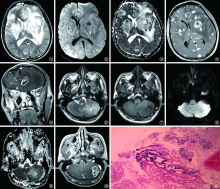
目的 总结HIV感染并发结核性脑膜炎(tuberculous meningitis,TBM)与弓形虫脑病患者在不同CD4 + T细胞水平时的MRI表现特征。方法 从2008年1月至2018年1月在首都医科大学附属北京地坛医院住院的HIV感染者中筛选中枢神经系统感染者46例,均进行了MRI检查。其中,弓形虫脑病21例(弓形虫组),89个病灶;TBM 25例(TBM组),174个病灶。 从病灶分布、形态、大小、磁共振扩散加权成像(DWI)和表观扩散系数(ADC)图像特征、周围水肿带程度等指标,评价两组在CD4 + T细胞<15和≥15个/μl水平时的上述MRI指标的差异。结果 弓形虫组、TBM组皮层及皮层下区病灶占所有病灶的比例:当CD4 + T细胞<15个/μl时,分别为83.0%(44/53)、45.8%(33/72),两组比较差异有统计学意义(χ 2=24.203,P=0.000);当CD4 + T细胞≥15个/μl时,分别为77.8%(28/36)、59.8%(61/102),两组比较差异无统计学意义(χ 2=5.076,P=0.079)。 当CD4 + T细胞<15个/μl时,弓形虫组、TBM组结节状病灶占所有病灶的比率分别为54.7%(29/53)、75.0%(54/72),两组比较差异有统计学意义(χ 2=5.629,P=0.018);当CD4 + T细胞≥15个/μl时,两组斑片状、环形病灶占所有病灶的比率分别为88.9%(32/36)、64.7%(66/102),两组比较差异有统计学意义(χ 2=7.560,P=0.006)。当CD4 + T细胞≥15个/μl时,DWI低、高信号病灶比率在弓形虫组分别为41.6%(15/36)、27.8%(10/36),在TBM组分别为7.8%(8/102)、41.2%(42/102),ADC图像低、高信号病灶比率在弓形虫组分别为22.2%(8/36)、41.7%(15/36),在TBM组分别为40.2%(41/102)、9.8%(10/102),两组比较差异均有统计学意义(DWI: χ 2=21.964,P=0.000;ADC: χ 2=18.440,P=0.000)。结论 不同CD4 + T细胞计数水平时,HIV感染并发TBM与弓形虫脑病患者MR扫描在病灶分布、形态、DWI和ADC图像信号特征上差异明显。

关键词: HIV感染, 结核, 脑膜, 弓形虫病, 脑, 磁共振成像, 对比研究

Abstract:

Objective To summarize the MR imaging characteristics of tuberculous meningitis (abbr. “TBM”)and toxoplasmosis encephalopathy(abbr. “TE”) in HIV infected individuals with different CD4 + T cell levels. Methods From January 2008 to January 2018, 46 patients with central nervous system infection were selected among HIV-infected patients admitted to Beijing Ditan Hospital, Capital Medical University. Among them, 21 cases were TE patients with a total of 89 lesions (TE group), 25 cases were TBM patients with a total of 174 lesions (TBM group). Differences in MR images between CD4 + T cells less than 15 and more than 15 cells/μl were evaluated, inclding lesion distribution, morphology, size, DWI and ADC characteristics, and degree of peripheral edema. Results The proportion of lesions in cortex and subcortex among all lesions within TE group and TBM group were: CD4 + T cells less than 15 cells/μl, 83.0% (44/53) and 45.8% (33/72), with statistically significant difference (χ 2=24.203, P=0.000); CD4 + T cells more than 15 cells/μl, 77.8% (28/36) and 59.8% (61/102), with no statistically difference (χ 2=5.076, P=0.079). When CD4 + T cells were less than 15 cells/μl, the proportion of nodular lesions among all lesions within TE group and TBM group were 54.7% (29/53) and 75.0% (54/72) respectively, with statistically significant difference between the two groups (χ 2=5.629, P=0.018). When CD4 + T cells were more than 15 cells/μl, the proportions of plaque-shaped circular lesions in those two groups were 88.9% (32/36) and 64.7% (66/102) respectively, with statistically significant difference between the two groups(χ 2=7.560, P=0.006). When CD4 + T cells were more than 15 cells/μl, the proportion of lesions with low and high DWI signal were 41.6% (15/36), 27.8% (10/36) within TE group, and 7.8% (8/102), 41.2% (42/102) within TBM group,difference of low, middle and high DWI signal proportion between TE and TBM group was statistically significant(χ 2=21.964, P=0.000). Additionally, proportion of lesions with low and high DWI signal were 22.2% (8/36), 41.7% (15/36) within TE group, and 40.2% (41/102), 9.8% (10/102) within TBM group, differences were statistically significant too(χ 2=18.440, P=0.000). Conclusion The MR images of TBM and TE patients in HIV infected individuals with different CD4 + T cell count levels are different in lesion distribution, morphology, DWI and ADC signal characteristics.

Key words: HIV infections, Tuberculosis, meningeal, Toxoplasmosis, cerebral, Magnetic resonance imaging, Comparative study