Email Alert | RSS    帮助

中国防痨杂志 ›› 2022, Vol. 44 ›› Issue (11): 1174-1179.doi: 10.19982/j.issn.1000-6621.20220169

• 论著 • 上一篇    下一篇

MRI结合扩散加权成像对颈部淋巴结结核药物治疗效果的预测价值

文小检(), 尹曲华, 凌杰, 姚其能   

  1. 湖南省结核病防治所/湖南省胸科医院放射科,长沙 410013
  • 收稿日期:2022-05-10 出版日期:2022-11-10 发布日期:2022-11-03
  • 通信作者: 文小检 E-mail:76565495@qq.com

Predictive value of MRI combined with diffusion-weighted imaging on the effectiveness of drug therapy for cervical lymph node tuberculosis

Wen Xiaojian(), Yin Quhua, Ling Jie, Yao Qineng   

  1. Department of Radiology, Hunan Institute for Tuberculosis Control/Hunan Chest Hospital, Changsha 410013,China
  • Received:2022-05-10 Online:2022-11-10 Published:2022-11-03
  • Contact: Wen Xiaojian E-mail:76565495@qq.com

摘要:

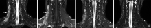
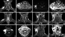
目的: 探讨MRI结合扩散加权成像(diffusion-weighted imaging,DWI)对颈部淋巴结结核药物治疗效果的预测价值。方法: 纳入2017年1月至2021年12月湖南省胸科医院收治住院进行标准化抗结核药物治疗的160例颈部淋巴结结核初治患者。收集患者MRI资料进行临床分型,根据复查结果将患者分为疗效良好组119例和疗效欠佳组41例。比较两组患者的临床分型和MRI影像学资料的差异。结果: 160例颈部淋巴结结核患者中结节增生型20例(12.50%),结节坏死型79例(49.38%),淋巴结周围炎型46例(28.75%),周围脓肿型15例(9.37%)。疗效良好组中结节增生型、结节坏死型、淋巴结周围炎型、周围脓肿型例数(构成比)分别为19(15.97%)、67(56.30%)、30(25.21%)、3(2.52%),而疗效欠佳组中对应各分型例数(构成比)分别为1(2.44%)、12(29.27%)、16(39.02%)、12(29.27%),两组之间差异有统计学意义(χ2=34.272,P<0.01);疗效良好组病变未突破包膜者占72.27%(86/119),明显高于疗效欠佳组(31.71%,13/41),差异有统计学意义(χ2=21.267,P<0.01)。表观扩散系数(apparent diffusion coefficient, ADC)图上为混杂信号且以高信号为主的患者96.3%(77/80)疗效良好,而ADC图为明显低信号的39例患者预后欠佳;疗效良好组的平均ADC值为(1.49±0.21)×10-3mm2/s,疗效欠佳组平均ADC值为(1.06±0.19)×10-3mm2/s,两组比较差异有统计学意义(t=11.576,P<0.01)。结论: MRI联合DWI对颈部淋巴结结核患者抗结核药物治疗的效果有一定的预测价值,病变突破淋巴结包膜或弥散受限多提示预后欠佳。

关键词: 结核,淋巴结, 磁共振成像, 弥散, 预测

Abstract:

Objective: To investigate the predictive value of MRI combined with diffusion-weighted imaging (DWI) on the effectiveness of drug therapy for cervical lymph node tuberculosis. Methods: A total of 160 cases of newly-treated patients with cervical lymph node tuberculosis who were admitted to Hunan Chest Hospital and received standardized anti-tuberculosis drug treatment from January 2017 to December 2021 were enrolled in the study.Their MRI data were collected for clinical classification. According to the re-examination results, the patients were divided into the good curative effect group (119 cases) and the poor curative effect group(41 cases).The differences of clinical classifications and MRI imaging data of the two groups were compared. Results: There were 20 cases (12.50%) with nodular hyperplasia, 79 cases (49.38%) with nodular necrosis, 46 cases (28.75%) with peri-lymph node inflammation, and 15 cases (9.37%) with peripheral abscess among 160 patients. The number (proportion) of nodular hyperplasia type, nodular necrosis type, peri-lymph node inflammation type and peripheral abscess type were 19 (15.97%), 67 (56.30%), 30 (25.21%) and 3 (2.52%) in the good curative effect group respectively, while the number (proportion) of corresponding subtypes were 1 (2.44%), 12 (29.27%), 16 (39.02%) and 12 (29.27%) in the poor curative effect group.There were statistical differences between the two groups (χ2=34.272, P<0.01). The lesions did not break through the capsule accounted for 72.27% (86/119) in the good curative effect group, which was significantly higher than that in the poor curative effect group (31.71%, 13/41), and the difference was statistically significant (χ2=21.267, P<0.01). The patients whose apparent diffusion coefficient (ADC) map showed mixed signals dominated by high signal (96.3%,77/80) usually got better curative effect, while the 39 cases whose ADC map showed obvious low signal had poor prognosis. The average ADC values of the good curative effect group and the poor curative effect group were (1.49±0.21)× 10-3mm2/s and (1.06±0.19)×10-3mm2/s respectively, and the difference was statistically significant (t=11.576, P<0.01). Conclusion: MRI combined with DWI had a certain predictive value for the effectiveness of anti-tuberculosis drug therapy in patients with cervical lymph node tuberculosis. Lesions that brook through the lymph node capsule or had limited diffusion were often indicative of poor prognosis.

Key words: Tuberculosis,lymph node, Diffusion magnetic resonance imaging, Forcasting

中图分类号: