Email Alert | RSS 帮助

中国防痨杂志 ›› 2026, Vol. 48 ›› Issue (2): 188-196.doi: 10.19982/j.issn.1000-6621.20250302
付旭文, 何维曜臻, 李翔( )
)
收稿日期:2025-07-25
出版日期:2026-02-10
发布日期:2026-02-03
通信作者:
李翔
E-mail:15887138804@163.com
基金资助:
Fu Xuwen, He Weiyaozhen, Li Xiang()
Received:2025-07-25
Online:2026-02-10
Published:2026-02-03
Contact:
Li Xiang
E-mail:15887138804@163.com
Supported by:摘要:
男性生殖系统结核(male genital tuberculosis,MGTB)可发生在男性生殖系统的各个器官,MGTB临床症状不典型或无临床症状,容易造成漏诊和误诊,随着医学影像学技术的发展,影像学表现在MGTB的诊断和鉴别诊断中有着重要的作用。作者旨在系统归纳男性生殖系统各个器官结核的超声、计算机断层扫描(computed tomography,CT)和磁共振成像(magnetic resonance imaging,MRI)的表现,提高对MGTB的认识和临床诊断能力。
中图分类号:
付旭文, 何维曜臻, 李翔. 男性生殖系统结核影像学表现[J]. 中国防痨杂志, 2026, 48(2): 188-196. doi: 10.19982/j.issn.1000-6621.20250302
Fu Xuwen, He Weiyaozhen, Li Xiang. Imaging manifestations of male reproductive system tuberculosis[J]. Chinese Journal of Antituberculosis, 2026, 48(2): 188-196. doi: 10.19982/j.issn.1000-6621.20250302
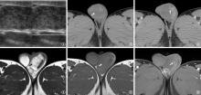
图1~6
患者,男性,24岁,睾丸结核。图1 超声提示左侧睾丸增大,弥漫性实质回声不均;图2 CT平扫提示左侧睾丸增大(白箭);图3 CT增强提示左侧睾丸外侧壁结节状强化(白箭);图4 MRI平扫 T2WI提示左侧睾丸增大,呈混杂信号(白箭);图5 MRI平扫T1WI提示左侧睾丸增大,呈稍低信号(白箭);图6 MRI T1WI增强提示左侧睾丸外侧壁结节状强化(白箭),另可见附睾环状强化(空心白箭)

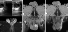
图7~12
患者,男性,24岁,与图1~6为同一患者,附睾结核。图7 超声提示左侧附睾尾部回声欠均结节(白箭);图8 CT平扫提示左侧附睾头部及尾部结节,头部结节较大(白箭);图9 CT增强提示左侧附睾头部结节不均匀强化(白箭);图10 MRI T2WI脂肪抑制相提示左侧附睾头部增大,信号不均匀(白箭);图11 MRI T1WI增强扫描附睾头部结节不均匀强化(白箭);图12 MRI T1WI增强扫描显示左侧精索明显强化(白箭)


图13
患者,男性,32岁,输精管结核。CT增强扫描门静脉期提示左侧输精管明显扩张,局部增厚管壁明显强化(白箭) 图14 患者,男性,23岁,输精管结核。CT增强扫描门静脉期提示双侧输精管明显扩张,增厚管壁明显强化,周围脂肪间隙模糊(白箭),另可见膀胱壁明显增厚
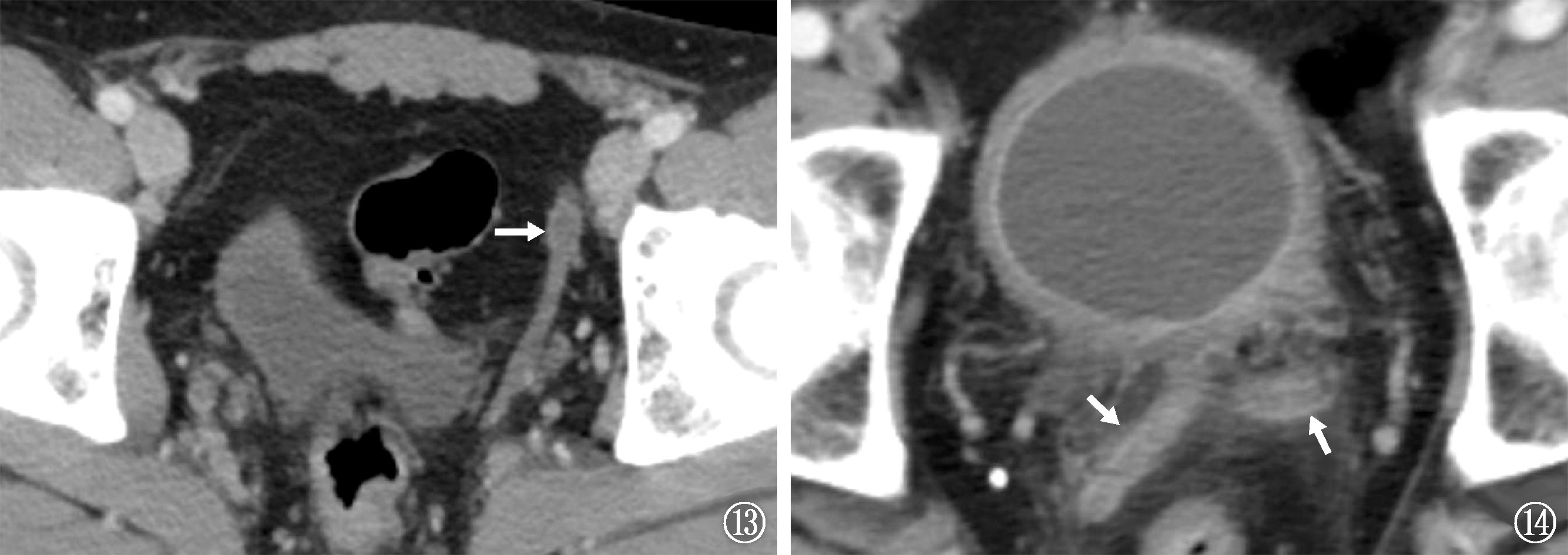
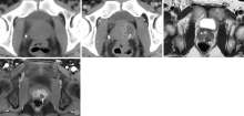
图15~16
患者,男性,22岁,前列腺结核。图15 CT平扫未见前列腺明显异常;图16 CT增强扫描可见前列腺左侧类圆形低密度影,边缘明显强化(白箭) 图17~18 患者,男性,32岁,前列腺结核。图17 MRI T2WI提示前列腺内低信号结节影(白箭);图18 MRI T1WI增强扫描提示前列腺结节周围环状强化(白箭)

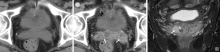
图19~21
患者,男性,58岁,精囊腺结核。图19 CT平扫提示精囊腺呈等密度;图20 CT增强提示双侧精囊腺不均匀强化(白箭);图21 MRI T2WI提示精囊腺内高信号精囊液信号减低(白箭),可见低信号结核性结节(空心白箭)

| [1] | 舒薇, 刘宇红. 笃志创新躬行致远:世界卫生组织《2023年全球结核病报告》结核病科学研究章节解读. 中国防痨杂志, 2024, 46(6):613-617. doi:10.19982/j.issn.1000-6621.20240159. |
| [2] | Merchant S, Bharati A, Merchant N. Tuberculosis of the genitourinary system-Urinary tract tuberculosis: Renal tuberculosis-Part I. Indian J Radiol Imaging, 2013, 23(1):46-63. doi:10.4103/0971-3026.113615. |
| [3] |
Kulchavenya E, Kim C, Bulanova O, et al. Male genital tuberculosis: epidemiology and diagnostic. World J Urol, 2012, 30(1):15-21. doi:10.1007/s00345-011-0695-y.
URL pmid: 21604018 |
| [4] |
Cherif S, Yoganathan K, Banks T, et al. Unmasking immune reconstitution inflammatory syndrome: a report of tuberculous epididymo-orchitis mimicking a testicular tumour in a Caucasian AIDS patient. Int J STD AIDS, 2017, 28(1):100-103. doi:10.1177/0956462416652536.
URL pmid: 27222288 |
| [5] |
Ramachandran A, Das CJ, Razik A. Male genital tract tuberculosis: A comprehensive review of imaging findings and differential diagnosis. Abdom Radiol (NY), 2021, 46(4):1677-1686. doi:10.1007/s00261-020-02811-0.
URL pmid: 33044653 |
| [6] | Farias LABG, Vitoriano FES, Ribeiro CKB, et al. Two cases of testicular tuberculosis in HIV patients with distinct clinical presentations. Trop Doct, 2024, 54(2):176-178. doi:10.1177/00494755231220400. |
| [7] | Sanches BDA, Tamarindo GH, Maldarine JDS, et al. Telocytes of the male urogenital system: Interrelationships, possible functions, and pathological implications. Cell Biol Int, 2021, 45(8):1613-1623. doi:10.1002/cbin.11612. |
| [8] |
Rodriguez-Takeuchi SY, Renjifo ME, Medina FJ. Extrapulmonary Tuberculosis: Pathophysiology and Imaging Findings. Radiographics, 2019, 39(7):2023-2037. doi:10.1148/rg.2019190109.
URL pmid: 31697616 |
| [9] |
Kimura M, Araoka H, Baba H, et al. First case of sexually transmitted asymptomatic female genital tuberculosis from spousal epididymal tuberculosis diagnosed by active screening. Int J Infect Dis, 2018, 73:60-62. doi:10.1016/j.ijid.2018.05.021.
URL pmid: 29879525 |
| [10] | Fu X, Bi Y, Qi M, et al. Computed tomography imaging analysis of hematogenous disseminated pulmonary tuberculosis cases combined with prostate tuberculosis. BMC Med Imaging, 2025, 25(1):212. doi:10.1186/s12880-025-01753-7. |
| [11] | 李翔, 马仲序, 付旭文, 等. 56例附睾结核CT影像表现分析. 中国防痨杂志, 2022, 44(10):1100-1103. doi:10.19982/j.issn.1000-6621.20220208. |
| [12] | Kumar R. Reproductive tract tuberculosis and male infertility. Indian J Urol, 2008, 24(3):392-395. doi:10.4103/0970-1591.42624. |
| [13] | Lachkar S, Diouri M, Ibrahimi A, et al. Isolated testicular tuberculosis: A case report. Urol Case Rep, 2024, 57:102869. doi:10.1016/j.eucr.2024.102869. |
| [14] | Harya SA, Nhungo CJ, Lori JM, et al. Isolated testicular tuberculosis mimicking testicular malignancy in a 45-year-old male treated at a tertiary hospital. Case report and literature review. Int J Surg Case Rep, 2024, 117:109511. doi:10.1016/j.ijscr.2024.109511. |
| [15] | Zhang X, Su D, Ni T, et al. Contrast-enhanced ultrasound for rare testicular tuberculosis. IDCases, 2024, 38:e2089. doi:10.1016/j.idcr.2024.e02089. |
| [16] | 杨博文, 艾洁尔古丽·麦合苏木, 安信, 等. 喀什地区睾丸/附睾结核10年单中心诊疗经验. 临床泌尿外科杂志, 2024, 39(9):832-838. doi:10.13201/j.issn.1001-1420.2024.09.016. |
| [17] | 张秀丽, 苏航, 陈争光, 等. 二维超声联合微血管血流成像对睾丸结核的诊断价值. 郑州大学学报(医学版), 2024, 59(5):715-718. doi:10.13705/j.issn.1671-6825.2024.04.061. |
| [18] | 边巴次仁, 次旦旺久, 任翠, 等. 睾丸肿瘤与结核的CT表现分析. 国际泌尿系统杂志, 2025, 45(1):77-80. doi:10.3760/cma.j.cn431460-20230712-00019. |
| [19] | 侯民羊, 苟杰. 睾丸结核的CT诊断. 中国医学影像学杂志, 2010, 18(4):381-383. doi:10.3969/j.issn.1005-5185.2010.04.016. |
| [20] | 闫瑞芳, 李学坤, 张改云, 等. MRI诊断附睾睾丸结核. 中国医学影像技术, 2020, 36(10):1517-1520. doi:10.13929/j.issn.1003-3289.2020.10.020. |
| [21] | 王唯伟, 寻静, 项梅, 等. 睾丸结核的MRI表现1例. 医学影像学杂志, 2021, 31(10):1780, 1784. doi:10.3760/cma.j.cmcr.2022.e05195. |
| [22] |
Yang DM, Kim HC, Kim SW, et al. Sonographic findings of tuberculous vasitis. J Ultrasound Med, 2014, 33(5):913-916. doi:10.7863/ultra.33.5.913.
URL pmid: 24764347 |
| [23] | 陈燕玲, 吴迪, 陈秀平, 等. 肺结核及并发肺外结核患者淋巴细胞亚群变化的研究及其临床意义. 结核与肺部疾病杂志, 2024, 5(4):294-304. doi:10.19983/j.issn.2096-8493.2024042. |
| [24] |
Cek M, Lenk S, Naber KG, et al. EAU guidelines for the management of genitourinary tuberculosis. Eur Urol, 2005, 48(3):353-362. doi:10.1016/j.eururo.2005.03.008.
URL pmid: 15982799 |
| [25] | 成瑞明, 刘罩明, 张铭, 等. 附睾结核的超声分型及其应用价值. 中华超声影像学杂志, 2016, 25(2):163-167. doi:10.3760/cma.j.issn.1004-4477.2016.02.018. |
| [26] | 杨高怡, 张文智, 蒋慧青, 等. 附睾结核超声造影表现分析. 中国超声医学杂志, 2015, 31(11):1048-1050. |
| [27] | 程洁, 邱小伟, 尤四峰, 等. CT及MRI对附睾结核诊断的特征性分析. 实用放射学杂志, 2023, 39(4):615-617, 638. doi:10.3969/j.issn.1002-1671.2023.04.024. |
| [28] | Man J, Cao L, Dong Z, et al. Diagnosis and treatment of epididymal tuberculosis: a review of 47 cases. PeerJ, 2020, 8:e8291. doi:10.7717/peerj.8291. |
| [29] |
Yang B, Zhou R, Wang X, et al. Magnetic resonance imaging features of epididymal and/or testicular tuberculosis: a case series. BMC Med Imaging, 2025, 25(1):157. doi:10.1186/s12880-025-01699-w.
URL pmid: 40355825 |
| [30] | Jung YY, Kim JK, Cho K. Genitourinary tuberculosis: comprehensive cross-sectional imaging. AJR Am J Roentgenol, 2005, 184(1):143-150. doi:10.2214/ajr.184.1.01840143. |
| [31] | Michaelides M, Sotiriadis C, Konstantinou D, et al. Tuberculous orchitis US and MRI findings. Correlation with histopathological findings. Hippokratia, 2010, 14(4):297-299. |
| [32] | 张文智, 杨高怡, 王大力, 等. 输精管结核的超声表现分析. 中国超声医学杂志, 2014, 30(8):737-739. doi:10.3969/j.issn.1002-0101.2014.08.022. |
| [33] | Jing J, Zhuang H, Luo Y, et al. Vas deferens sonographic appearances of tuberculosis lesions of 19 cases of male genital systemic tuberculosis. Medicine (Baltimore), 2019, 98(11):e14843. doi:10.1097/MD.0000000000014843. |
| [34] |
Qi M, Zhang L, Gan W, et al. The Imaging Features and Diagnostic Value of Computerised Tomography in Seminal Duct Tuberculosis. J Multidiscip Healthc, 2023, 16:1395-1402. doi:10.2147/JMDH.S401660.
URL pmid: 37223245 |
| [35] |
Pak BK, Kang DM, Kim SH. Vas Deferens Abscess Rupture: A Case Report. J Belg Soc Radiol, 2022, 106(1):70. doi:10.5334/jbsr.2840.
URL pmid: 35974891 |
| [36] | Ye D, Liu X, Yang Y, et al. Case Report: Epididymal NK/T-cell lymphoma and adrenal diffuse large B-cell lymphoma are misdiagnosed as tuberculosis: two case reports and literature review. Front Oncol, 2025, 15:1529049. doi:10.3389/fonc.2025.1529049. |
| [37] |
Yadav S, Singh P, Hemal A, et al. Genital tuberculosis: current status of diagnosis and management. Transl Androl Urol, 2017, 6(2):222-233. doi:10.21037/tau.2016.12.04.
URL pmid: 28540230 |
| [38] | Mishra KG, Ahmad A, Singh G, et al. Tuberculosis of the prostate gland masquerading prostate cancer; five cases expe-rience at IGIMS. Urol Ann, 2019, 11(4):389-392. doi:10.4103/UA.UA_119_18. |
| [39] | 卫宜锐, 王浩, 杨朴深, 等. 前列腺结核与前列腺癌临床误诊分析. 临床误诊误治, 2024, 37(9):1-5. doi:10.3969/j.issn.1002-3429.2024.09.001. |
| [40] | Baral S, Chhetri RK, Gyawali M, et al. Prostate tuberculosis complicated by huge prostatic abscess: A rare case report from Nepal. Int J Surg Case Rep, 2020, 77:152-156. doi:10.1016/j.ijscr.2020.10.045. |
| [41] | Yang G, Ruan L. Imaging findings of prostate tuberculosis by transrectal contrast-enhanced ultrasound and comparison with 2D ultrasound and pathology. Br J Radiol, 2022, 95(1129):20210713. doi:10.1259/bjr.20210713. |
| [42] | Li Y, Dan S, Yang F, et al. Prostate tuberculosis mimicking prostate cancer: Case report and literature review. Medicine (Baltimore), 2023, 102(47):e36172. doi:10.1097/MD.0000000000036172. |
| [43] |
Joshi PV, Shewalkar B, George T, et al. The Great Mimicker-Tuberculosis Involving Prostate and Vertebrae Posing as Metastatic Prostate Carcinoma on FDG PET/CT. Clin Nucl Med, 2020, 45(3):206-208. doi:10.1097/RLU.0000000000002789.
URL pmid: 31652162 |
| [44] |
Cheng Y, Huang L, Zhang X, et al. Multiparametric Magnetic Resonance Imaging Characteristics of Prostate Tuberculosis. Korean J Radiol, 2015, 16(4):846-852. doi:10.3348/kjr.2015.16.4.846.
URL pmid: 26175584 |
| [45] | Bour L, Schull A, Delongchamps N, et al. Multiparametric MRI features of granulomatous prostatitis and tubercular prostate abscess. Diagn Interv Imaging, 2013, 94(1):84-90. doi:10.1016/j.diii.2012.09.001. |
| [46] | 王向东, 王亚丽, 高跃丽. 前列腺结核的磁共振弥散加权成像及超声表现. 新发传染病电子杂志, 2020, 5(3):188-190. doi:10.19871/j.cnki.xfcrbzz.2020.03.010. |
| [47] |
Suzuki T, Takeuchi M, Naiki T, et al. MRI findings of granu-lomatous prostatitis developing after intravesical Bacillus Calmette-Guerin therapy. Clin Radiol, 2013, 68(6):595-599. doi:10.1016/j.crad.2012.12.005.
URL pmid: 23384503 |
| [48] | Engin G, Acunas B, Acunas G, et al. Imaging of extrapulmonary tuberculosis. Radiographics, 2000, 20(2):471-488,529-530, 532. doi:10.1148/radiographics.20.2.g00mc07471.quiz. |
| [49] |
Chen Y, Liu M, Guo Y. Proton magnetic resonance spectroscopy in prostate tuberculosis. Urology, 2010, 75(5):1065-1066. doi:10.1016/j.urology.2009.06.069.
URL pmid: 19781746 |
| [50] | William Y, Sugiono M, Diana Prasetiyo P, et al. A Case Report and Literature Review of Prostatic Tuberculosis Masquerading as Prostate Cancer: A Diagnostic Challenge in a Tuberculosis-Endemic Region. Trop Med Infect Dis, 2025, 10(5):145. doi:10.3390/tropicalmed10050145. |
| [51] | Lee S, Oh YT, Kim HM, et al. Imaging Patterns of Bacillus Calmette-Guerin-Related Granulomatous Prostatitis Based on Multiparametric MRI. Korean J Radiol, 2022, 23(1):60-67. doi:10.3348/kjr.2020.1369. |
| [52] | 刘盼丽, 欧舒斐, 欧陕兴, 等. 35例腹盆腔结核的CT诊断分析. 结核病与肺部健康杂志, 2016, 5(3):180-185. doi:10.3969/j.issn.2095-3755.2016.03.005. |
| [53] |
Reddy MN, Verma S. Lesions of the Seminal Vesicles and their MRI Characteristics. J Clin Imaging Sci, 2014, 4:61. doi:10.4103/2156-7514.143734.
URL pmid: 25396077 |
| [54] | Ocal O, Karaosmanoglu AD, Karcaaltincaba M, et al. Imaging findings of congenital anomalies of seminal vesicles. Pol J Radiol, 2019, 84:e25-e31. doi:10.5114/pjr.2019.82711. |
| [55] | 李翔, 付旭文, 魏佳璐, 等. 精囊腺结核的MRI影像特征分析——附7例精囊腺结核MRI影像表现. 中国防痨杂志, 2023, 45(5):526-530. doi:10.19982/j.issn.1000-6621.20220538. |
| [56] |
Gan W, Bi Y, Fu X, et al. Magnetic Resonance Imaging Manifestations in 13 Cases of Seminal Vesicle Tuberculosis. Infect Drug Resist, 2023, 16:6871-6879. doi:10.2147/IDR.S427561.
URL pmid: 37908784 |
| [57] | Venyo AK. Tuberculosis of the Penis: A Review of the Litera-ture. Scientifica (Cairo), 2015, 2015:601624. doi:10.1155/2015/601624. |
| [58] | Kawashima A, Sandler CM, Wasserman NF, et al. Imaging of urethral disease: a pictorial review. Radiographics, 2004,24 Suppl 1:S195-S216. doi:10.1148/rg.24si045504. |
| [59] | Mayilvaganan KR, Naren Satya Srinivas M, Reddy VN, et al. Tuberculosis Penis with ‘Watering Can Penis’ Appearance: Report of a Rare Case with Retrograde Urethrography and Voiding Cystourethrography Findings. Pol J Radiol, 2016, 81:454-457. doi:10.12659/PJR.897943. |
| [60] | Gangalakshmi C, Sankaramahalingam. Tuberculosis of Glans Penis-A Rare Presentation. J Clin Diagn Res, 2016, 10(12):PD5-PD6. doi:10.7860/JCDR/2016/19163.9057. |
| [61] | Naeem M, Zulfiqar M, Siddiqui MA, et al. Imaging Manifestations of Genitourinary Tuberculosis. Radiographics, 2021, 41(4):1123-1143. doi:10.1148/rg.2021200154. |
| [62] | 胡玉敬, 边艳珠. 18F-氟代脱氧葡萄糖正电子发射计算机体层摄影-CT在结核病诊治中的应用价值. 中国防痨杂志, 2023, 45(3):318-322. doi:10.19982/j.issn.1000-6621.20220500. |
| [63] | 李红岩. 肺外结核18F-FDGPET/CT影像表现分析及临床价值探讨. 武汉:华中科技大学, 2019. |
| [64] |
Ran P, Liang X, Zhang Y, et al. FDG PET/CT in a Case of Bilateral Tuberculous Epididymo-orchitis. Clin Nucl Med, 2019, 44(9):757-760. doi:10.1097/RLU.0000000000002606.
URL pmid: 31107741 |
| [65] | Bai M, Yu Q, Yuan L, et al. Prostate tuberculosis mimicking malignancy on 18F-FDG PET/CT in a patient with diffuse large B-cell lymphoma: A case report. Medicine (Baltimore), 2024, 103(22):e38296. doi:10.1097/MD.0000000000038296. |
| [66] | 莫纯威, 徐万帮, 唐刚华. 靶向成纤维细胞活化蛋白PET显像剂的研究进展. 国际放射医学核医学杂志, 2022, 46(12):735-741. doi:10.3760/cma.j.cn121381-202203029-00251. |
| [67] | Yu Q, Xie Q, Zhu X, et al. Increase 68 Ga-FAPI Uptake in Urogenital Tuberculosis. Clin Nucl Med, 2025, 50(1):98-100. doi:10.1097/RLU.0000000000005418. |
| [68] | 梁逸宁, 侯代伦. 结核病影像学诊断年度进展2024. 中华结核和呼吸杂志, 2025, 48(2):175-180. doi:10.3760/cma.j.cn112147-20241031-00650. |
| [69] | 李汶翰, 杨静, 李春华. 人工智能在肺结核影像诊断及耐药性预测中的研究进展. 中国防痨杂志, 2024, 46(9):1098-1103. doi:10.19982/j.issn.1000-6621.20240123. |
| [70] | 王雪静, 黄智, 曾嘉宾, 等. 新一代人工智能在前列腺癌临床诊断的研究进展. 临床外科杂志, 2025, 33(2):118-122. doi:10.3969/j.issn.1005-6483.20242018. |
| [1] | 宁锋钢, 房坤, 王珏, 吕岩, 贺伟, 侯代伦. 磁共振三维颅脑容积成像增强扫描在颅内结核影像诊断中的应用[J]. 中国防痨杂志, 2025, 47(11): 1489-1494. |
| [2] | 胡婷. 超声检查在结核性胸腔积液诊断中的应用价值[J]. 中国防痨杂志, 2024, 46(S2): 55-57. |
| [3] | 杨敏, 王新文. 间质性肺病影像学诊断研究进展[J]. 中国防痨杂志, 2024, 46(S1): 316-318. |
| [4] | 李翔, 浦英, 付旭文, 杞敏, 魏佳璐, 寸新华. 误诊为脊柱结核的真菌性脊柱炎临床及影像特征分析[J]. 中国防痨杂志, 2024, 46(9): 1109-1114. |
| [5] | 胡蔷, 金林原, 周晓晖, 张倩榕, 邓雨杭, 唐贤朋, 周海依, 张敏. 超声检查对儿童胸部结核诊断及治疗效果评估的价值[J]. 中国防痨杂志, 2024, 46(11): 1350-1355. |
| [6] | 刘馨, 于千会. 不同b值弥散加权成像对肺结核分型诊断及对耐多药风险评估的预测[J]. 中国防痨杂志, 2024, 46(11): 1356-1364. |
| [7] | 曹兵生, 张更臣, 傅莉媛, 王金山. 超声引导下经皮穿刺活检在髋关节结核诊断中的应用价值[J]. 中国防痨杂志, 2023, 45(7): 669-672. |
| [8] | 温立旻, 侯代伦. 肺结核合并肺癌影像学评估方法现状及进展[J]. 中国防痨杂志, 2023, 45(6): 620-624. |
| [9] | 李翔, 杞敏, 姜建杰, 魏佳璐, 付旭文, 李海雯, 张乐. 颅内结核合并急性脑梗死的危险因素分析[J]. 中国防痨杂志, 2023, 45(5): 499-506. |
| [10] | 李翔, 付旭文, 魏佳璐, 干玮, 李海雯, 代敏, 和进堂. 精囊腺结核的MRI影像特征分析——附7例精囊腺结核MRI影像表现[J]. 中国防痨杂志, 2023, 45(5): 526-530. |
| [11] | 胡玉敬, 边艳珠. 18F-氟代脱氧葡萄糖正电子发射计算机体层摄影-CT在结核病诊治中的应用价值[J]. 中国防痨杂志, 2023, 45(3): 318-322. |
| [12] | 杜艳妮, 薛明, 关春爽, 邢玉雪, 陈步东, 谢汝明. 艾滋病合并肺结核患者胸部CT不典型影像表现分析[J]. 中国防痨杂志, 2023, 45(2): 159-164. |
| [13] | 李翔, 魏佳璐, 付旭文, 杞敏, 干玮, 寸新华. 脊髓结核MRI影像表现及分型[J]. 中国防痨杂志, 2023, 45(12): 1186-1192. |
| [14] | 李翔, 付旭文, 干玮, 魏佳璐, 杞敏, 寸新华. MRI表现为椎体内弥漫分布结节的脊柱结核二例[J]. 中国防痨杂志, 2023, 45(10): 1007-1010. |
| [15] | 中华医学会结核病学分会超声专业委员会, 中国医师协会介入医师分会超声介入专业委员会单位. 结核性胸膜炎超声诊断、分型及介入治疗专家共识(2022年版)[J]. 中国防痨杂志, 2022, 44(9): 880-897. |
| 阅读次数 | ||||||
|
全文 |
|
|||||
|
摘要 |
|
|||||
 京公网安备11010202007215号
ip访问总数: ip当日访问总数: 当前在线人数:
京公网安备11010202007215号
ip访问总数: ip当日访问总数: 当前在线人数:
 本作品遵循Creative Commons Attribution 3.0 License授权许可