Email Alert | RSS    帮助

中国防痨杂志 ›› 2018, Vol. 40 ›› Issue (11): 1152-1158.doi: 10.3969/j.issn.1000-6621.2018.11.002

• 论著 • 上一篇    下一篇

肺结核患者肺组织微生物组特征研究

王宇轩,王冲,董宇杰,杜伟丽,宋婧,刘子臣,李琨,刘树库(),车南颖()   

  1. 101149 首都医科大学附属北京胸科医院 北京市结核病胸部肿瘤研究所病理科 耐药结核病研究北京市重点实验室(王宇轩、董宇杰、杜伟丽、宋婧、刘子臣、李琨、车南颖),微创诊疗中心(王冲、刘树库);
  • 收稿日期:2018-08-22 出版日期:2018-11-10 发布日期:2018-12-04
  • 通信作者: 刘树库,车南颖 E-mail:liushuku@aliyun.com;cheny0448@163.com
  • 基金资助:
    北京市科技计划医药协同科技创新研究(Z181100001918027);北京市科学技术委员会重点项目(D181100000418003);北京市医院管理局登峰计划(DFL20151501,DFL20181601);北京市科学技术委员会重点项目(D14117005214003);北京市卫生与健康科技成果和适宜技术推广项目(2018-TG-41)

Characteristics of the microbiome in lung tissue of patients with pulmonary tuberculosis

WANG Yu-xuan,WANG Chong,DONG Yu-jie,DU Wei-li,SONG Jing,LIU Zi-chen,LI Kun,LIU Shu-ku(),CHE Nan-ying()   

  1. Department of Pathology, Beijing Key Laboratory for Drug Resistant Tuberculosis Research, Beijing Chest Hospital, Capital Medical University, Beijing Tuberculosis and Thoracic Tumor Research Institute, Beijing 101149, China
  • Received:2018-08-22 Online:2018-11-10 Published:2018-12-04
  • Contact: Shu-ku LIU,Nan-ying CHE E-mail:liushuku@aliyun.com;cheny0448@163.com

摘要:

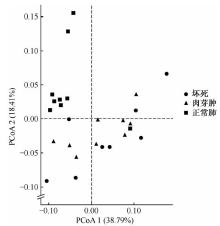
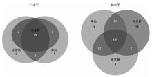
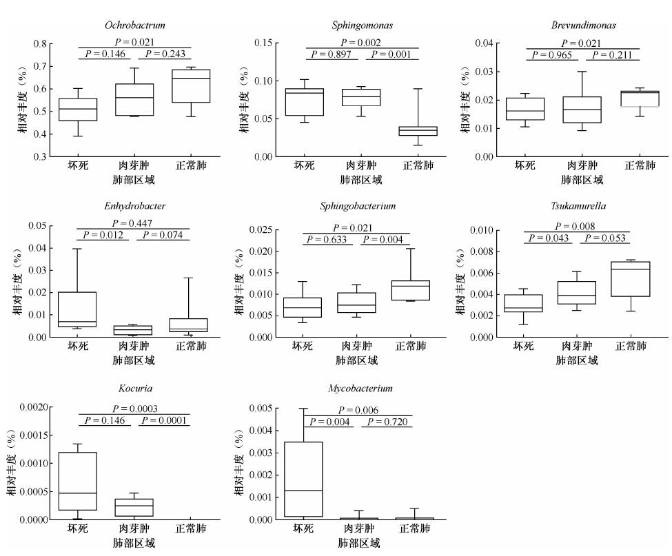
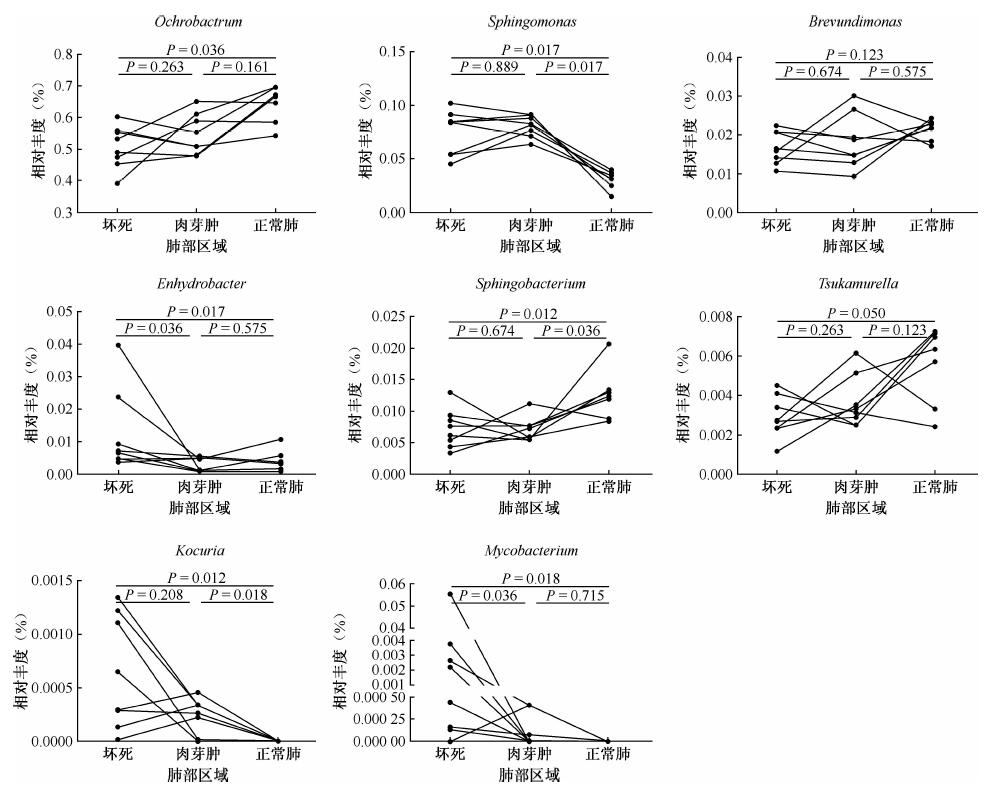
目的 探讨肺结核患者肺部病灶坏死区、肉芽肿区和正常肺区的微生物组基本特征。方法 选取2016—2017年首都医科大学附属北京胸科医院10例临床及病理确诊为肺结核患者的福尔马林固定石蜡包埋(FFPE)的组织标本,通过显微切割技术分别获得FFPE组织的坏死区、肉芽肿区和正常肺区。采用高通量测序的方法,检测其微生物组特征。结果 肺结核患者肺部切除标本中主要的微生物类群(门水平,相对丰度>1%)有ProteobacteriaActinobacteriaFirmicutesTM7Bacteroidetes。在门水平上,坏死区、肉芽肿区和正常肺区的微生物群落组成未见差异。在属水平上,主要有7个类群(相对丰度>1%):坏死区、肉芽肿区、正常肺区相对丰度Ochrobactrum分别为(50.70±6.76)%、(56.18±7.50)%、(61.31±7.95)%;Sphingomonas分别为(7.50±2.07)%、(7.69±1.29)%、(3.81±2.08)%;Corynebacterium分别为(5.39±2.39)%、(5.23±1.39)%、(7.43±3.56)%;Brevundimonas分别为(1.67±0.42)%、(1.75±0.67)%、(2.09±0.34)%;Brevibacterium分别为(2.38±2.23)%、(2.60±2.60)%、(2.29±0.64)%;Sphingobacterium分别为(0.72±0.31)%、(0.78±0.26)%、(1.20±0.38)%和Enhydrobacter分别为(1.25±1.28)%、(0.31±0.19)%、(0.67±0.80)%。基于Bray-Curtis距离的Adonis分析结果表明,在属水平上,坏死区和正常肺区,肉芽肿区和正常肺区中微生物群落组成差异有统计学意义(F值分别为3.94和4.76,P值分别为0.005和0.002)。Mycobacterium的相对丰度非常低,主要存在于肺结核病灶的坏死区[(0.81±1.92)%]。配对样品检验结果表明Mycobacterium的相对丰度在坏死区[(0.81±1.92)%]和肉芽肿区[(0.01±0.01)%]、坏死区和正常肺区[(0.01±0.02)%]中差异有统计学意义(Z值分别为2.10和-2.37,P值分别为0.036和0.018)。结论 结核病患者肺部自坏死区、肉芽肿区到正常肺的组织变化中,SphingomonasEnhydrobacterKocuriaMycobacterium的相对丰度整体呈下降趋势,而OchrobactrumSphingobacterium的相对丰度总体呈上升趋势。肺结核患者肺部菌群的变化特征可能与肺结核的发病有重要关系。

关键词: 结核, 肺, 活组织检查, 石蜡包埋, 高通量核苷酸测序, 微生物组

Abstract:

Objective To explore the characteristics of the microbiome in the lung including necrosis, granuloma and normal lung areas of patients with pulmonary tuberculosis.Methods A total of 10 matched-pairs formalin-fixed and paraffin-embedded (FFPE) specimens were selected from 10 patients who were diagnosed as pulmonary tuberculosis in Beijing Chest Hospital from 2016 to 2017. The tissues were divided into necrotic, granuloma and normal lung areas using Leica Microsystems. Characteristics of microbiome was investigated using next-generation sequencing (NGS) technology.Results The major phyla in lung samples of pulmonary tuberculosis patients (relative abundance >1%) were Proteobacteria, Actinobacteria, Firmicutes, TM7 and Bacteroidetes. Adonis analysis based on bray_curtis distance showed that microbial communities were not significantly different at the phylum level. In genus level, seven mainly groups were detected (relative abundance >1%): Ochrobactrum (relative abundance of necrosis, granuloma and normal lung areas: (50.70±6.76)%, (56.18±7.50)%, (61.31±7.95)%), Sphingomonas ((7.50±2.07)%, (7.69±1.29)%, (3.81±2.08)%), Corynebacterium ((5.39±2.39)%, (5.23±1.39)%, (7.43±3.56)%), Brevundimonas ((1.67±0.42)%, (1.75±0.67)%, (2.09±0.34)%), Brevibacterium ((2.38±2.23)%, (2.60±2.60)%, (2.29±0.64)%), Sphingobacterium ((0.72±0.31)%, (0.78±0.26)%, (1.20±0.38)%) and Enhydrobacter ((1.25±1.28)%, (0.31±0.19)%, (0.67±0.80)%). Adonis analysis based on Bray-Curtis distance showed that the microbial community in normal area was significantly different from those in necrotic area (F=3.94, P=0.005) and granulomatous area (F=4.76, P=0.002). The relative abundance of Mycobacterium was very low and mainly existed in the necrotic areas ((0.81±1.92)%). Wilcoxon matched-pairs signed-ranks test showed that relative abundances of Mycobacterium were significantly different between necrotic and granulomatous areas ((0.81±1.92)% vs (0.01±0.01)%; Z=2.10, P=0.036), necrotic areas and normal lung areas ((0.81±1.92)% vs (0.01±0.02)%; Z=-2.37, P=0.018).Conclusion The relative abundances of Sphingomonas, Enhydrobacter, Kocuria and Mycobacterium decreased gradually from necrotic area, granuloma area to normal lung area, while the relative abundance of Ochrobactrum and Sphingobacterium showed the reverse trend. The characteristics of microbiome in lungs from pulmonary tuberculosis patients might be related to the changes of histomorphology in pulmonary.

Key words: Tuberculosis, pulmonary, Biopsy, Paraffin embedding, High-throughput nucleotide sequencing, Microbiome