Email Alert | RSS 帮助

中国防痨杂志 ›› 2023, Vol. 45 ›› Issue (5): 446-453.doi: 10.19982/j.issn.1000-6621.20220535
李姗姗1, 王玉峰2, 舒薇3, 逄宇1( )
)
收稿日期:2023-01-19
出版日期:2023-05-10
发布日期:2023-04-25
通信作者:
逄宇
E-mail:pangyupound@163.com
基金资助:
Li Shanshan1, Wang Yufeng2, Shu Wei3, Pang Yu1()
Received:2023-01-19
Online:2023-05-10
Published:2023-04-25
Contact:
Pang Yu
E-mail:pangyupound@163.com
Supported by:摘要:
结核病的精准防控需要快速、准确的实验室诊断技术。自1882年罗伯特·科赫发现结核分枝杆菌以来,结核病诊断技术经历了从传统的病原学诊断到免疫学和分子生物学诊断的巨大飞跃,然而这种诊断技术的发展仍然与实现2035年终止结核病的宏伟目标存在一定差距。作者在梳理既往结核病诊断技术研发进展的基础上,围绕结核病防控的核心诊断需求,探究未来实验室诊断的重要发展管线。
中图分类号:
李姗姗, 王玉峰, 舒薇, 逄宇. 结核病实验室诊断技术研发新进展[J]. 中国防痨杂志, 2023, 45(5): 446-453. doi: 10.19982/j.issn.1000-6621.20220535
Li Shanshan, Wang Yufeng, Shu Wei, Pang Yu. Progress and reflections on development of laboratory diagnostic technology for tuberculosis[J]. Chinese Journal of Antituberculosis, 2023, 45(5): 446-453. doi: 10.19982/j.issn.1000-6621.20220535
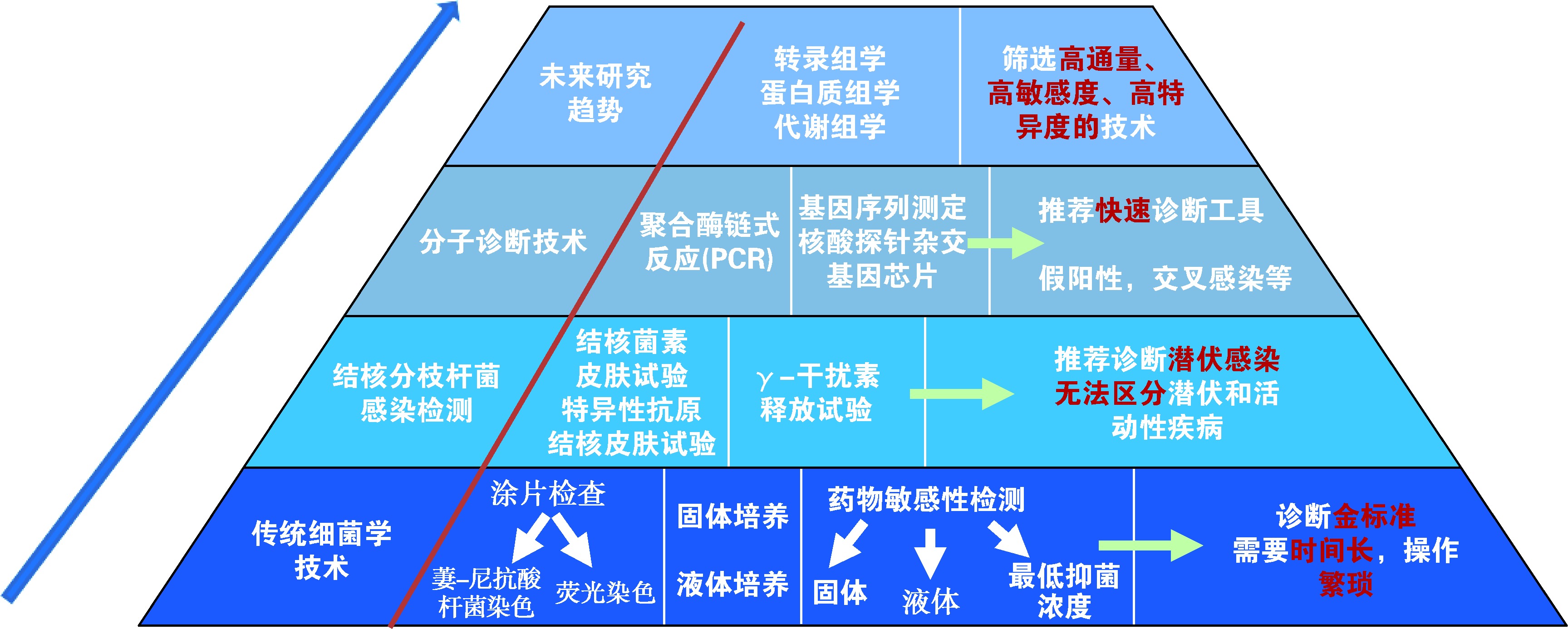
图1
结核病实验室诊断技术研发管线
| [1] | World Health Organization.Global tuberculosis report 2022. Geneva: World Health Organization, 2022. |
| [2] | World Health Organization. WHO operational handbook on tuberculosis. Module 3: diagnosis-rapid diagnostics for tuberculosis detention, 2021 update. Geneva: World Health Organi-zation, 2021. |
| [3] |
逄宇, 王玉峰, 高兴辉, 等. 结核病实验室检测产品和技术应用进展. 中国临床新医学, 2021, 14(1):23-34. doi:10.3969/j.issn.1674-3806.2021.01.05.
doi: 10.3969/j.issn.1674-3806.2021.01.05 |
| [4] | 中华人民共和国国家卫生和计划生育委员会.WS 288—2017 肺结核诊断. 2017-11-09. |
| [5] |
Azman AS, Golub JE, Dowdy DW. How much is tuberculosis screening worth? Estimating the value of active case finding for tuberculosis in South Africa, China, and India. BMC Med, 2014, 12:216. doi:10.1186/s12916-014-0216-0.
doi: 10.1186/s12916-014-0216-0 pmid: 25358459 |
| [6] |
Chen JO, Qiu YB, Rueda ZV, et al. Role of community-based active case finding in screening tuberculosis in Yunnan province of China. Infect Dis Poverty, 2020, 9(1):7. doi:10.1186/s40249-020-0625-6.
doi: 10.1186/s40249-020-0625-6 |
| [7] |
Rangaka MX, Cavalcante SC, Marais BJ, et al. Controlling the seedbeds of tuberculosis: diagnosis and treatment of tuberculosis infection. Lancet, 2015, 386(10010): 2344-2353. doi:10.1016/S0140-6736(15)00323-2.
doi: 10.1016/S0140-6736(15)00323-2 pmid: 26515679 |
| [8] |
Steingart KR, Henry M, Ng V, et al. Fluorescence versus conventional sputum smear microscopy for tuberculosis: a systematic review. Lancet Infect Dis, 2006, 6:570-581. doi:10.1016/S1473-3099(06)70578-3.
doi: 10.1016/S1473-3099(06)70578-3 pmid: 16931408 |
| [9] |
Anthony RM, Kolk AH, Kuijper S, et al. Light emitting diodes for auramine O fluorescence microscopic screening of Mycobacterium tuberculosis. Int J Tuberc Lung Dis, 2006, 10:1060-1062.
pmid: 16964802 |
| [10] |
Bennedsen J, Larsen SO. Examination for tubercle bacili by fluorescence microscopy. Scand J Respir Dis, 1966, 47:114-120.
pmid: 4161476 |
| [11] |
Middlebrook G, Cohn ML. Bacteriology of tuberculosis: Laboratory Methods. Am J Pub Health, 1958, 48(7):844-853. doi:10.2105/ajph.48.7.844.
doi: 10.2105/ajph.48.7.844 |
| [12] |
DeLand FH, Wagner RN Jr. Early detection of bacterial growth with carbon-14 labeled glucose. Radiology, 1969, 92(1):154-155. doi:10.1148/92.1.154.
doi: 10.1148/92.1.154 pmid: 5762072 |
| [13] |
Wilson SM, McNerney R, Nye PM, et al. Progress toward a simplified polymerase chain reaction and its application to diagnosis of tuberculosis. J Clin Microbiol, 1993, 31(4):776-782. doi:10.1128/jcm.31.4.776-782.
doi: 10.1128/jcm.31.4.776-782.1993 pmid: 8463386 |
| [14] |
Noordhoek GT, Kolk AH, Bjune G, et al. Sensitivity and specificity of PCR for detection of Mycobacterium tuberculosis: a blind comparison study among seven laboratories. J Clin Microbiol, 1994, 32(2):277-284. doi:10.1128/jcm.32.2.277-284.1994.
doi: 10.1128/jcm.32.2.277-284.1994 pmid: 8150935 |
| [15] | World Health Organization. The use of loop-mediated isothermal amplification (TB-LAMP) for the diagnosis of pulmonary tuberculosis. Policy guidance. Geneva: World Health Organization, 2016. |
| [16] | World Health Organization. Molecular line probe assay for rapid screening of patients at risk of multidrug-resistant tuberculosis (MDR-TB). Policy statement. Geneva: World Health Organization, 2008. |
| [17] | World Health Organization. Policy statement: automated real-time nucleic acid amplification technology for rapid and simultaneous detection of tuberculosis and rifampicin resistance: Xpert MTB/RIF system. Geneva: World Health Organization, 2011. |
| [18] | World Health Organization. WHO meeting report of a technical expert consultation:non-inferiority analysis of Xpert MTF/RIF Ultra compared to Xpert MTB/RIF. Geneva: World Health Organization, 2017. |
| [19] | World Health Organization. Molecular assays intended as initial tests for the diagnosis of pulmonary and extrapulmonary TB and rifampicin resistance in adults and children: rapid communication. Policy update. Geneva: World Health Organization, 2020. |
| [20] | World Health Organization. Commercial sero-diagnostic tests for diagnosis of tuberculosis: policy statement. Geneva: World Health Organization, 2011. |
| [21] | World Health Organization. WHO warns against the use of inaccurate blood tests for active tuberculosis. Geneva: World Health Organization, 2011. |
| [22] | World Health Organization. The use of lateral flow urine lipoarabinomannan assay (LF-LAM) for the diagnosis and screening of active tuberculosis in people living with HIV. Policy guidance. Geneva: World Health Organization, 2015. |
| [23] | World Health Organization. Latent tuberculosis infection: Updated and consolidated guidelines for programmatic management. Geneva: World Health Organization, 2018. |
| [24] | World Health Organization. WHO consolidated guidelines on tuberculosis. Module 3: Diagnosis—Tests for tuberculosis infection. Geneva: World Health Organization, 2022. |
| [25] |
Heifets L, Sanchez T. New agar medium for testing susceptibility of Mycobacterium tuberculosis to pyrazinamide. J Clin Microbiol, 2000, 38(4):1498-1501. doi:10.1128/JCM.38.4.1498-1501.2000.
doi: 10.1128/JCM.38.4.1498-1501.2000 pmid: 10747133 |
| [26] | World Health Organization. WHO consolidated guidelines on tuberculosis. Module 3: diagnosis-rapid diagnostics for tuberculosis detection, 2021 update. Geneva: World Health Organi-zation, 2021. |
| [27] |
Yan LP, Tang SJ, Yang Y, et al. A Large Cohort Study on the Clinical Value of Simultaneous Amplification and Testing for the Diagnosis of Pulmonary Tuberculosis. Medicine (Baltimore), 2016, 95(4):e2597. doi:10.1097/MD.0000000000002597.
doi: 10.1097/MD.0000000000002597 URL |
| [28] |
Zhang ZM, Du J, Liu T, et al. EasyNAT MTC assay: A simple, rapid, and low-cost cross-priming amplification method for the detection of Mycobacterium tuberculosis suitable for point-of-care testing. Emerg Microbes Infect, 2021, 10(1):1530-1535. doi:10.1080/22221751.2021.1959271.
doi: 10.1080/22221751.2021.1959271 URL |
| [29] |
Quan ST, Jiang TT, Jiao WW, et al. A Novel Cross-Priming Amplification-Based Assay for Tuberculosis Diagnosis in Children Using Gastric Aspirate. Front Microbiol, 2022, 13:819654. doi:10.3389/fmicb.2022.819654.
doi: 10.3389/fmicb.2022.819654 URL |
| [30] | 马晓光, 李辉, 石洁, 等. 荧光PCR探针熔解曲线法检测结核分枝杆菌耐异烟肼突变. 现代预防医学, 2013, 40(22):4201-4207. |
| [31] |
王峰, 崔运勇, 胡思玉, 等. 实时聚合酶联反应熔解曲线法快速检测耐药多结核病分枝杆菌. 中华结核和呼吸杂志, 2011, 34(12):888-892. doi:10.3760/cma.j.jssn.1001-0939.2011.12.003.
doi: 10.3760/cma.j.jssn.1001-0939.2011.12.003 |
| [32] |
Sun Y, Gao L, Xia H, et al. Accuracy of molecular diagnostic tests for drug-resistant tuberculosis detection in China: a systematic review. Int J Tuberc Lung Dis, 2019, 23(8):931-942. doi:10.5588/ijtld.18.0550.
doi: 10.5588/ijtld.18.0550 pmid: 31533884 |
| [33] | World Health Organization. Use of alternative interferon-gamma release assays for the diagnosis of TB infection: WHO policy statement. Geneva: World Health Organization, 2022. |
| [34] |
You E, Kim MH, Lee WI, et al. Evaluation of IL-2, IL-10, IL-17 and IP-10 as potent discriminative markers for active tuberculosis among pulmonary tuberculosis suspects. Tuberculosis, 2016, 99:100-108. doi:10.1016/j.tube.2016.04.009.
doi: 10.1016/j.tube.2016.04.009 pmid: 27450011 |
| [35] |
Pankhurst LJ, Del Ojo Elias C, Votintseva AA, et al. Rapid, comprehensive, and afordable mycobacterial diagnosis with whole-genome sequencing: a prospective study. Lancet Respir Med, 2016, 4(1):49-58. doi:10.1016/S2213-2600(15)00466-X.
doi: 10.1016/S2213-2600(15)00466-X pmid: 26669893 |
| [36] |
Finci I, Albertini A, Merker M, et al. Investigating resistance in clinical Mycobacterium tuberculosis complex isolates with genomic and phenotypic antimicrobial susceptibility testing: a multicentre observational study. Lancet Microbe, 2022, 3(9): e672-e682. doi:10.1016/S2666-5247(22)00116-1.
doi: 10.1016/S2666-5247(22)00116-1. URL |
| [37] |
Walker TM, Miotto P, Köser CU, et al. The 2021 WHO catalogue of Mycobacterium tuberculosis complex mutations associated with drug resistance: A genotypic analysis. Lancet Microbe, 2022, 3(4):e265-e273. doi:10.1016/S2666-5247(21)00301-3.
doi: 10.1016/S2666-5247(21)00301-3. URL |
| [38] |
Broger T, Nicol MP, Sigal GB, et al. Diagnostic accuracy of 3 urine lipoarabinomannan tuberculosis assays in HIV-negative outpatients. J Clin Invest, 2020, 130(11):5756-5764. doi:10.1172/JCI140461.
doi: 10.1172/JCI140461 URL |
| [39] |
Liu C, Zhao Z, Fan J, et al. Quantification of circulating Mycobacterium tuberculosis antigen peptides allows rapid diagnosis of active disease and treatment monitoring. Proc Natl Acad Sci U S A, 2017, 114(15):3969-3974. doi:10.1073/pnas.1621360114.
doi: 10.1073/pnas.1621360114 URL |
| [40] |
Liu C, Lyon CJ, Bu Y, et al. Clinical Evaluation of a Blood Assay to Diagnose Paucibacillary Tuberculosis via Bacterial Antigens. Clin Chem, 2018, 64(5):791-800. doi:10.1373/clinchem.2017.273698.
doi: 10.1373/clinchem.2017.273698 pmid: 29348166 |
| [41] |
Seifert M, Vargas E, Ruiz-Valdepeñas Montiel V, et al. Detection and quantification of Mycobacterium tuberculosis antigen CFP 10 in serum and urine for the rapid diagnosis of active tuberculosis disease. Sci Rep, 2021, 11(1):19193. doi:10.1038/s41598-021-98471-1.
doi: 10.1038/s41598-021-98471-1 pmid: 34584117 |
| [42] |
Phunpae P, Chanwong S, Tayapiwatana C, et al. Rapid diagnosis of tuberculosis by identification of Antigen 85 in mycobacterial culture system. Diagn Microbiol Infect Dis, 2014, 78 (3): 242-248. doi:10.1016/j.diagmicrobio.2013.11.028.
doi: 10.1016/j.diagmicrobio.2013.11.028 URL |
| [43] |
Peláez EC, Estevez MC, Mongui A, et al. Detection and Quantification of HspX Antigen in Sputum Samples Using Plasmonic Biosensing: Toward a Real Point-of-Care (POC) for Tuberculosis Diagnosis. ACS Infec Dis, 2020, 6(5):1110-1120. doi:10.1021/acsinfecdis.9b00502.
doi: 10.1021/acsinfecdis.9b00502 URL |
| [44] |
Cao XJ, Li YP, Wang JY, et al. MPT 64 assays for the rapid detection of Mycobacterium tuberculosis. BMC Infect Dis, 2021, 21:336. doi:10.1186/s12879-021-06022-w.
doi: 10.1186/s12879-021-06022-w |
| [45] |
Gupta RK, Turner CT, Venturini C, et al. Concise whole blood transcriptional signatures for incipient tuberculosis: a systematic review and patient-level pooled meta-analysis. Lancet Respir Med, 2020, 8(4):395-406. doi:10.1016/s2213-2600(19)30282-6.
doi: 10.1016/S2213-2600(19)30282-6 pmid: 31958400 |
| [46] |
Sutherland SJ, Spuy VG, Gindeh A, et al. Diagnostic Accuracy of the Cepheid 3-gene Host Response Fingerstick Blood Test in a Prospective, Multi-site Study: Interim Results. Clin Infect Dis, 2022, 74(12): 2136-2141. doi:10.1093/cid/ciab839.
doi: 10.1093/cid/ciab839 URL |
| [47] |
Ahmad R, Xie L, Pyle M, et al. A rapid triage test for active pulmonary tuberculosis in adult patients with persistent cough. Sci Transl Med, 2019, 11(515): eaaw8287. doi:10.1126/scitranslmed.aaw8287.
doi: 10.1126/scitranslmed.aaw8287 URL |
| [48] |
Zetola NM, Modongo C, Matsiri O, et al. Diagnosis of pulmonary tuberculosis and assessment of treatment response through analyses of volatile compound patterns in exhaled breath samples. J Infect, 2017, 74(4):367-376. doi:10.1016/j.jinf.2016.12.006.
doi: S0163-4453(16)30336-X pmid: 28017825 |
| [49] |
Coronel Teixeira R, Rodríguez M, Jiménez de Romero N, et al. The potential of a portable, point-of-care electronic nose to diagnose tuberculosis. J Infect, 2017, 75(5):441-447. doi:10.1016/j.jinf.2017.08.003.
doi: S0163-4453(17)30260-8 pmid: 28804027 |
| [1] | 廖艺璇, 方创森, 李燕明. 加强结核病非定点医疗机构能力建设提高结核病诊断水平[J]. 中国防痨杂志, 2023, 45(5): 437-441. |
| [2] | 王大宽, 张彬, 刘自森, 段伟焘, 高磊. 基层结核病预防性治疗门诊建设的探索与展望[J]. 中国防痨杂志, 2023, 45(5): 442-445. |
| [3] | 宋瑞雪, 魏荣荣, 董静, 贾红彦, 杜博平, 孙琦, 邢爱英, 李自慧, 朱传智, 张宗德, 潘丽萍. γ-干扰素诱导蛋白-10 mRNA检测技术对结核病辅助诊断的价值[J]. 中国防痨杂志, 2023, 45(5): 471-476. |
| [4] | 李红娜, 毛昕, 徐碧宇, 吴晨阳, 董丽儒, 宋旭东. 常见原位病理染色技术诊断肺结核的临床价值研究[J]. 中国防痨杂志, 2023, 45(5): 477-482. |
| [5] | 岳永宁, 范大鹏, 商学钗, 李浩, 李奂谕, 周锦阳, 蔡龙. BS-400全自动分枝杆菌液体培养系统对分枝杆菌培养的效果评价[J]. 中国防痨杂志, 2023, 45(5): 483-487. |
| [6] | 闫晓婧, 王庆枫, 杨扬, 初乃惠, 聂文娟. 支气管肺泡灌洗液纳米孔测序对涂片阴性肺结核诊断价值的验证性研究[J]. 中国防痨杂志, 2023, 45(5): 487-492. |
| [7] | 段鸿飞. 及时掌握研究进展切实提高非结核分枝杆菌病治疗水平[J]. 中国防痨杂志, 2023, 45(4): 329-332. |
| [8] | 赵爱华, 都伟欣, 王国治, 徐苗. 重组结核分枝杆菌变态反应原皮肤试验用制剂命名探讨[J]. 中国防痨杂志, 2023, 45(4): 333-335. |
| [9] | 鲍生娟, 邵玲玲, 王敬, 韩喜琴, 黄海荣, 段鸿飞. 脓肿分枝杆菌脓肿亚种肺病6个月治疗效果及影响因素分析[J]. 中国防痨杂志, 2023, 45(4): 349-354. |
| [10] | 韩喜琴, 王敬, 谭秋清, 李晓华, 张立群, 邵玲玲, 段鸿飞. 93例非结核分枝杆菌肺病临床分析[J]. 中国防痨杂志, 2023, 45(4): 355-361. |
| [11] | 陈华, 陈品儒, 李艳阳, 邓政先, 许柳清, 梁锋, 胡锦兴. 靶向高通量测序鉴定非结核分枝杆菌菌种的应用价值[J]. 中国防痨杂志, 2023, 45(4): 362-366. |
| [12] | 张沛, 赵阿会, 高敏, 甄新安, 要玉霞, 蒋建国, 孙建伟, 孙定勇. 2016—2021年河南省MTB/HIV双重感染流行特征分析[J]. 中国防痨杂志, 2023, 45(4): 377-382. |
| [13] | 朱玉梅, 夏子涵, 李金莉, 何慧珊, 王峰. 多色探针熔解曲线分析技术检测准广泛耐药肺结核的应用价值[J]. 中国防痨杂志, 2023, 45(4): 406-411. |
| [14] | 周伟东, 张正冬, 林梅, 李同霞. 鸟-胞内分枝杆菌复合群脊柱疾病的诊断与治疗进展[J]. 中国防痨杂志, 2023, 45(4): 420-425. |
| [15] | 邹远妩, 李静, 王彪, 王卓, 屈少毅, 袁志敏, 仵倩红. 结核分枝杆菌RNA恒温扩增实时荧光检测技术检测肺泡灌洗液诊断肺结核的价值[J]. 中国防痨杂志, 2023, 45(4): 434-436. |
| 阅读次数 | ||||||
|
全文 |
|
|||||
|
摘要 |
|
|||||
 京公网安备11010202007215号
ip访问总数: ip当日访问总数: 当前在线人数:
京公网安备11010202007215号
ip访问总数: ip当日访问总数: 当前在线人数:
 本作品遵循Creative Commons Attribution 3.0 License授权许可