Email Alert | RSS    帮助

中国防痨杂志 ›› 2018, Vol. 40 ›› Issue (7): 689-695.doi: 10.3969/j.issn.1000-6621.2018.07.005

• 论著 • 上一篇    下一篇

获得性免疫缺陷综合征并发颅内结核的MRI特征及与CD4 +T淋巴细胞计数的关系

李晶晶,闫铄,薛明,魏连贵,吕志彬,谢汝明()   

  1. 100015 首都医科大学附属北京地坛医院放射科
  • 收稿日期:2018-03-13 出版日期:2018-07-10 发布日期:2018-09-07
  • 通信作者: 谢汝明 E-mail:13911320739@163.com

MR imaging features of acquired immunodeficiency syndrome patients complicated with intracranial tuberculosis and its relationship with CD4 + T cell count

Jing-jing LI,Shuo YAN,Ming XUE,Lian-gui WEI,Zhi-bin LYU,Ru-ming XIE()   

  1. Department of Radiology, Beijing Ditan Hospital of Capital Medical University, Beijing 100015,China
  • Received:2018-03-13 Online:2018-07-10 Published:2018-09-07
  • Contact: Ru-ming XIE E-mail:13911320739@163.com

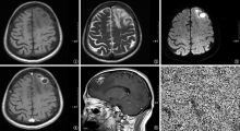
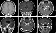
摘要:

目的 探讨获得性免疫缺陷综合征(AIDS)并发颅内结核的磁共振成像(MRI)特征及与CD4 +T淋巴细胞计数的关系。 方法 回顾性分析2014年1月至2017年12月首都医科大学附属北京地坛医院收治的46例AIDS并发颅内结核患者的临床资料,总结其MRI特点。采用Mann-Whitney检验脑实质结核与脑膜结核患者CD4 +T淋巴细胞计数的差异;采用χ 2检验比较CD4 +T淋巴细胞计数≥100个/μl和<100个/μl患者间MRI特征的差异,包括病灶部位、范围、大小、形态、强化方式,以及并发其他脏器结核情况等,并统计CD4 +T淋巴细胞计数与病变大小和强化方式之间的关系。 结果 46例AIDS并发颅内结核患者中,脑实质型结核患者的CD4 +T淋巴细胞计数[47(20.5,131.5)个/μl]低于脑膜型结核[153(130.5,228.5)个/μl](Z=-2.37,P=0.018)。CD4 +T淋巴细胞计数≥100个/μl的患者脑膜型结核(19.6%,9/46)、累及基底池(17.4%,8/46)、外侧裂池(13.0%,6/46)和脑沟(13.6%,6/46)较<100个/μl[分别为4.3%(2/46),2.2%(1/46), 2.2%(1/46), 2.2%(1/46)]更常见(χ 2=7.62,P=0.006; Fisher精确概率检验,P值分别为0.001、0.008和0.008),而分布于大脑皮层下(19.6%,9/46)较<100个/μl(47.8%,22/46)更少见(Fisher精确概率检验,P=0.037)。病变直径3~5mm(136个病灶,47.1%)者呈点状强化,>5mm(89个病灶,30.8%)者呈环形强化,差异有统计学意义(χ 2=105.36,P<0.001)。脑实质结核组病变多位于大脑皮层下,增强扫描呈点状或环形强化(88.6%,31/35);脑膜结核以基底池脑膜增厚强化为主(81.8%,9/11);且脑实质[17.4%(8/46)]及脑膜结核灶[21.7%(10/46)]均易呈簇集状分布,差异有统计学意义(χ 2=4.13,P=0.042)。 结论 AIDS并发颅内结核的MRI表现与患者的CD4 +T淋巴细胞计数密切相关。当AIDS患者CD4 +T淋巴细胞计数<100个/μl时,颅内结核以脑实质型为主,病灶多分布于大脑皮层下;当CD4 +T淋巴细胞计数≥100个/μl时,以脑膜结核为主,病变易累及基底池、外侧裂池和脑沟;颅内病灶直径<5mm时以点状强化为主,≥5mm时以环形强化为主。

关键词: 获得性免疫缺陷综合征, 结核, 中枢神经系统, 磁共振成像, CD4淋巴细胞计数, 因果律

Abstract:

Objective To investigate the features of magnetic resonance imaging (MRI) of the acquired immunodeficiency syndrome (AIDS) patients complicated with cerebral tuberculosis (TB) and its relationship with CD4 + T-cell count. Methods A retrospective analysis was conducted. The related clinical information and data were collected from 46 AIDS patients complicated with cerebral TB, who were hospitalized at Beijing Ditan Hospital from January 2014 to December 2017. Those information and data were analyzed and the patients’ MRI features were summarizes; the difference of CD4 + T-cell count between the patient groups of brain parenchymal TB and meningeal TB was compared by using Mann-Whitney test; the difference of MRI features in two groups of the patients with CD4 + T cells ≥100/μl and <100/μl were compared by using χ 2 test, such as the lesion location, scope, size, shape, the way of enhancement, as well as the situation combined with other organ TB, etc.; the relationship between the CD4 + T-cell count and the lesion size, the way of enhancement was also analyzed. Results Among 46 enrolled AIDS patients with cerebral TB, the CD4 + T-cell count was significantly lower in the patients with brain parenchymal TB (47 (20.5,131.5)/μl) than that in the patients with meningeal TB (153 (130.5,228.5)/μl) (Z=-2.37, P=0.018). In the patients with CD4 + T cells ≥100/μl, the proportion of meningeal TB was 19.6% (9/46) and the proportions that the lesions located in cerebral basal cistern, cerebral lateral fissure cistern and cerebral sulcus were 17.4% (8/46), 13.0% (6/46) and 13.6% (6/46) respectively, which were much higher than those (4.3% (2/46), 2.2% (1/46), 2.2% (1/46), 2.2% (1/46) respectively) in the patients with CD4 + T cells <100/μl (χ 2=7.62, P=0.006; Fisher’s exat test: P=0.001, 0.008 and 0.008, respectively); the proportion that the lesions located in cerebral cortex was 19.6% (9/46) which was much lower than that (47.8%, 22/46) in the patients with CD4 + T cells <100/μl (Fisher’s exat test, P=0.037). When the lesion diameter was 3-5 mm, the point enhancement was found on MRI (136 lesions, 47.1%), while the ring enhancement was found on the MRI when the lesion diameter was more than 5 mm (89 lesions, 30.8%); the difference was statistically significant (χ 2=105.36, P<0.001). In the patient group with brain parenchymal TB, most of their lesions located in cerebral cortex and the point or ring enhancements were found (88.6%, 31/35); in the patient group with meningeal TB, the meningeal thickening enhancement was found on MRI (81.8%, 9/11); the lesions were all in cluster distributions in both brain parenchyma TB (17.4%, 8/46) and meningeal TB (21.7%, 10/46), the difference was statistically significant (χ 2=4.13,P=0.042). Conclusion The features of MRI in AIDS patients complicated with cerebral TB are closely related to their CD4 + T-cell counts. When the number of CD4 + T-cell is <100/μl, the brain parenchymal TB is the major type and the lesions are more likely to locate in cerebral cortex; when the number of CD4 T-cell is ≥100个/μl, the meningeal TB is the major type and the lesions are more likely to locate in cerebral basal cistern, cerebral lateral fissure cistern and cerebral sulcus; the point enhancement is observed when the lesion diameter is <5 mm, while the ring enhancement is found when the lesion diameter is >5 mm.

Key words: Acquired immunodeficiency syndrome, Tuberculosis, central nervous system, Magnetic resonance imaging, CD4 lymphocyte count, Causality