Email Alert | RSS    帮助

中国防痨杂志 ›› 2019, Vol. 41 ›› Issue (8): 828-832.doi: 10.3969/j.issn.1000-6621.2019.08.005

• 论著 • 上一篇    下一篇

超声造影引导下经皮肺穿刺活检在菌阴疑似肺结核患者诊断中的价值

张怡,毕珂,朱惠铭,丛阳,沈梦君,陈宏伟,王茵()   

  1. 同济大学附属同济医院病理科(毕珂)
  • 收稿日期:2019-05-15 出版日期:2019-08-10 发布日期:2019-08-13
  • 通信作者: 王茵 E-mail:lpbbl@aliyun.com
  • 基金资助:
    上海市科学技术委员会2018年度医学引导类(中、西医)科技支撑项目(18411966700)

Diagnostic value of contrast-enhanced ultrasound-guided percutaneous lung puncture biopsy in culture negative presumptive tuberculosis patients

Yi ZHANG,Ke BI,Hui-ming ZHU,Yang CONG,Meng-jun SHEN,Hong-wei CHEN,Yin WANG()   

  1. *Department of Ultrasound, Shanghai Pulmonary Hospital, Tongji University, Shanghai 200433, China
  • Received:2019-05-15 Online:2019-08-10 Published:2019-08-13
  • Contact: Yin WANG E-mail:lpbbl@aliyun.com

摘要:

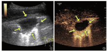
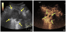
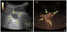
目的 探讨超声造影引导下经皮肺穿刺活检对菌阴疑似肺结核诊断的应用价值。方法 选取2018年1—12月同济大学附属上海市肺科医院收治的菌阴疑似肺结核、且行超声造影引导下经皮肺穿刺活检的患者56例。归纳总结常规超声及超声造影表现、坏死的检出率、穿刺活检的阳性率及术后并发症情况。结果 56例患者超声造影引导下经皮肺穿刺活检阳性率为78.6%(44/56),其中确诊肺结核35例(79.5%,35/44),非结核分枝杆菌病2例,肺癌2例,肺炎5例,未得到阳性结果患者12例(经临床诊断性抗结核药物治疗确诊为肺结核)。超声造影对肺结核病灶内部坏死的检出率(68.1%,32/47)高于常规超声(36.2%,17/47),两者检出率比较差异有统计学意义(χ2=9.592,P=0.002)。超声造影引导下经皮肺穿刺活检过程中发生咯血1例(1.8%,1/56),咯血量约20ml,留院观察30min,症状缓解,无继续咯血。结论 对于菌阴疑似肺结核患者,超声造影引导下经皮肺穿刺活检是一种有效而安全的检查方法,具有较好的临床应用价值。

关键词: 结核, 超声检查, 介入性, 活组织检查, 针吸, 评价研究, 超声造影

Abstract:

Objective To explore the diagnostic value of percutaneous lung biopsy guided by contrast-enhanced ultrasound for culture negative tuberculosis.Methods A total of 56 patients were recruited who were admitted to Shanghai Pulmonary Hospital Affiliated to Tongji University from Jan.2018 to Dec.2018 and were diagnosed to be bacterial negative presumptive pulmonary tuberculosis. All patients underwent contrast-enhanced ultrasound and ultrasound-guided percutaneous lung biopsy. The results of conventional ultrasound and contrast-enhanced ultrasound, the detection rate of necrosis, the positive rate of puncture biopsy, and the complications were analyzed.Results The positive rate of ultrasound-guided percutaneous lung biopsy among 56 patients was 78.6% (44/56), including 35 cases of tuberculosis(79.5%, 35/44), 2 cases of non-tuberculosis mycobacterium, 2 cases of lung cancer, 5 cases of pneumonia, 12 cases of negative results(confirmed by diagnostic anti-tuberculosis treatment).The detection rate of intralesional necrosis of pulmonary tuberculosis by contrast-enhanced ultrasonography (68.1%,32/47) was higher than that by conventional ultrasonography (36.2%,17/47). There was a significant difference between those two methods (χ 2=9.592, P=0.002). Complications occurred only in 1 case (1.8%, 1/56) as hemoptysis during ultrasound-guided percutaneous lung biopsy, while the amount of blood was around 20 ml, symptom relived after 30 minutes, no hemoptysis thereafter. Conclusion For culture negative presumptive pulmonary tuberculosis cases, contrast-enhanced ultrasound-guided percutaneous lung biopsy is an effective and safe method for the diagnosis of negative tuberculosis, which has high clinical application value.

Key words: Tuberculosis, Ultrasonography, interventional, Biopsy, needle, Evaluation studies, Contrast-enhanced ultrasound