Email Alert | RSS    帮助

中国防痨杂志 ›› 2019, Vol. 41 ›› Issue (1): 64-68.doi: 10.3969/j.issn.1000-6621.2019.01.014

• 论著 • 上一篇    下一篇

以磨玻璃样密度影为表现的早期肺癌并发肺结核患者的诊治分析

刘大伟,朱建坤,金锋(),王成,张运曾,乔高锋,赵彬   

  1. 250013 济南,山东大学附属山东省胸科医院胸外科
  • 收稿日期:2018-10-16 出版日期:2019-01-10 发布日期:2019-01-09

Analysis of the diagnosis and treatment of early lung cancer complicated with pulmonary tuberculosis manifested as ground-glass opacities

Da-wei LIU,Jian-kun ZHU,Feng JIN(),Cheng WANG,Yun-zeng ZHANG,Gao-feng QIAO,Bin ZHAO   

  1. Department of Thoracic Surgery,Shandong Provincial Chest Hospital Affiliated to Shandong University,Ji’nan 250013,China
  • Received:2018-10-16 Online:2019-01-10 Published:2019-01-09

摘要:

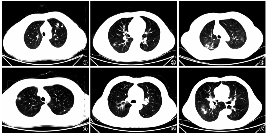
目的 分析以磨玻璃样密度影(ground-glass opacity,GGO)为表现的早期肺癌并发肺结核患者的临床特点,以达到早期识别及治疗的目的。方法 对山东大学附属山东省胸科医院自2013年1月至2018年2月确诊的14例以GGO为首要表现的肺癌并发肺结核患者的临床表现、CT扫描征象、手术方式、病理类型等进行回顾性分析。结果 以GGO为表现的早期肺癌并发肺结核以查体时发现多见(9/14),CT表现为陈旧性结核病灶并发混合密度GGO(mGGO) 12例;GGO与结核病灶位于同侧同叶4例。术前对患者进行规范抗结核药物治疗9例,术后继续行规范抗结核药物治疗6例。术前有3例患者行CT引导下肺穿刺活检确诊肺癌,其余11例为术中冰冻切片病理检查证实。行肺叶切除加纵隔淋巴结清扫10例,肺叶(GGO病灶所在处)切除加同侧异叶肺结核瘤局部切除2例,肺段切除1例,楔形切除1例。肺结核病灶标本经病理检查确诊10例,另4例依据病史及影像学表现符合陈旧性肺结核诊断;表现为GGO的早期肺癌病灶标本经病理检查确诊腺癌11例,鳞癌1例,腺鳞癌1例,大细胞癌1例。纵隔淋巴结病理检查均未见转移。本组患者术后均顺利康复,短期随访未见肿瘤复发、转移及结核复燃等。结论 GGO为表现的早期肺癌并发肺结核患者其临床表现无特异性,薄层CT扫描加动态观察有利于诊断。手术方式需在肺癌切除的基础上兼顾结核病灶的处理,规范的抗结核药物治疗加手术切除治疗效果满意。

关键词: 肺肿瘤, 结核, 肺, 共病现象, 诊断显像, 早期诊断, 综合疗法, 评价研究

Abstract:

Objective To analyze the clinical characteristics of early lung cancer complicated with pulmonary tuberculosis manifested as ground-glass opacity (GGO) in order to achieve the purpose of early identification and treatment.Methods We analyzed retrospectively the clinical manifestation, CT imaging features, operative method and pathological types in 14 cases of lung cancer complicated with pulmonary tuberculosis manifested with GGO diagnosed in Shandong Provincial Chest Hospital Affiliated to Shandong University during Jan. 2013 to Feb. 2018.Results Of 14 cases of early lung cancer complicated with pulmonary tuberculosis manifested with GGO, there were found by health examination in 9 cases, CT signs with obsolete tuberculous lesion manifested as mixed density GGO (mGGO) in 12 cases, GGO and tuberculous lesion located at the ipsilateral lung and same lobe in 4 cases. Nine patients were received standard chemotherapy before operation and 6 patients continued to chemotherapy after operation. Lung cancer was diagnosed by CT-guided puncture lung biopsy in 3 cases before operation, while the other 11 cases were confirmed by intraoperative frozen section pathological examination.Lung lobectomy and mediastinal lymph node dissection were performed in 10 cases, lobectomy (GGO location) plus local resection for tuberculoma located at ipsilateral lung different lobe in 2 cases, segmentectomy in 1 case, wedge resection in 1 case.Pulmonary tuberculosis was diagnosed by pathological examination in 10 cases, and by composite reference standard (CRS) based on medical history and CT imaging features in 4 cases. Samples from early lung cancer manifested GGO were diagnosed by pathological examination as adenocarcinoma in 11 cases, squamous cell carcinoma, adenosquamous squamous cell carcinoma and large cell carcinoma in each one cases. No metastasis was found by pathological examination in mediastinal lymph nodes. All the patients recovered well after operation. Norecurrence, metastasis of tumor and tuberculosis relapse occurred in the short term follow-up.Conclusion The clinical manifestations of early lung cancer patients complicated with pulmonary tuberculosis manifested by GGO are not specific. Thin-slice CT scanning and dynamic observation are helpful for diagnosis. Surgical method needs to give consideration to tumor resection and tuberculous foci management. It is satisfactory for efficacy that standard chemotherapy and surgical resection be performed in these patients,

Key words: Lung neoplasms, Tuberculosis, pulmonary, Comorbidity, Diagnostic imaging, Early diagnosis, Combined modality therapy, Evaluation studies