Email Alert | RSS    帮助

中国防痨杂志 ›› 2018, Vol. 40 ›› Issue (10): 1046-1050.doi: 10.3969/j.issn.1000-6621.2018.10.004

• 论著 • 上一篇    下一篇

三种细胞因子在肺结核与肺癌胸腔积液中的诊断价值

刘浩然1,张亚莉2,任卫聪1,赵玲娟1,李传友1,王伟1,(),高孟秋3,()   

  1. 1. 101149 首都医科大学附属北京胸科医院 北京市结核病胸部肿瘤研究所 耐药结核病研究北京市重点实验室细菌免疫室
    2. 药剂科
    3. 结核科
  • 收稿日期:2018-09-03 出版日期:2018-10-10 发布日期:2018-10-18
  • 通信作者: 王伟,高孟秋 E-mail:wangwei010@aliyun.com;gaomqwdm@aliyun.com
  • 基金资助:
    北京市医院管理局“登峰”计划(DFL20151501);北京市科技重大专项(D181100000418004)

Differential expression and diagnostic value of three cytokines in pleural effusion between pulmonary tuberculosis and lung cancer

Hao-ran LIU1,Ya-li ZHANG2,Wei-cong REN1,Ling-juan ZHAO1,Chuan-you LI1,Wei WANG1,(),Meng-qiu GAO3,()   

  1. 1. Department of Bacteriology and Immunology, Beijing Key Laboratory for Drug Resistance Tuberculosis Research, Beijing Chest Hospital, Capital Medical University, Beijing Tuberculosis and Thoracic Tumor Research Institute, Beijing 101149, China
  • Received:2018-09-03 Online:2018-10-10 Published:2018-10-18
  • Contact: Wei WANG,Meng-qiu GAO E-mail:wangwei010@aliyun.com;gaomqwdm@aliyun.com

摘要:

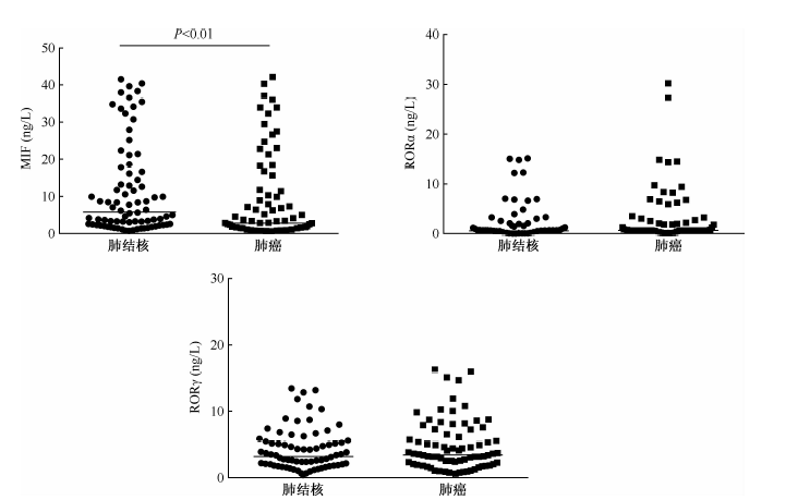
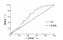
目的 分析肺结核患者(TB)和肺癌患者胸腔积液中巨噬细胞迁移抑制因子(MIF)、孤核受体α(RORα)和孤核受体γ(RORγ)水平的差异,探讨其在肺结核与肺癌鉴别诊断中的价值。 方法 通过简单随机抽样,将已确诊为肺结核和肺癌的冻存胸腔积液样本进行编号,抽取样本各80例,总结研究对象的基本信息,采用酶联免疫吸附法检测各样本中的MIF、RORα及RORγ水平。采用SPSS 20.0软件进行统计学对比分析,本研究中两组计量资料不符合正态分布,采用“Mann-Whitney U检验”,以P<0.05为差异有统计学意义。 结果 肺结核组和肺癌组胸腔积液中的MIF水平中位数(四分位数)[M(Q1,Q3)]分别为5.83(2.41,16.43)、2.79(0.91,11.12)ng/L,差异有统计学意义(U=2314.50,P<0.01);肺结核组和肺癌组胸腔积液中RORα水平[M(Q1,Q3)]分别为0.63(0.37,1.66)、0.63(0.57,2.16)ng/L,差异无统计学意义(U=3525.00,P>0.05);肺结核组和肺癌组胸腔积液中RORγ水平[M(Q1,Q3)]分别为3.23(1.82,5.26)、3.44(1.94,7.11)ng/L,差异无统计学意义(U=3431.50,P>0.05)。 结论 肺结核患者胸腔积液中MIF水平高于肺癌患者, 检测胸腔积液中MIF水平对肺结核与肺癌的鉴别有一定诊断价值;而胸腔积液中RORα及RORγ水平检测的价值仍有待进一步验证。

关键词: 结核, 肺, 肺肿瘤, 胸腔积液, 巨噬细胞游走抑制因子, 孤儿核受体类, 酶联免疫吸附测定, 诊断, 鉴别

Abstract:

Objective To analyze the difference of macrophage migration inhibitory factor (MIF), retinoid acid related orphan receptor alpha (RORα) and retinoid acid related orphan receptor gamma (RORγ) in the pleural effusion of patients with pulmonary tuberculosis (TB) and lung cancer, and to explore their values in the differential diagnosis. Methods By simple random sampling, the frozen pleural effusion samples of pulmonary tuberculosis and lung cancer were numbered, and 80 samples were collected, the basic information of the subjects was summarized. The levels of MIF, RORα and RORγ in the pleural effusion were detected by enzyme-linked immunosorbent assay (ELISA). The application software SPSS 20.0 was used for comparison and analysis, and the measurement data in this study do not conform to normal distribution, so the Mann-Whitney U test has been used, the difference was statistically significant when P<0.05. Results The medians of MIF level (quartile) [M (Q1,Q3)] in the pleural effusion of the tuberculosis group and lung cancer group were 5.83 (2.41, 16.43) and 2.79 (0.91, 11.12) ng/L respectively, there was significant difference between the groups (U=2314.50,P<0.01). The RORα levels [M (Q1, Q3)] in the pleural effusion of the tuberculosis group and lung cancer group were 0.63 (0.37, 1.66) and 0.63 (0.57, 2.16) ng/L respectively, there was no significant difference between the two groups (U=3525.00, P>0.05); the levels of RORγ in the pleural effusion of the tuberculosis group and lung cancer group were 3.23 (1.82, 5.26) and 3.44 (1.94, 7.11) ng/L respectively, with no significant difference (U=3431.50, P>0.05). Conclusion The level of MIF in the pleural effusion of patients with tuberculosis is higher than the patients with lung cance, the detection of MIF level in pleural effusion is certainly diagnostically valuable in differentiating pulmonary tuberculosis from lung cancer. And the diagnostic value of RORα and RORγ in pleural effusion remains to be verified.

Key words: Tuberculosis, pulmonary, Lung neoplasms, Pleural effusion, Macrophage migration-inhibitory factors, Orphan nuclear receptors, Enzyme-linked immunosorbent assay, Diagnosis, differential