Email Alert | RSS    帮助

中国防痨杂志 ›› 2018, Vol. 40 ›› Issue (9): 1003-1006.doi: 10.3969/j.issn.1000-6621.2018.09.019

• 短篇论著 • 上一篇    下一篇

儿童继发性肺结核的CT表现分析

辛丹1,2,辛军1,()   

  1. 1. 110004 中国医科大学附属盛京医院放射科
    2. 沈阳市第十人民医院 沈阳市胸科医院放射影像科
  • 收稿日期:2018-02-22 出版日期:2018-09-10 发布日期:2018-10-17
  • 通信作者: 辛军 E-mail:xinj@sj-hospital.org

Analysis of CT images of secondary pulmonary tuberculosis in children

Dan XIN1,2,Jun XIN1,()   

  1. 1. Department of Radiolo-gy, Shengjing Hospital of China Medical University,Shenyang 110004, China
    2. Radiologist of Shenyang Tenth People’s Hospital, Shenyang Chest Hospital;
  • Received:2018-02-22 Online:2018-09-10 Published:2018-10-17
  • Contact: Jun XIN E-mail:xinj@sj-hospital.org

摘要:

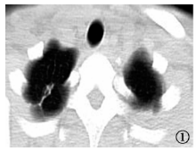
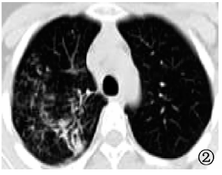
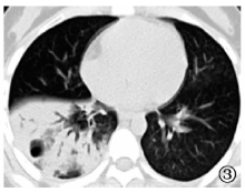
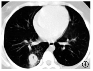
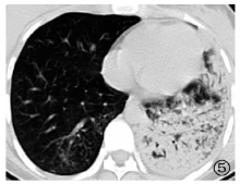
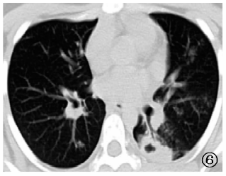
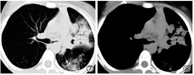
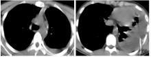
回顾性分析2012年8月至2017年9月沈阳市第十人民医院(沈阳市胸科医院)临床确诊的75例儿童继发性肺结核患者的临床资料及CT表现。75例患儿病灶好发于上叶尖后段[右肺上叶后段为64.0%(48/75)、左肺上叶尖后段为62.7%(47/75)、右肺上叶尖段为56.0%(42/75)]、下叶背段[右肺下叶背段为61.3%(46/75)、左肺下叶背段为42.7%(32/75)]。病变以多种形态并存于肺内,其中以纤维条索状影[92.0%(69/75)]、小叶中心腺泡样结节及结节状融合影[91.7%(68/75)]、斑片状影[86.7%(65/75)]、淡片状影[80.0%(60/75)]为最常见的病灶形态,而结核球的发生率很低[5.3%(4/75)]。病灶密度改变中,钙化[58.7%(44/75)]的发生率稍高,空洞多表现为厚壁[22.7%(17/75)]及无壁空洞[10.7%(8/75)]。结核性胸膜炎[77.3%(58/75)]、支气管结核[36.0%(27/75)]的并发率高。

关键词: 儿童, 结核, 肺, 体层摄影术, 螺旋计算机, 诊断显像, 疾病特征

Abstract:

The clinical data/information and CT findings of 75 children with secondary pulmonary tuberculosis, who were diagnosed at Shenyang Tenth People Hospital from August 2012 to September 2017, were retrospectively analyzed. Among 75 cases, their lesions mainly occurred in the posterior or apical segment of the upper lobe of the lung (proportion occurred in the posterior segment of the right upper lobe was 64.0% (48/75), in the posterior or apical segment of the left upper lobe was 62.7% (47/75), in the apical segment of the right upper lobe was 56.0% (42/75)) and the back segment of the lower lobe of the lung (proportion occurred in the back segment of the right lower lobe was 61.3% (46/75), in the back segment of the left lower lobe was 42.7% (32/75)). The lesions in the lungs were in a variety of forms. The most common images of the lesions included fibrous strips (92.0% (69/75)), acinar-like nodules in the lobules & nodular fusion (91.7% (68/75)), patchy shadow (86.7% (65/75)) and light film (80.0% (60/75)); the incidence of tuberculoma was very low (5.3% (4/75)). In the changes of lesion density, the incidence of calcification (58.7% (44/75)) was slightly higher; the most cavities were thick-walled (22.7% (17/75))) or wall-less (10.7% (8/75)). The rate complicated with tuberculous pleurisy (77.3% (58/75)) or bronchial tuberculosis (36.0% (27/75)) was high.

Key words: Child, Tuberculosis, pulmonary, Tomography, spiral computed, Diagnostic imaging, Disease attributes