Email Alert | RSS    帮助

中国防痨杂志 ›› 2025, Vol. 47 ›› Issue (10): 1254-1260.doi: 10.19982/j.issn.1000-6621.20250181

• 论著 • 上一篇    下一篇

结核病病理诊断质控蜡块的制备及其性能评估

黄晓洁1, 杜伟丽1, 赵宏1, 苏丹1, 刘子臣1, 赵颖丽1, 刘毅2(), 车南颖1()   

  1. 1首都医科大学附属北京胸科医院病理科/北京市结核病胸部肿瘤研究所,北京 101149
    2首都医科大学附属北京胸科医院生物样本与数据资源库/北京市结核病胸部肿瘤研究所,北京 101149
  • 收稿日期:2025-05-06 出版日期:2025-10-10 发布日期:2025-09-29
  • 通信作者: 车南颖,Email:cheny0448@163.com;刘毅,Email:18910250336@163.com
  • 基金资助:
    国家自然科学基金(82072381);国家自然科学基金(82472378);北京市属医学科研院所公益发展改革试点项目(JYY2023-15);北京研究型病房卓越计划(BRWEP2024W042160103);北京市医院管理中心“登峰”人才培养计划(DFL20241601)

Preparation and performance evaluation of quality control paraffin blocks for the pathological diagnosis of tuberculosis

Huang Xiaojie1, Du Weili1, Zhao Hong1, Su Dan1, Liu Zichen1, Zhao Yingli1, Liu Yi2(), Che Nanying1()   

  1. 1Department of Pathology, Beijing Chest Hospital, Capital Medical University/Beijing Tuberculosis and Thoracic Tumor Research Institute, Beijing 101149, China
    2Biobank of Beijing Chest Hospital, Capital Medical University/Beijing Tuberculosis and Thoracic Tumor Research Institute, Beijing 101149, China
  • Received:2025-05-06 Online:2025-10-10 Published:2025-09-29
  • Contact: Che Nanying, Email: cheny0448@163.com;Liu Yi, Email: 18910250336@163.com
  • Supported by:
    National Natural Science Foundation of China(82072381);National Natural Science Foundation of China(82472378);Beijing Municipal Public Welfare Development and Reform Pilot Project for Medical Research(JYY2023-15);Beijing Research Ward Excellence Program(BRWEP2024W042160103);Dengfeng Talents Project of Beijing Hospitals Authority(DFL20241601)

摘要:

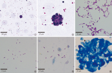
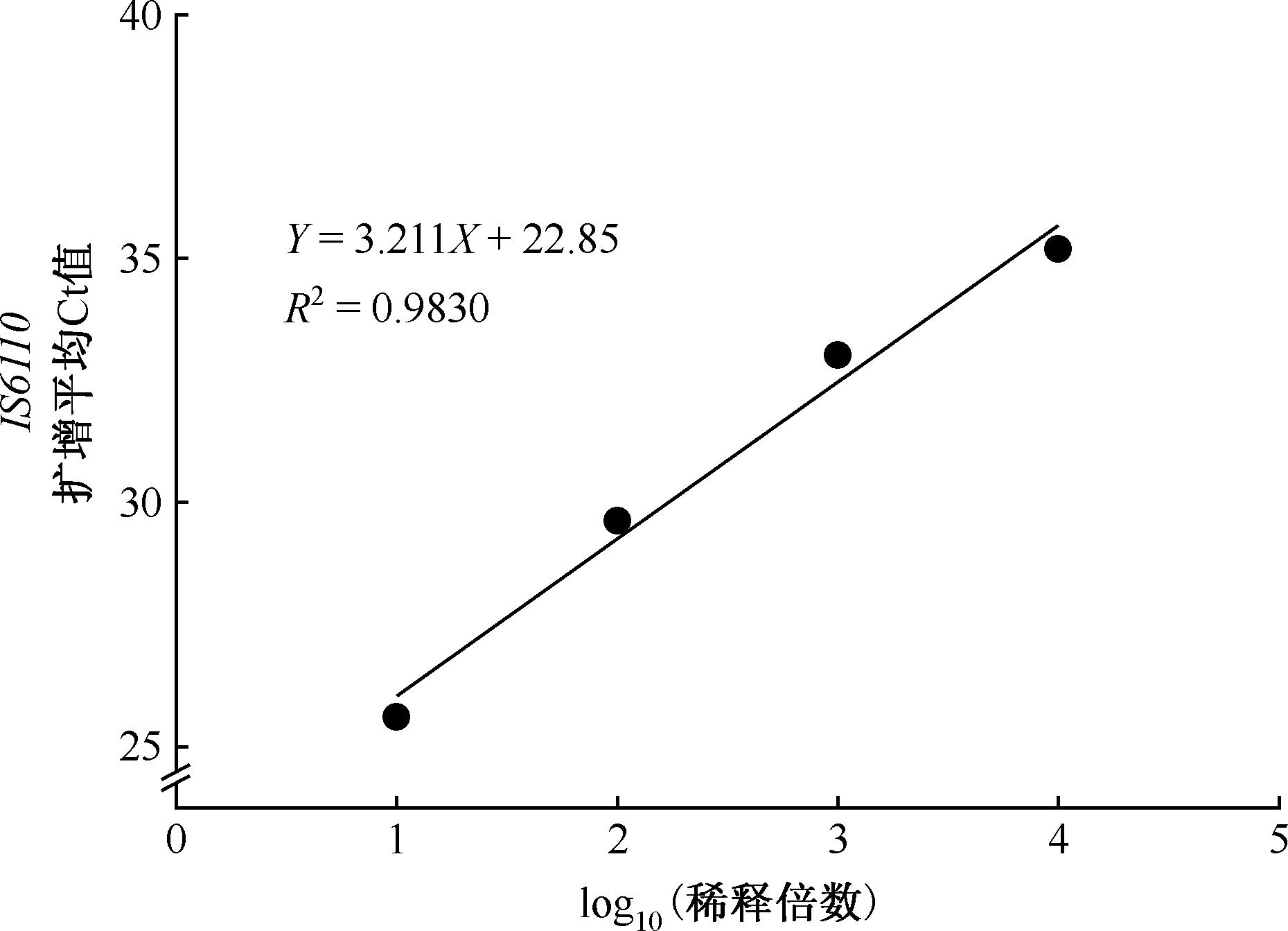
目的: 建立结核病病理质控蜡块制备方法,并对质控蜡块进行细胞形态、结核分枝杆菌(Mycobacterium tuberculosis, MTB)分布及MTB DNA分布方面的性能评估。方法: 用灭活MTB H37Rv悬液和恶性胸腔积液细胞沉淀混合,以2%琼脂为基质制备MTB浓度不同的蜡块,磷酸盐缓冲液代替MTB悬液制作阴性蜡块。每个圆柱体琼脂块沿纵轴等距分为4层(每层厚约3mm),每层制成1个蜡块,形成分层样本组。通过HE 染色评估细胞形态完整性及空间分布均匀性,抗酸染色和荧光定量PCR(real-time PCR,qPCR)分析各组MTB数量、IS6110拷贝数及其对应琼脂块纵轴方向MTB分布的均一性。结果: 成功制备了适用于MTB抗酸染色和MTB DNA检测的蜡块质控品。HE染色显示蜡块中各种细胞形态完整且空间分布均匀。抗酸染色显示,理论MTB量为0.0336、0.336、3.36个/切片的分层样本组中,每组蜡块对应的4张切片各有0、1和2张切片阳性,检测结果不稳定,不宜作为抗酸染色阳性质控品;理论MTB量为33.6、336个/切片的分层样本组中,每个蜡块对应的切片中均找到MTB,为合格抗酸染色质控品,MTB平均密度分别为(21.50±9.26)条/300视野和(169.25±40.25)条/300视野。qPCR结果显示,理论MTB量为0.0336个/切片的分层样本组中,有3个蜡块检测为阳性,1个为阴性,结果存在波动,为不合格分子检测质控品;理论MTB量为0.336、3.36、33.6、336个/切片的分层样本组中,检测均阳性,IS6110基因拷贝数分布为(6.48±3.54)拷贝/5张切片、(26.53±6.65)拷贝/5张切片、(283.93±98.51)拷贝/5张切片、(4446.75±833.84)拷贝/5张切片,扩增Ct值变异系数均<5%,各组对应琼脂块纵轴方向上MTB均匀分布,为合格的分子检测质控品。阴性质控品抗酸染色均为阴性、qPCR IS6110均无扩增。结论: 本研究成功建立了结核病病理诊断质控品制备方案。推荐使用至少33.6个/切片的MTB制备抗酸染色的质控品,至少0.336个/切片的MTB用于制备分子检测质控品,并根据检测方法的检出限及不同应用场景选择起始MTB量。

关键词: 分枝杆菌,结核, 诊断, 病理学, 质量控制, 评价研究

Abstract:

Objective: To develop a standardized method for preparing quality control (QC) paraffin blocks for pathological diagnosis of tuberculosis, and to evaluate their performance with respect to cellular morphology, spatial distribution of Mycobacterium tuberculosis (MTB), and MTB DNA. Methods: QC paraffin blocks containing different concentrations of MTB were prepared by combining inactivated MTB H37Rv suspension with sedimented cells from malignant pleural effusion, using 2% agar as the embedding matrix. Negative control blocks were generated by replacing the MTB suspension with phosphate-buffered saline (PBS). Each cylindrical agar block was sectioned longitudinally into four equal layers (approximately 3 mm thick), yielding stratified sample groups. Hematoxylin and eosin (HE) staining was used to assess the integrity of cellular morphology and the uniformity of spatial distribution. Acid-fast staining (AFB) and quantitative real-time PCR (qPCR) were employed to evaluate MTB load, IS6110 copy number, and the homogeneity of MTB distribution along the longitudinal axis of the blocks. Results: Standardized QC paraffin blocks compatible with both AFB staining and MTB DNA detection were successfully developed. HE staining confirmed the preservation of cellular morphology and a uniform spatial distribution of cells within the blocks. In AFB staining, stratified sample groups containing theoretical MTB loads of 33.6 and 336 bacilli per section consistently exhibited detectable bacilli across all slices, qualifying them as reliable positive QC materials for staining. The corresponding average MTB densities were (21.50±9.26) and (169.25±40.25) bacilli per 300 fields, respectively. Quantitative PCR analysis demonstrated consistent positivity in all blocks with theoretical MTB concentrations of 0.336, 3.36, 33.6, and 336 bacilli per section. The IS6110 gene copy numbers in these groups were (6.48±3.54), (26.53±6.65), (283.93±98.51), and (4446.75±833.84) copies per five sections, respectively. The coefficient of variation for Ct values remained below 5% in all groups, and the longitudinal distribution of MTB DNA within the blocks was uniform, supporting their qualification as standardized molecular QC materials. Negative controls were consistently negative in both AFB staining and qPCR, with no IS6110 amplification observed. Conclusion: We developed a standardized protocol for the preparation of QC materials tailored to the pathological diagnosis of tuberculosis. Based on assay sensitivity and clinical application scenarios, we recommend a minimum MTB load of 33.6 bacilli per section for AFB staining QC and 0.336 bacilli per section for molecular diagnostic QC. The selection of initial MTB concentration should be guided by the detection limits of the assay and the intended diagnostic context.

Key words: Mycobacterium tuberculosis, Diagnosis, Pathology, Quality control, Evaluation studies

中图分类号: