Email Alert | RSS 帮助

中国防痨杂志 ›› 2025, Vol. 47 ›› Issue (9): 1126-1134.doi: 10.19982/j.issn.1000-6621.20250195
国家感染性疾病临床医学研究中心, 北京大学深圳医院, 深圳市炎症与免疫性疾病重点实验室, 深圳市第三人民医院, 结节性红斑病因诊断的专家共识小组
收稿日期:2025-05-13
出版日期:2025-09-10
发布日期:2025-08-27
基金资助:National Clinical Research Center for Infectious Diseases , Peking University Shenzhen Hospital , Shenzhen Key Laboratory of Immunity and Inflammatory Diseases , The Third People’s Hospital of Shenzhen , Expert Consensus Group on the Etiology and Diagnosis of Erythema Nodosum
Received:2025-05-13
Online:2025-09-10
Published:2025-08-27
Supported by:摘要:
结节性红斑的病因复杂,可能与感染、自身免疫疾病、肿瘤、药物及特发性等多个因素相关。如何明确结节性红斑的病因及其诊断策略是一个亟待解决的临床问题。由国家感染性疾病临床医学研究中心、深圳市第三人民医院联合北京大学深圳医院、深圳市炎症与免疫性疾病重点实验室,共同制定了我国第一部关于结节性红斑病因诊断的专家共识。本共识详细阐明结节性红斑的病因分类及发生机制,并对结节性红斑病因诊断流程、病理表现及鉴别诊断,给出了基于循证的推荐,旨在规范结节性红斑的诊断流程,提高结节性红斑的诊断和治疗水平,从而改善患者的生活质量和预后。
中图分类号:
国家感染性疾病临床医学研究中心, 北京大学深圳医院, 深圳市炎症与免疫性疾病重点实验室, 深圳市第三人民医院, 结节性红斑病因诊断的专家共识小组. 结节性红斑病因诊断的专家共识(2025版)[J]. 中国防痨杂志, 2025, 47(9): 1126-1134. doi: 10.19982/j.issn.1000-6621.20250195
National Clinical Research Center for Infectious Diseases , Peking University Shenzhen Hospital , Shenzhen Key Laboratory of Immunity and Inflammatory Diseases , The Third People’s Hospital of Shenzhen , Expert Consensus Group on the Etiology and Diagnosis of Erythema Nodosum . Expert consensus on the etiology and diagnosis of erythema nodosum (2025 version)[J]. Chinese Journal of Antituberculosis, 2025, 47(9): 1126-1134. doi: 10.19982/j.issn.1000-6621.20250195
表1
GRADE证据质量分级
| 证据级别 | 具体描述 | 研究类型 | 表达字母 |
|---|---|---|---|
| 高质量证据 | 非常确信真实的效应值接近效应估计,未来研究几乎不可能改变现有评价结果的可信度 | 随机对照试验;质量升高二级的观察性研究 | A |
| 中质量证据 | 对效应估计值有中等程度的信心:真实值有可能接近估计值,但仍存在二者大小相同的可能性;未来研究可能对现有评估有重要影响,可能改变评价结果的可信度 | 质量降低一级的随机对照试验;质量升高一级的观察性研究 | B |
| 低质量证据 | 对效应估计值的确信程度有限:真实值可能与估计值大不相同;未来研究很有可能对现有评估有重要影响,改变评估结果可信度的可能性较大 | 质量降低二级的随机对照试验;观察性研究 | C |
| 极低质量证据 | 对效应估计值几乎没有信心:真实值很可能与估计值大不相同;任何评估都很不确定 | 质量降低三级的随机对照试验;质量降低一级的观察性研究;系列病例观察;个案报道 | D |
表2
GRADE证据推荐强度分级
| 推荐强度 | 具体描述 | 表达数字 |
|---|---|---|
| 支持使用某项干预措施的强推荐 | 评价者确信干预措施利大于弊 | 1 |
| 支持使用某项干预措施的弱推荐 | 利弊不确定或无论高低质量的证据均显示利弊相当 | 2 |
| 反对使用某项干预措施的弱推荐 | 证据均显示利弊相当 | 2 |
| 反对使用某项干预措施的强推荐 | 评价者确信干预措施弊大于利 | 1 |
表3
结节性红斑的常见病因
| 病因 | 分类 |
|---|---|
| 感染 | 链球菌感染、结核分枝杆菌感染、其他(如细菌、真菌和病毒等)感染 |
| 自身免疫 | 白塞综合征、炎症性肠病、系统性红斑狼疮、大动脉炎、结节病、其他(如干燥综合征、类风湿关节炎等) |
| 肿瘤 | 霍奇金淋巴瘤、白血病、淋巴瘤(如B细胞淋巴瘤、Lennert淋巴瘤、非霍奇金淋巴瘤、T细胞丰富的B细胞淋巴瘤) |
| 药物 | 化学制剂、生物制剂及免疫检查点抑制剂、疫苗等 |
| 特发性 | 未明确病因及分类 |
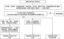
图1
感染所致结节性红斑的诊断流程

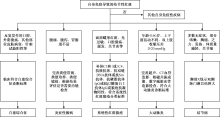
图2
自身免疫导致结节性红斑诊断流程
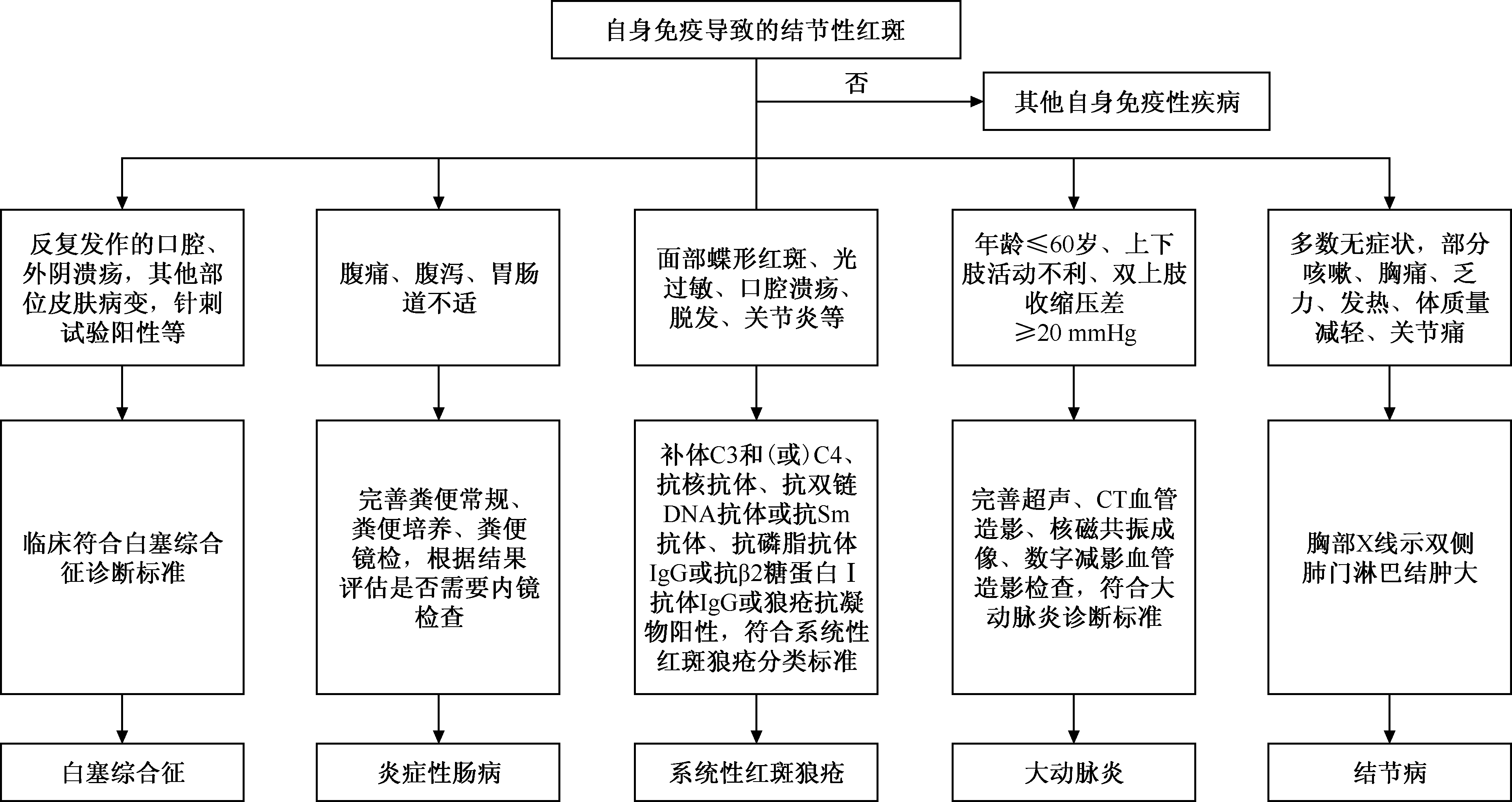
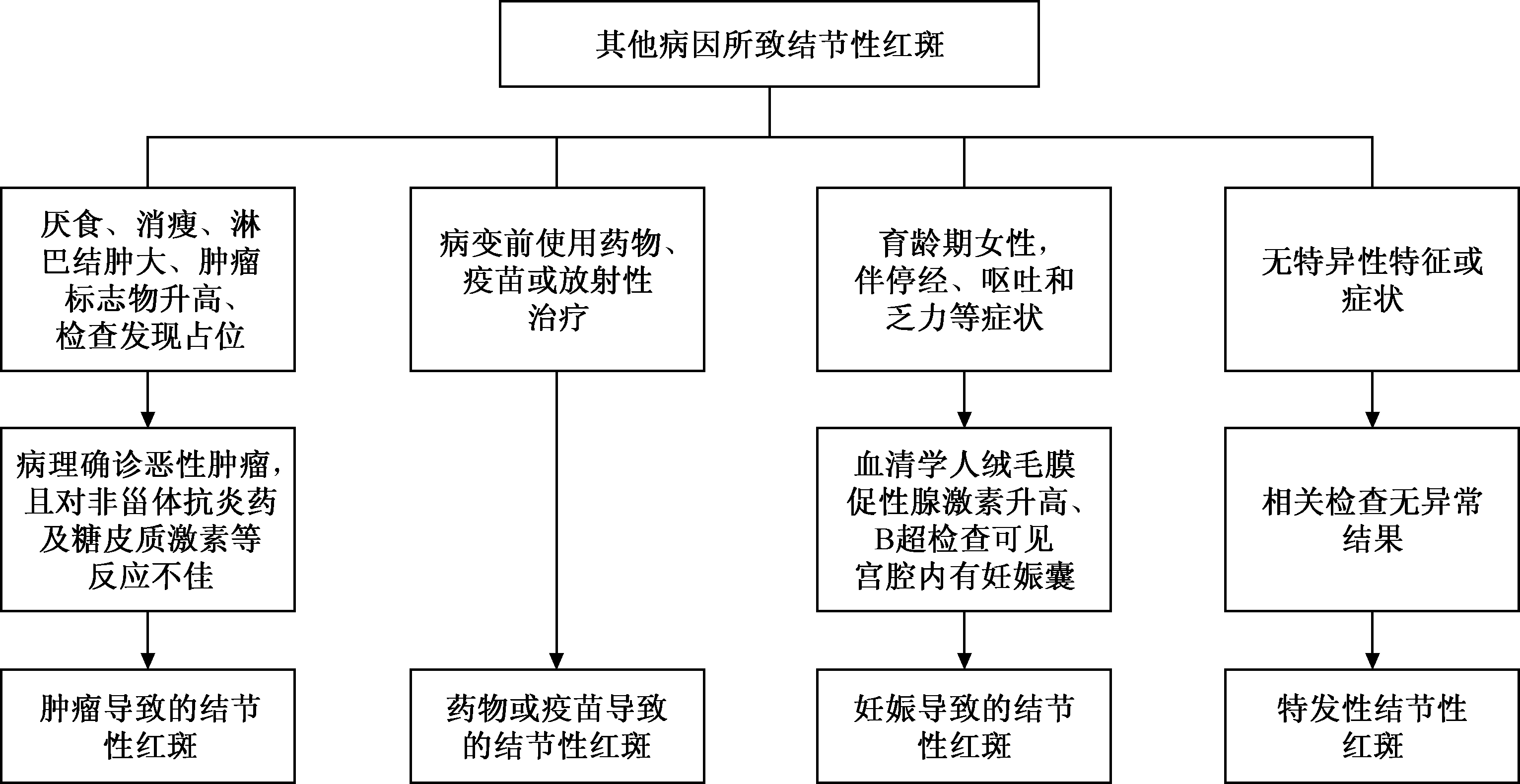
图3
其他病因所致结节性红斑诊断流程
| [1] |
Pérez-Garza DM, Chavez-Alvarez S, Ocampo-Candiani J, et al. Erythema Nodosum: A Practical Approach and Diagnostic Algorithm. Am J Clin Dermatol, 2021, 22(3): 367-378. doi:10.1007/s40257-021-00592-w.
pmid: 33683567 |
| [2] |
Leung AKC, Leong KF, Lam JM. Erythema nodosum. World J Pediatr, 2018, 14(6): 548-554. doi:10.1007/s12519-018-0191-1.
pmid: 30269303 |
| [3] | Laborada J, Cohen PR. Tuberculosis-Associated Erythema Nodosum. Cureus, 2021, 13(12): e20184. doi:10.7759/cureus.20184. |
| [4] |
Thurber S, Kohler S. Histopathologic spectrum of erythema nodosum. J Cutan Pathol, 2006, 33(1): 18-26. doi:10.1111/j.0303-6987.2006.00402.x.
pmid: 16441407 |
| [5] |
Shavit E, Marzano AV, Alavi A. Ulcerative versus non-ulcerative panniculitis: is it time for a novel clinical approach to panniculitis? Int J Dermatol, 2021, 60(4): 407-417. doi:10.1111/ijd.15224.
pmid: 33040341 |
| [6] | Borges T, Silva S. Panniculitis: A Cardinal Sign of Autoinflammation. Curr Rheumatol Rev, 2024, 20(4): 350-360. doi:10.2174/0115733971254702231020060633. |
| [7] | De Simone C, Caldarola G, Scaldaferri F, et al. Clinical, histopathological, and immunological evaluation of a series of patients with erythema nodosum. Int J Dermatol, 2016, 55(5): e289-94. doi:10.1111/ijd.13212. |
| [8] | Bertola EA, Simonetti GD, Del Giorno R, et al. Extrarenal Immune-Mediated Disorders Linked with Acute Poststreptococcal Glomerulonephritis: a Systematic Review. Clin Rev Allergy Immunol, 2019, 57(2): 294-302. doi:10.1007/s12016-019-08761-w. |
| [9] | Chen S, Chen J, Chen L, et al. Mycobacterium tuberculosis infection is associated with the development of erythema nodosum and nodular vasculitis. PLoS One, 2013, 8(5): e62653. doi:10.1371/journal.pone.0062653. |
| [10] | Bjorn-Mortensen K, Ladefoged K, Simonsen J, et al. Erythema nodosum and the risk of tuberculosis in a high incidence setting. Int J Circumpolar Health, 2016, 75: 32666. doi:10.3402/ijch.v75.32666. |
| [11] | 杨松, 严晓峰. 结节性红斑的临床特点及其对结核病的诊断价值. 中国防痨杂志, 2019, 41(11): 1227-1230. doi:10.3969/j.issn.1000-6621.2019.11.015. |
| [12] | 颜韵灵, 郑宝庆, 王晓华. 与结核相关的脂膜炎研究进展. 中华皮肤科杂志, 2020, 53(11): 944-947. doi:10.35541/cjd.20190499. |
| [13] | 杨勤宇, 李惠, 赵恒光. 结核相关结节性红斑的研究进展. 中国皮肤性病学杂志, 2010, 24(3): 280-282. |
| [14] |
Giordano N, Corallo C, Miracco C, et al. Erythema nodosum associated with Staphylococcus xylosus septicemia. J Microbiol Immunol Infect, 2016, 49(1): 134-137. doi:10.1016/j.jmii.2012.10.003.
pmid: 23266237 |
| [15] | Ozbagcivan O, Akarsu S, Avci C, et al. Examination of the Microbial Spectrum in the Etiology of Erythema Nodosum: A Retrospective Descriptive Study. J Immunol Res, 2017, 2017: 8139591. doi:10.1155/2017/8139591. |
| [16] | Negera E, Walker SL, Bobosha K, et al. T-cell regulation in Erythema Nodosum Leprosum. PLoS Negl Trop Dis, 2017, 11(10): e0006001. doi:10.1371/journal.pntd.0006001. |
| [17] |
Braun-Falco M, Ring J. Nodular erythema as early sign of systemic aspergillosis. J Eur Acad Dermatol Venereol, 2006, 20(5): 610-612. doi:10.1111/j.1468-3083.2006.01493.x.
pmid: 16684298 |
| [18] | 李丹, 刘洪艳, 王妍, 等. 新型冠状病毒肺炎并发结节性红斑1例. 中国感染与化疗杂志, 2022, 22(2): 223-225. doi:10.16718/j.1009-7708.2022.02.021. |
| [19] | Kordeva S, Ivanov L, Broshtilova V, et al. Erythema nodosum as first clinical sign of acute Borrelia burgdorferi infection. Braz J Infect Dis, 2024, 28(6): 103877. doi:10.1016/j.bjid.2024.103877. |
| [20] | 张琛, 高炳爱, 陈玉欣, 等. 结节性红斑的病因及发病机制. 中国麻风皮肤病杂志, 2015, 31(7): 408-410. |
| [21] |
Ilkit M, Durdu M, Karakaş M. Cutaneous id reactions: a comprehensive review of clinical manifestations, epidemiology, etiology, and management. Crit Rev Microbiol, 2012, 38(3): 191-202. doi:10.3109/1040841X.2011.645520.
pmid: 22300403 |
| [22] | Nascimento LS, de Castro YS, Figueira JA, et al. Toxoplasma gondii infection and high levels of IgE are associated to erythema nodosum leprosy (ENL). PLoS One, 2024, 19(6): e0300704. doi:10.1371/journal.pone.0300704. |
| [23] | Alpsoy E, Donmez L, Onder M, et al. Clinical features and natural course of Behçet’s disease in 661 cases: a multicentre study. Br J Dermatol, 2007, 157(5): 901-906. doi:10.1111/j.1365-2133.2007.08116.x. |
| [24] | Davatchi F, Chams-Davatchi C, Shams H, et al. Behcet’s disease: epidemiology, clinical manifestations, and diagnosis. Expert Rev Clin Immunol, 2017, 13(1): 57-65. doi:10.1080/1744666X.2016.1205486. |
| [25] | 刘白, 姜祎群. 结节性红斑的病因学研究进展. 国际皮肤性病学杂志, 2016, 42(1): 30-32. |
| [26] | Cheng Y, Zhao X, Chen Y, et al. Circulating immune complexome analysis identified anti-tubulin-α-1c as an inflammation associated autoantibody with promising diagnostic value for Behcet’s Disease. PLoS One, 2018, 13(6): e0199047. doi:10.1371/journal.pone.0199047. |
| [27] | Greuter T, Navarini A, Vavricka SR. Skin Manifestations of Inflammatory Bowel Disease. Clin Rev Allergy Immunol, 2017, 53(3): 413-427. doi:10.1007/s12016-017-8617-4. |
| [28] |
Nguyen GC, Torres EA, Regueiro M, et al. Inflammatory bowel disease characteristics among African Americans, Hispanics, and non-Hispanic Whites: characterization of a large North American cohort. Am J Gastroenterol, 2006, 101(5): 1012-1023. doi:10.1111/j.1572-0241.2006.00504.x.
pmid: 16696785 |
| [29] | Farhi D, Cosnes J, Zizi N, et al. Significance of erythema nodosum and pyoderma gangrenosum in inflammatory bowel diseases: a cohort study of 2402 patients. Medicine (Baltimore), 2008, 87(5): 281-293. doi:10.1097/MD.0b013e318187cc9c. |
| [30] | Chowaniec M, Starba A, Wiland P. Erythema nodosum-review of the literature. Reumatologia, 2016, 54(2): 79-82. doi:10.5114/reum.2016.60217. |
| [31] |
Rogler G, Singh A, Kavanaugh A, et al. Extraintestinal Manifestations of Inflammatory Bowel Disease: Current Concepts, Treatment, and Implications for Disease Management. Gastroenterology, 2021, 161(4): 1118-1132. doi:10.1053/j.gastro.2021.07.042.
pmid: 34358489 |
| [32] |
Vavricka SR, Schoepfer A, Scharl M, et al. Extraintestinal Manifestations of Inflammatory Bowel Disease. Inflamm Bowel Dis, 2015, 21(8): 1982-1992. doi:10.1097/MIB.0000000000000392.
pmid: 26154136 |
| [33] | He R, Zhao S, Cui M, et al. Cutaneous manifestations of inflammatory bowel disease: basic characteristics, therapy, and potential pathophysiological associations. Front Immunol, 2023, 14: 1234535. doi:10.3389/fimmu.2023.1234535. |
| [34] | 王泽芳. 结节性红斑283例临床分析. 四川医学, 2010, 31(9): 1286-1288. doi:10.3969/j.issn.1004-0501.2010.09.032. |
| [35] | Chasset F, Francès C. Cutaneous Manifestations of Medium- and Large-Vessel Vasculitis. Clin Rev Allergy Immunol, 2017, 53(3): 452-468. doi:10.1007/s12016-017-8612-9. |
| [36] | 李贤光, 王立, 张璇, 等. 大动脉炎合并皮肤黏膜损害的临床分析. 中华风湿病学杂志, 2013, 17(8), 549-551. doi:10.3760/cma.j.issn.1007-7480.2013.08.011. |
| [37] | Koneti J, Cherukuri SP, Gadde S, et al. Sarcoidosis and Its Dermatological Manifestations: A Narrative Review. Cureus, 2022, 14(8): e28053. doi:10.7759/cureus.28053. |
| [38] |
Caplan A, Rosenbach M, Imadojemu S. Cutaneous Sarcoidosis. Semin Respir Crit Care Med, 2020, 41(5): 689-699. doi:10.1055/s-0040-1713130.
pmid: 32593176 |
| [39] | Statement on sarcoidosis. Joint Statement of the American Thoracic Society (ATS), the European Respiratory Society (ERS) and the World Association of Sarcoidosis and Other Granulomatous Disorders (WASOG) adopted by the ATS Board of Directors and by the ERS Executive Committee, February 1999. Am J Respir Crit Care Med, 1999, 160(2):736-755. doi:10.1164/ajrccm.160.2.ats4-99. |
| [40] |
Afacan Yıldırım E, Aladagˇ Öztürk P, Adışen E, et al. The relationship between erythema nodosum and prognosis in systemic sarcoidosis: a retrospective cohort study. An Bras Dermatol, 2022, 97(5): 606-611. doi:10.1016/j.abd.2021.09.011.
pmid: 35811196 |
| [41] | Vingopoulos F, Karagiotis T, Palioura S. Bilateral interstitial keratitis, erythema nodosum and atrial fibrillation as presenting signs of polyarteritis nodosa. Am J Ophthalmol Case Rep, 2020, 18: 100619. doi:10.1016/j.ajoc.2020.100619. |
| [42] | Elbendary A, Abdel-Halim MRE, Ragab G. Updates in cutaneous manifestations of systemic vasculitis. Curr Opin Rheumatol, 2022, 34(1): 25-32. doi:10.1097/BOR.0000000000000847. |
| [43] | Zhao Y, Shao Y, Zhou J, et al. Erythema nodosum, malignant melanoma and non-melanoma skin cancer in relation to inflammatory bowel disease: a Mendelian randomization study. Sci Rep, 2025, 15(1): 1369. doi:10.1038/s41598-025-85249-y. |
| [44] | Machado RA, Paranaíba LMR, Martins L, et al. Variable expressivity and novel PTEN mutations in Cowden syndrome. Oral Surg Oral Med Oral Pathol Oral Radiol, 2019, 127(1): 55-61. doi:10.1016/j.oooo.2018.08.016. |
| [45] |
Bonci A, Di Lernia V, Merli F, et al. Erythema nodosum and Hodgkin’s disease. Clin Exp Dermatol, 2001, 26(5): 408-411. doi:10.1046/j.1365-2230.2001.00847.x.
pmid: 11488828 |
| [46] | Sun D, Zheng S, Hong YX, et al. Subcutaneous panniculitis-like T cell lymphoma presented as erythema nodosum: A case report. Dermatol Ther, 2021, 34(1): e14572. doi:10.1111/dth.14572. |
| [47] |
Lee JK, Hong D, Seo YJ, et al. A case of extranodal natural killer/T-cell lymphoma, initially misdiagnosed as erythema nodosum. Indian J Dermatol Venereol Leprol, 2020, 86(6): 715-718. doi:10.4103/ijdvl.IJDVL_583_19.
pmid: 33106459 |
| [48] |
Hamzaoui A, Gassab E, Kochteli I, et al. Erythema nodosum revealing parathyroid carcinoma. Eur Ann Otorhinolaryngol Head Neck Dis, 2011, 128(5): 272-274. doi:10.1016/j.anorl.2011.02.003.
pmid: 21514266 |
| [49] |
Patel RR, Kirkland EB, Nguyen DH, et al. Erythema nodosum in association with newly diagnosed hairy cell leukemia and group C streptococcus infection. Am J Dermatopathol, 2008, 30(2): 160-162. doi:10.1097/DAD.0b013e3181618a8a.
pmid: 18360121 |
| [50] | 王丽玮, 徐浩翔, 崔盘根. 结节性红斑的诊疗进展. 中华皮肤科杂志, 2017, 50(3): 225-228. doi:10.3760/cma.j.issn.0412-4030.2017.03.020. |
| [51] |
Choi ME, Lee KH, Won CH, et al. A case of erythema nodosum-like panniculitis induced by nivolumab in a patient with oesophageal cancer. Australas J Dermatol, 2019, 60(2): 154-156. doi:10.1111/ajd.12970.
pmid: 30656640 |
| [52] | Damevska K, Simeonovski V. Covid-19 vaccine associated erythema nodosum: Factors to consider. Dermatol Ther, 2022, 35(5): e15410. doi:10.1111/dth.15410. |
| [53] |
Mössner R, Zimmer L, Berking C, et al. Erythema nodosum-like lesions during BRAF inhibitor therapy: Report on 16 new cases and review of the literature. J Eur Acad Dermatol Venereol, 2015, 29(9): 1797-1806. doi:10.1111/jdv.13039.
pmid: 25752368 |
| [54] | Saito M, Fujii K, Banno H, et al. Development of Erythema Nodosum After Olaparib Treatment in a Patient With Recurrent Breast Cancer and BRCA 2 Mutation: A Case Report. Cureus, 2023, 15(9): e44864. doi:10.7759/cureus.44864. |
| [55] | Aly MH, Alshehri AA, Mohammed A, et al. First Case of Erythema Nodosum Associated With Pfizer Vaccine. Cureus, 2021, 13(11): e19529. doi:10.7759/cureus.19529. |
| [56] | Yokoi S, Iwata Y, Sugiura K. A retrospective clinicopathological study of erythema nodosum. J Dermatol, 2024, 51(7): 985-990. doi:10.1111/1346-8138.17195. |
| [57] |
Acosta KA, Haver MC, Kelly B. Etiology and therapeutic management of erythema nodosum during pregnancy: an update. Am J Clin Dermatol, 2013, 14(3): 215-222. doi:10.1007/s40257-013-0024-x.
pmid: 23625180 |
| [1] | 杜润泽, 艾尔帕提·玉素甫, 董士铭, 徐韬, 蔡晓宇, 王婷, 牙克甫·阿卜力孜, 盛伟斌, 买尔旦·买买提. Lasso-logistic回归在感染性脊柱炎鉴别诊断中的应用[J]. 中国防痨杂志, 2025, 47(S1): 1-4. |
| [2] | 杜润泽, 董士铭, 艾尔帕提·玉素甫, 徐韬, 蔡晓宇, 王婷, 牙克甫·阿卜力孜, 盛伟斌, 买尔旦·买买提. 布鲁氏菌性脊柱炎的精确诊断:多因素Logistic回归预测模型的构建与验证[J]. 中国防痨杂志, 2025, 47(S1): 17-20. |
| [3] | 李春晶, 李莉, 梁雪薇, 袁嘉瑞, 林静. CT三维重建在骨结核术后效果评估中的价值研究[J]. 中国防痨杂志, 2025, 47(S1): 47-49. |
| [4] | 周巍, 任鑫. 探讨多层螺旋CT在肺结核诊断和鉴别诊断中的临床应用价值[J]. 中国防痨杂志, 2025, 47(S1): 71-73. |
| [5] | 刘艳芳, 孙维颖, 许佳, 付跃峰, 王洋, 史华伟. 肺痹汤2号治疗感染后肺纤维化气虚血瘀型的临床疗效观察[J]. 中国防痨杂志, 2025, 47(S1): 74-77. |
| [6] | 鞠梅洁, 苏凤梅. 盐酸氨溴索治疗老年慢阻肺合并肺部感染的效果分析[J]. 中国防痨杂志, 2025, 47(S1): 81-83. |
| [7] | 邢霄, 芮爱菊. 加速康复外科护理联合呼吸功能锻炼预防肝内胆管结石术后肺部感染的作用[J]. 中国防痨杂志, 2025, 47(S1): 96-100. |
| [8] | 王娜平. 急性上消化道出血患者发生肺部感染的临床特点及危险因素分析[J]. 中国防痨杂志, 2025, 47(S1): 101-104. |
| [9] | 郭召东. 老年腹股沟疝患者术后肺部感染的危险因素分析[J]. 中国防痨杂志, 2025, 47(S1): 110-112. |
| [10] | 袁俊明. 胰岛素注射对糖尿病并发肺部感染患者血糖指标和感染治愈率影响分析[J]. 中国防痨杂志, 2025, 47(S1): 113-116. |
| [11] | 李松柏. PLT和HCT对腹腔镜胆囊切除术患者术后并肺部感染的预测价值[J]. 中国防痨杂志, 2025, 47(S1): 121-124. |
| [12] | 暴建萍. 二维高频超声对颈部淋巴结结核的诊断价值评价[J]. 中国防痨杂志, 2025, 47(S1): 141-143. |
| [13] | 胡松. 多层螺旋CT在早期肺癌诊断中的应用价值[J]. 中国防痨杂志, 2025, 47(S1): 157-159. |
| [14] | 张蕾. 慢性阻塞性肺疾病伴下呼吸道感染患者血清NT-proBNP及PCT水平变化[J]. 中国防痨杂志, 2025, 47(S1): 181-183. |
| [15] | 洪华林, 孟鸿鑫, 王飞, 邵伟芳. 基于肺炎支原体感染患儿临床资料构建预后风险列线图模型[J]. 中国防痨杂志, 2025, 47(S1): 212-216. |
| 阅读次数 | ||||||
|
全文 |
|
|||||
|
摘要 |
|
|||||
 京公网安备11010202007215号
ip访问总数: ip当日访问总数: 当前在线人数:
京公网安备11010202007215号
ip访问总数: ip当日访问总数: 当前在线人数:
 本作品遵循Creative Commons Attribution 3.0 License授权许可