Email Alert | RSS    帮助

中国防痨杂志 ›› 2024, Vol. 46 ›› Issue (12): 1448-1458.doi: 10.19982/j.issn.1000-6621.20240275

• 论著 • 上一篇    下一篇

活性氧/蛋白激酶RNA样内质网激酶信号轴对卡介苗诱导RAW264.7小鼠巨噬细胞铁死亡的调控作用

陈飞飞1(), 郑永智2, 吴素芳1, 康乾2, 晋春阳2   

  1. 1河南省胸科医院结核病科,郑州 450000
    2河南省中医院关节科,郑州 450000
  • 收稿日期:2024-07-03 出版日期:2024-12-10 发布日期:2024-12-03
  • 通信作者: 陈飞飞,Email:cff15890688270@126.com
  • 基金资助:
    河南省教育厅项目(23A360023)

Regulating effect of reactive oxygen species/protein kinase RNA-like endoplasmic reticulum kinase signaling axis on BCG-induced ferroptosis in mouse macrophages

Chen Feifei1(), Zheng Yongzhi2, Wu Sufang1, Kang Qian2, Jin Chunyang2   

  1. 1Department of Tuberculosis, Henan Provincial Chest Hospital, Zhengzhou 450000, China
    2Department of Cartilage, Henan Provincial Hospital of Traditional Chinese Medicine, Zhengzhou 450000, China
  • Received:2024-07-03 Online:2024-12-10 Published:2024-12-03
  • Contact: Chen Feifei, Email: cff15890688270@126.com
  • Supported by:
    Henan Provincial Department of Education(23A360023)

摘要:

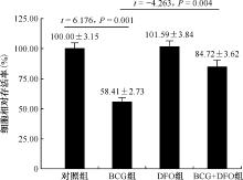
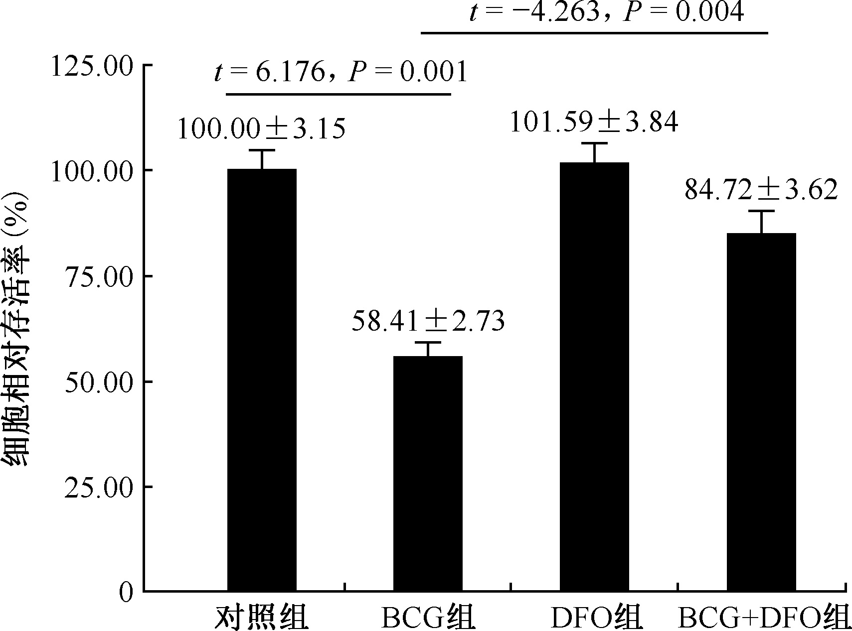
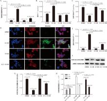
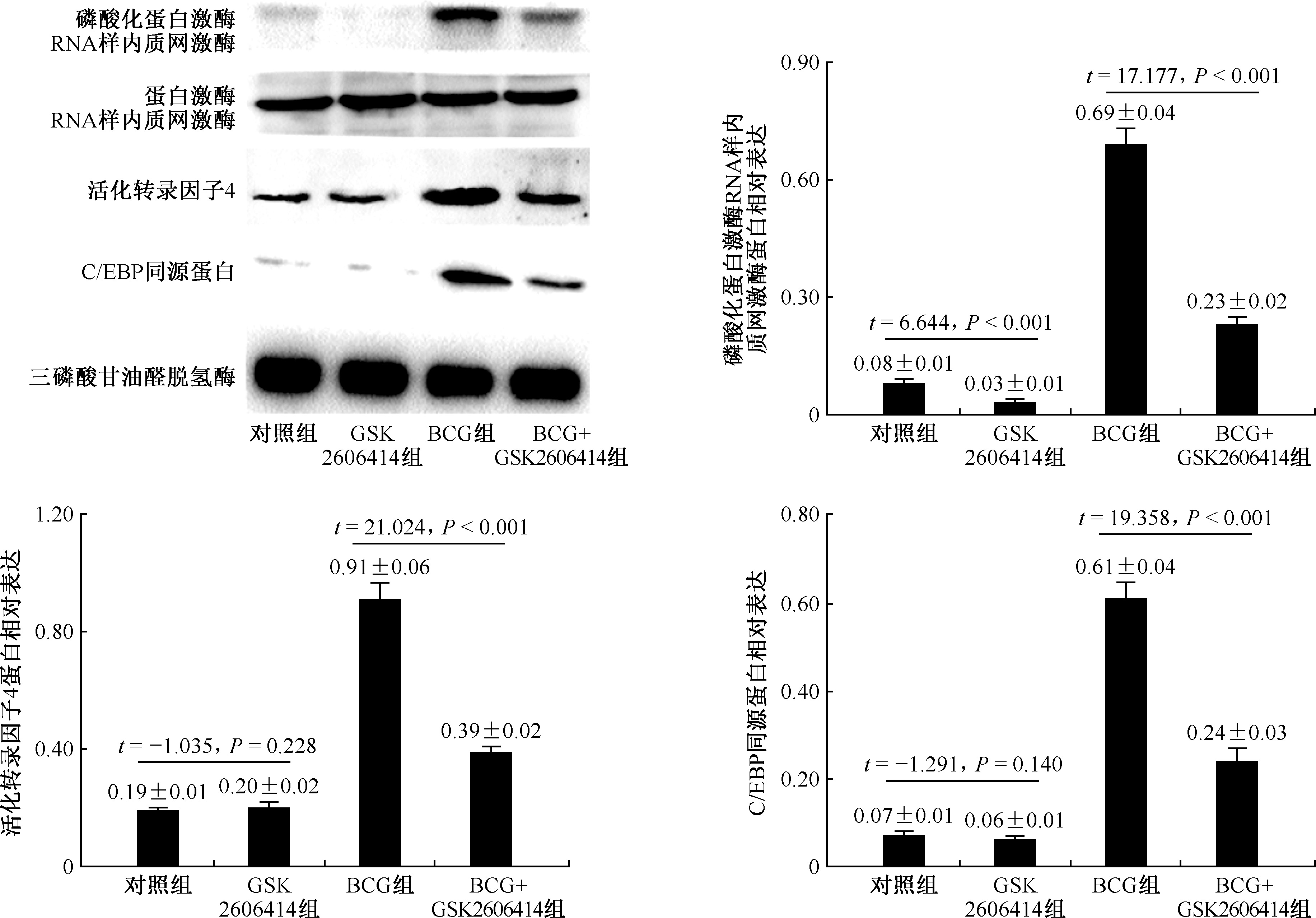
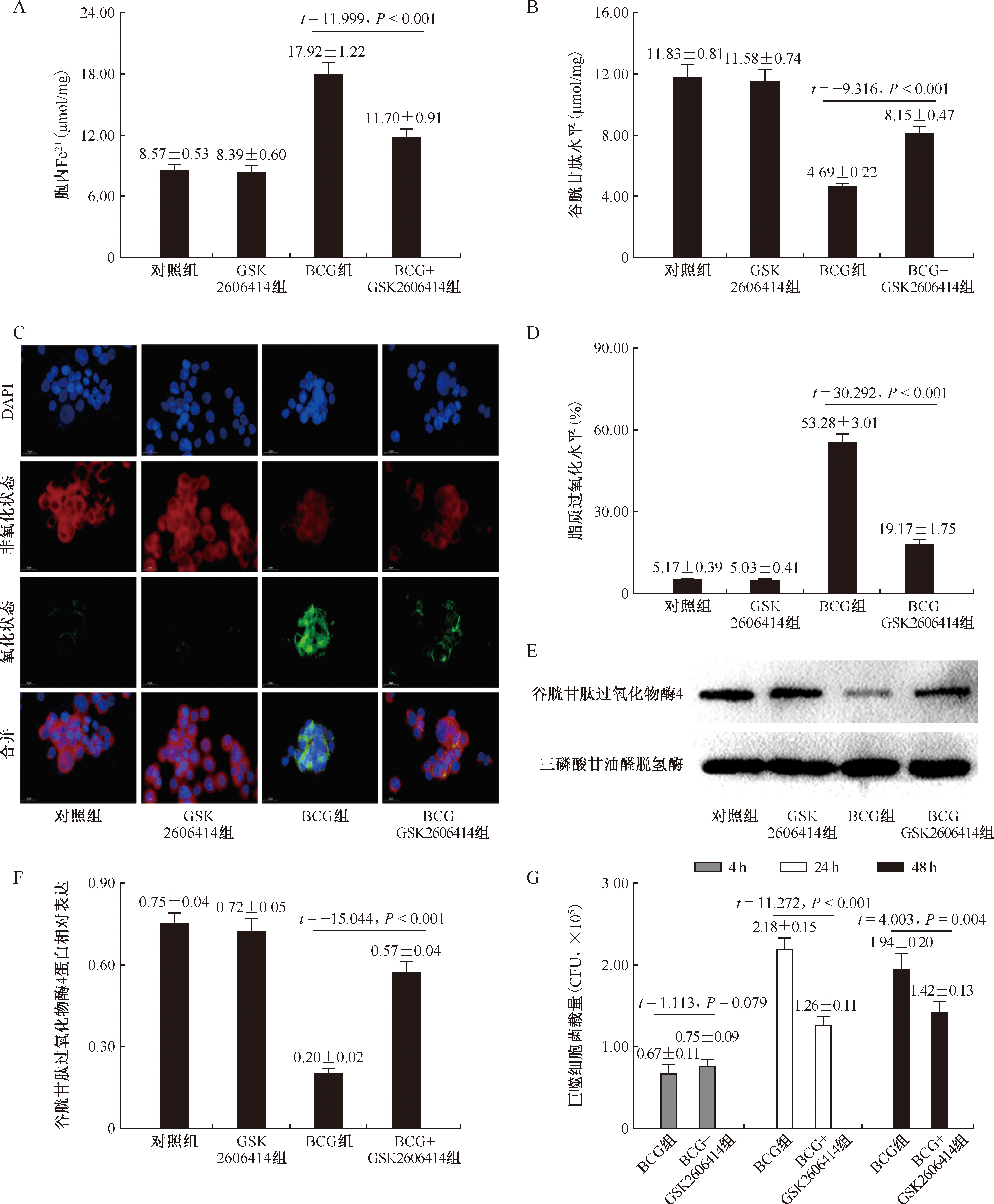
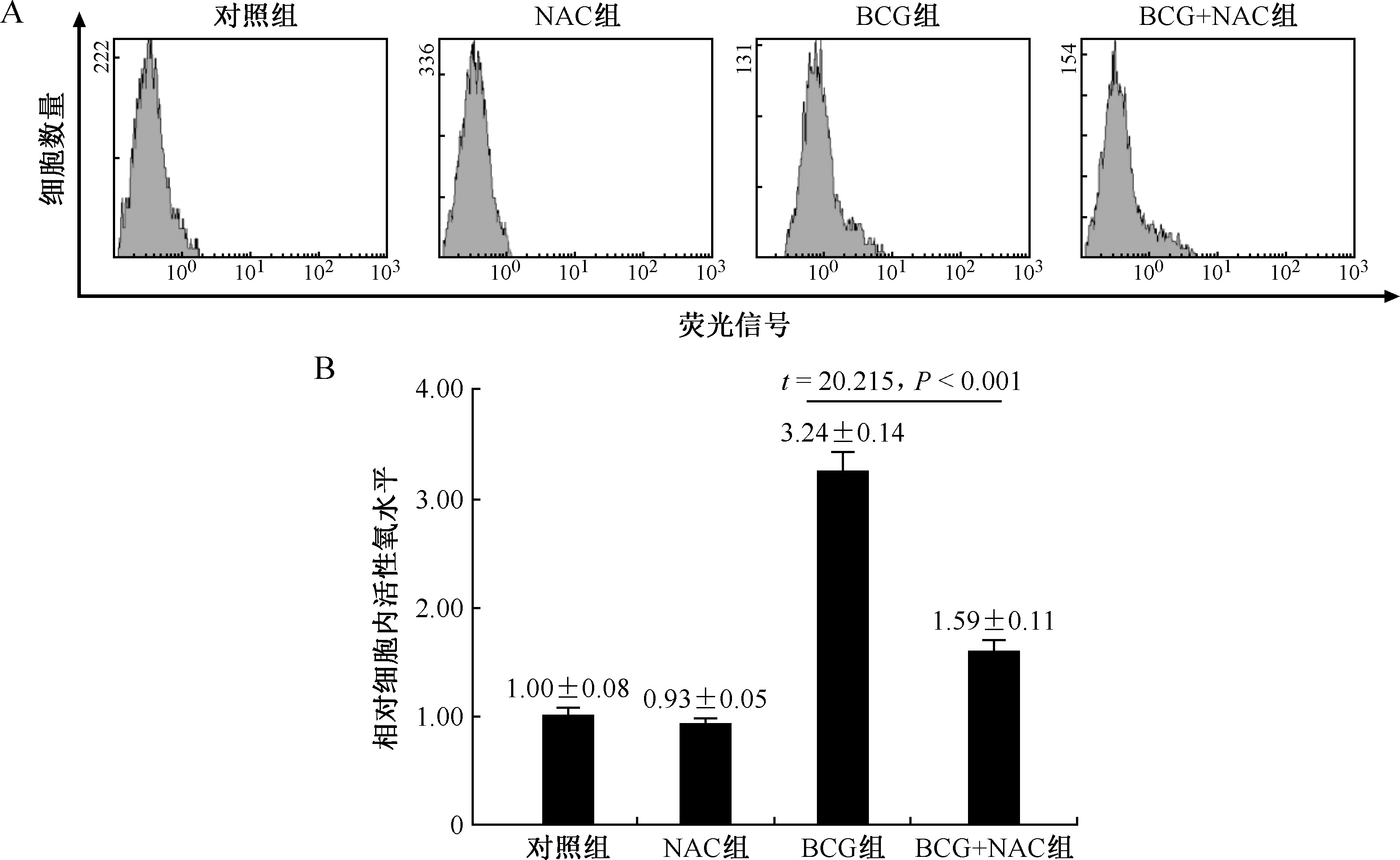
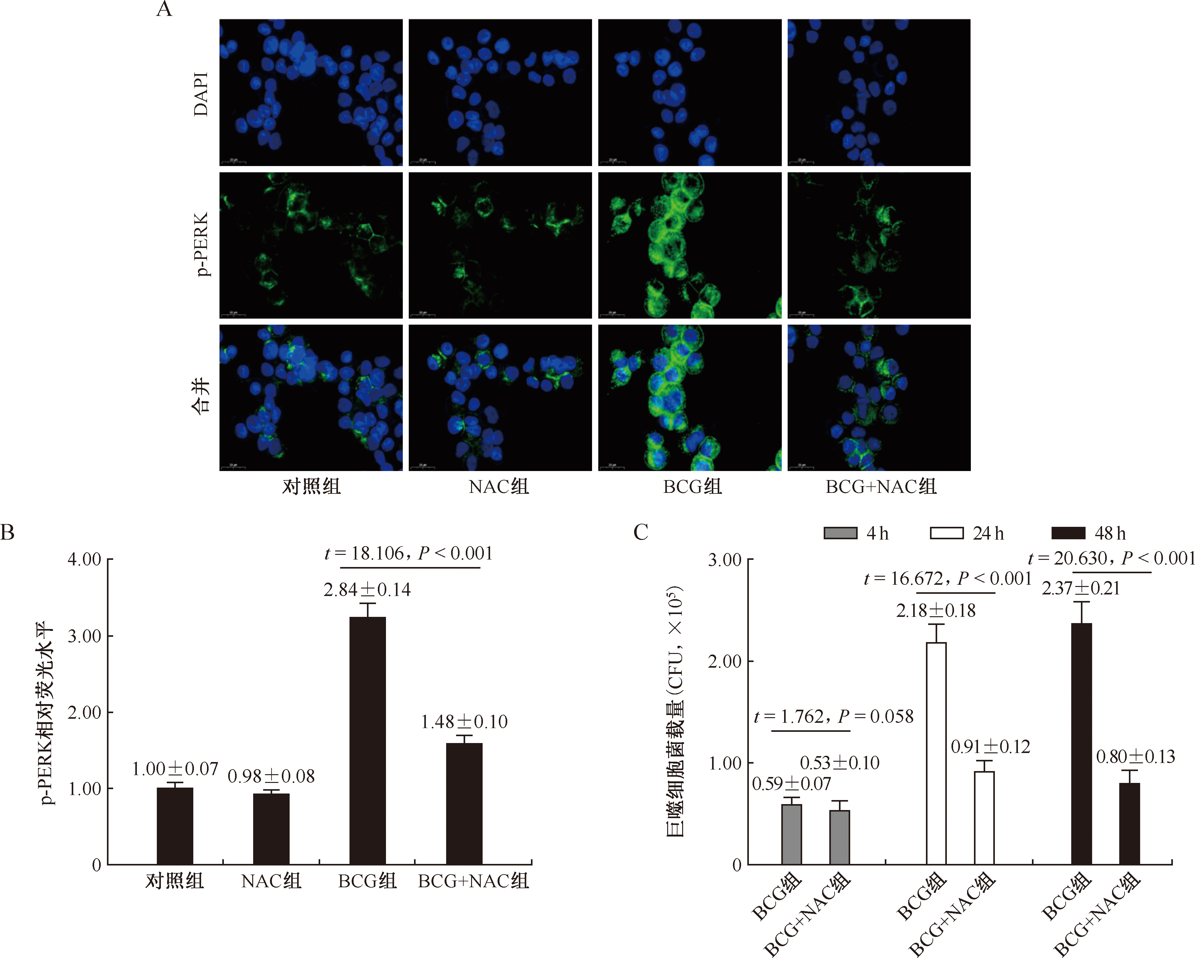
目的: 探讨活性氧(reactive oxygen species, ROS)/蛋白激酶RNA样内质网激酶(protein kinase RNA-like endoplasmic reticulum kinase, PERK)信号通路对卡介苗(BCG)感染引起的RAW264.7小鼠巨噬细胞铁死亡的调控作用。方法: 采用铁死亡螯合剂去铁胺(deferoxamine, DFO)研究铁死亡在BCG感染诱导细胞死亡中的作用,将RAW264.7细胞分为对照组、BCG组、DFO组、BCG+DFO组;采用PERK抑制剂GSK2606414研究PERK信号通路在BCG感染诱导细胞铁死亡中的作用,将RAW264.7细胞分为对照组、BCG组、GSK2606414组、BCG+GSK2606414组;采用ROS清除剂N-乙酰半胱氨酸(N-acetylcysteine, NAC)观察ROS在调控PERK中的作用,将RAW264.7细胞分为对照组、BCG组、NAC组和BCG+NAC组。通过比色法检测细胞乳酸脱氢酶(lactate dehydrogenase, LDH)释放率、Fe2+、谷胱甘肽(glutathione, GSH)及脂质过氧化水平;通过蛋白免疫印迹法检测谷胱甘肽过氧化物酶4(glutathione peroxidase 4, GPX4)、磷酸化PERK(p-PERK)、活化转录因子4(activating transcription factor 4, ATF4)及C/EBP同源蛋白(C/EBP-homologous protein, CHOP)表达水平;通过流式细胞术检测ROS含量;通过免疫荧光法检测p-PERK蛋白水平;通过菌落形成单位(colony-forming unit, CFU)实验计算BCG在细胞内的存活。结果: 与BCG组比较,BCG+DFO组细胞存活率[(84.72±3.62)% vs. (58.41±2.73)%]、GSH水平[(8.85±0.54)μmol/mg vs. (4.81±0.36)μmol/mg]和GPX4蛋白相对表达水平(0.82±0.06 vs. 0.33±0.03)升高(t=-4.263,P=0.004;t=-10.116,P<0.001;t=-10.519,P<0.001),LDH释放率[(15.70±3.18)% vs. (56.24±4.98)%]、Fe2+含量[(8.15±0.64)μmol/mg vs. (18.68±1.27)μmol/mg]和脂质过氧化水平[(22.18±2.24)% vs. (58.13±4.47)%]均明显降低(t=35.982,P<0.001;t=20.203,P<0.001;t=32.528,P<0.001)。与BCG组比较,BCG+GSK2606414组细胞中p-PERK蛋白相对表达水平(0.23±0.02 vs. 0.69±0.04)、ATF4蛋白相对表达水平(0.39±0.02 vs. 0.91±0.06)和CHOP蛋白相对表达水平(0.24±0.03 vs. 0.61±0.04),以及Fe2+[(11.70±0.91)μmol/mg vs. (17.92±1.22)μmol/mg]和脂质过氧化[(19.17±1.75)% vs. (53.28±3.01)%]水平均明显降低(t=17.177,P<0.001;t=21.024,P<0.001;t=19.358,P<0.001;t=11.999,P<0.001;t=30.292,P<0.001),细胞中GPX4蛋白相对表达水平(0.57±0.04 vs. 0.20±0.02)和GSH水平[(8.15±0.47)μmol/mg vs. (4.69±0.22)μmol/mg]则明显升高(t=-15.044,P<0.001;t=-9.316,P<0.001)。此外,BCG感染GSK2606414处理后的巨噬细胞,其24h和48h的菌载量[(1.72±0.15)×105CFU和(1.48±0.12)×105CFU]较BCG组[(3.51±0.28)×105CFU和(2.94±0.21)×105CFU]明显降低(t=17.576,P<0.001;t=15.225,P<0.001)。与BCG组(3.24±0.14)比较,BCG+NAC组细胞中的相对ROS水平(1.59±0.11)明显降低(t=20.215,P<0.001)。BCG感染NAC处理后的巨噬细胞,其24h和48h的菌载量[(0.91±0.12)×105CFU和(0.80±0.13)×105CFU]较BCG组[(2.18±0.18)×105CFU和(2.37±0.21)×105CFU]明显降低(t=16.672,P<0.001;t=20.630,P<0.001)。结论: BCG感染诱导的巨噬细胞铁死亡与ROS/PERK信号的激活有关。

关键词: 分枝杆菌, 结核, 巨噬细胞, 铁死亡, 活性氧

Abstract:

Objective: To investigate the role of reactive oxygen species (ROS)/protein kinase RNA-like endoplasmic reticulum kinase (PERK) signaling pathway in the regulation of ferroptosis in RAW264.7 macrophages induced by BCG infection. Methods: The ferroptosis inhibitor deferoxamine (DFO) was used to investigate the role of ferroptosis in BCG infection-induced apoptosis by dividing RAW264.7 cells into control, BCG, DFO, and BCG+DFO groups; the PERK inhibitor GSK2606414 was used to investigate the role of PERK signaling pathway in BCG infection-induced cell ferroptosis by dividing RAW264.7 cells into control, BCG, GSK2606414, and BCG+GSK2606414 groups. The ROS scavenger N-acetylcysteine (NAC) was used to investigate the role of ROS in the regulation of PERK by dividing RAW264.7 cells into control, BCG, NAC and BCG+NAC groups. Colorimetric assay was used to detect cellular lactate dehydrogenase (LDH) release rate, Fe2+, glutathione (GSH) and lipid peroxidation concentration. Immunoblotting was used to detect expression of glutathione peroxidase 4 (GPX4), phosphorylated PERK (p-PERK), activating transcription factor 4 (ATF4), and C/EBP-homologous protein (CHOP); flow cytometry was used to detect ROS concentration. Immunofluorescence was used to detect p-PERK protein concentration. Colony-forming unit (CFU) assay was used to calculate the survival of BCG in cells. Results: Compared with the BCG group, the BCG+DFO group had elevated cell survival rate ((84.72±3.62)% vs. (58.41±2.73)%, t=-4.263, P=0.004), GSH level ((8.85±0.54) μmol/mg vs. (4.81±0.36) μmol/mg, t=-10.116, P<0.001), and relative GPX4 protein expression level (0.82±0.06 vs. 0.33±0.03, t=-10.519, P<0.001); LDH release rate ((15.70±3.18)% vs. (56.24±4.98)%), Fe2+ ((8.15±0.64) μmol/mg vs. (18.68±1.27) μmol/mg) and lipid peroxidation level ((22.18±2.24)% vs. (58.13±4.47)%) were significantly reduced (t=35.982, P<0.001; t=20.203, P<0.001; t=32.528, P<0.001). Compared with the BCG group, the BCG+ GSK2606414 group had p-PERK (0.23±0.02 vs. 0.69±0.04), ATF4 (0.39±0.02 vs. 0.91±0.06), CHOP protein (0.24±0.03 vs. 0.61±0.04), Fe2+ ((11.70±0.91) μmol/mg vs. (17.92±1.22) μmol/mg) and lipid peroxidation ((19.17±1.75)% vs. (53.28±3.01)%) all significantly reduced (t=17.177, P<0.001; t=21.024, P<0.001; t=19.358, P<0.001; t=11.999, P<0.001; t=30.292, P<0.001, respectively), while GPX4 protein relative expression (0.57±0.04 vs. 0.20±0.02) and GSH level ((8.15±0.47) μmol/mg vs. (4.69±0.22) μmol/mg) were significantly elevated in cells (t=-15.044, P<0.001; t=-9.316, P<0.001). In addition, BCG-infected GSK2606414-treated macrophages had significantly lower bacterial loads at 24 h and 48 h ((1.72±0.15)×105CFU and (1.48±0.12)×105CFU) compared with those in the BCG group ((3.51±0.28)×105CFU and (2.94±0.21)×105CFU; t=17.576, P<0.001; t=15.225, P<0.001). The relative level of ROS in the cells of the BCG+NAC group (1.59±0.11) was significantly lower compared to the BCG group (3.24±0.14, t=20.215, P<0.001). The bacterial loads of BCG-infected NAC-treated macrophages were significantly lower at 24 and 48 h ((0.91±0.12)×105CFU and (0.80±0.13)×105CFU) compared to those of the BCG group ((2.18±0.18)×105CFU and (2.37±0.21)×105CFU; t=16.672, P<0.001; t=20.630, P<0.001). Conclusion: BCG infection-induced macrophage ferroptosis is associated with activation of ROS/PERK signaling.

Key words: Mycobacterium tuberculosis, Macrophages, Ferroptosis, Reactive oxygen species

中图分类号: