Email Alert | RSS    帮助

中国防痨杂志 ›› 2024, Vol. 46 ›› Issue (6): 687-698.doi: 10.19982/j.issn.1000-6621.20240127

• 论著 • 上一篇    下一篇

新型冠状病毒感染合并肺结核患者的临床特征和预后情况分析

李瑶, 方喆, 罗丹霖, 胡艳梅, 唐蜜, 唐志冈, 文新民, 张勇, 姚碧波, 王起, 易恒仲()   

  1. 湖南省胸科医院,长沙 410013
  • 收稿日期:2024-04-03 出版日期:2024-06-10 发布日期:2024-06-03
  • 通信作者: 易恒仲,Email:46498321@qq.com
  • 基金资助:
    湖南省科技厅临床医疗技术创新引导项目(2020SK50702);长沙市自然科学基金(kq2208097);长沙市自然科学基金(kq2208096);湖南省自然科学基金面上项目(2023JJ30335);湖南省科技创新平台与人才计划(2018SK7003)

Clinical characteristics and prognosis of patients with novel coronavirus infection complicated with pulmonary tuberculosis

Li Yao, Fang Zhe, Luo Danlin, Hu Yanmei, Tang Mi, Tang Zhigang, Wen Xinmin, Zhang Yong, Yao Bibo, Wang Qi, Yi Hengzhong()   

  1. Hunan Chest Hospital, Changsha 410013, China
  • Received:2024-04-03 Online:2024-06-10 Published:2024-06-03
  • Contact: Yi Hengzhong, Email: 46498321@qq.com
  • Supported by:
    Clinical Medical Technology Innovation Guidance Project of Hunan Provincial Department of Science and Technology(2020SK50702);Changsha Municipal Natural Science Foundation(kq2208097);Changsha Municipal Natural Science Foundation(kq2208096);Natural Science Foundation of Hunan Province(2023JJ30335);Science and Technology Innovation Platform and Talent Project of Hunan Province(2018SK7003)

摘要:

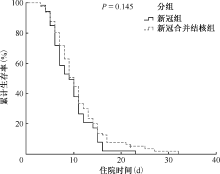
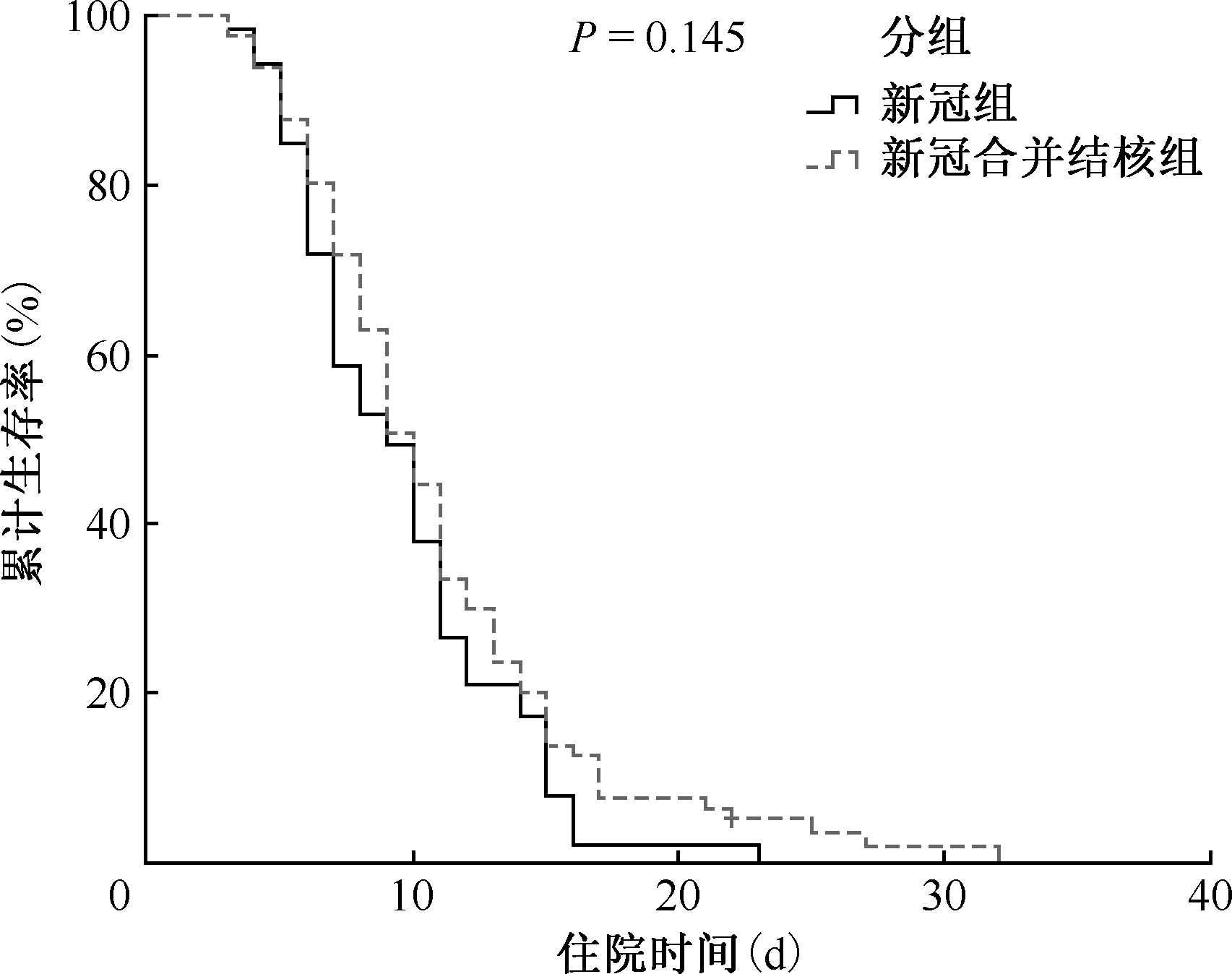
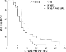
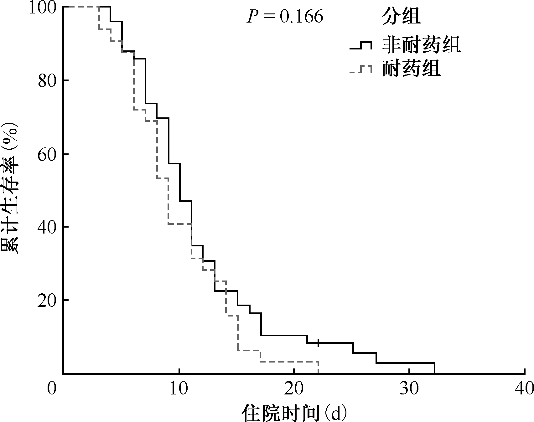
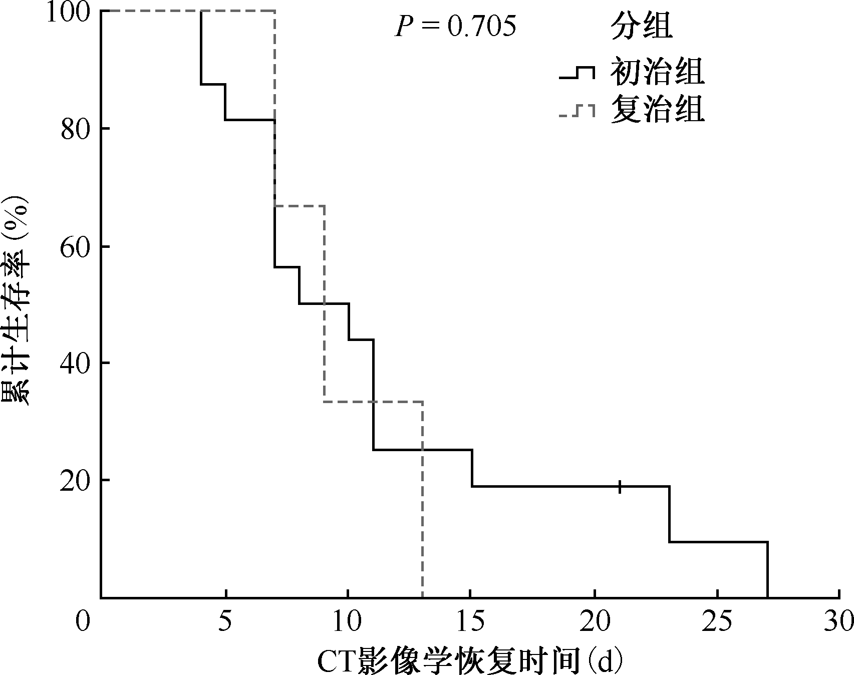
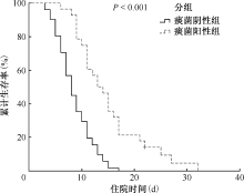
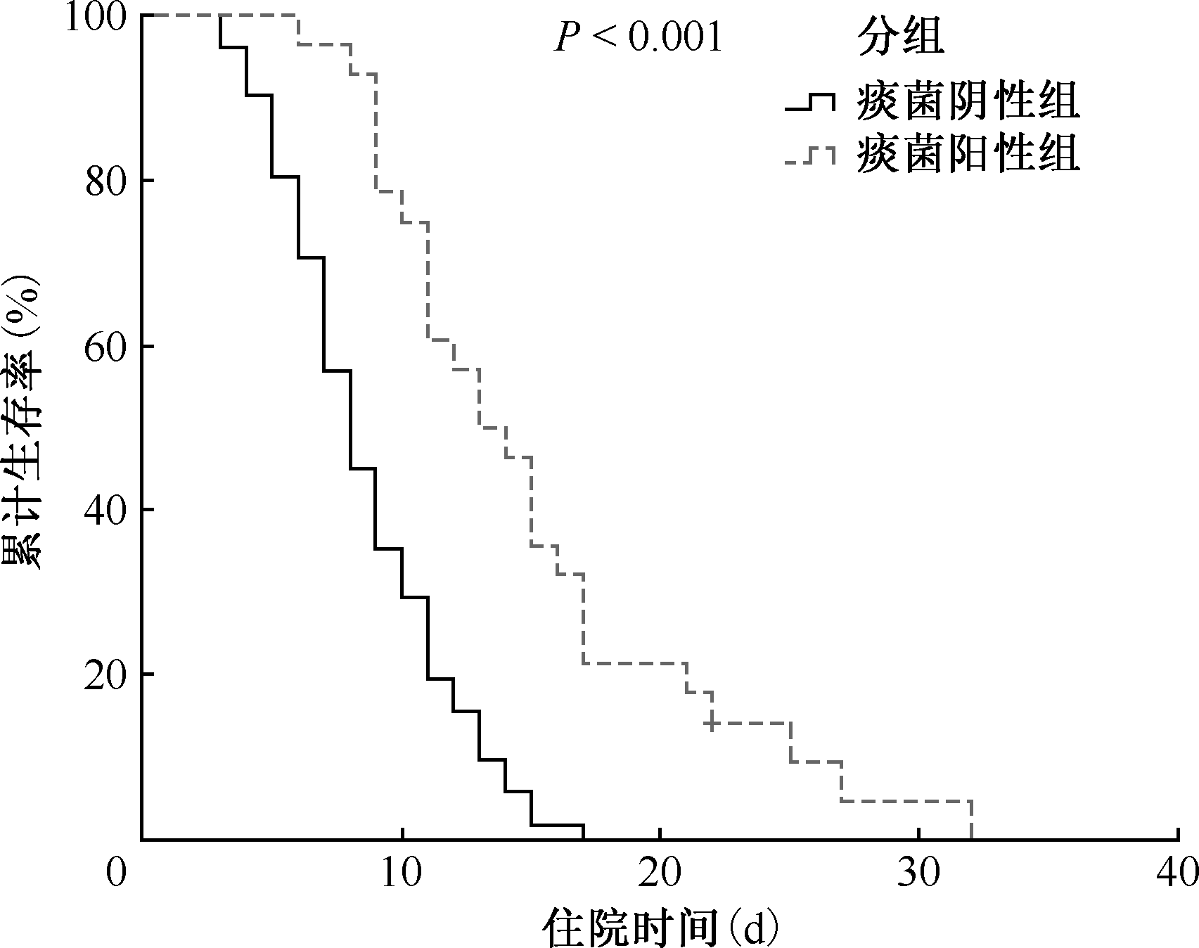
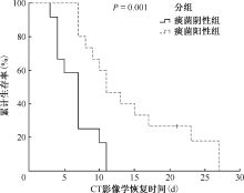
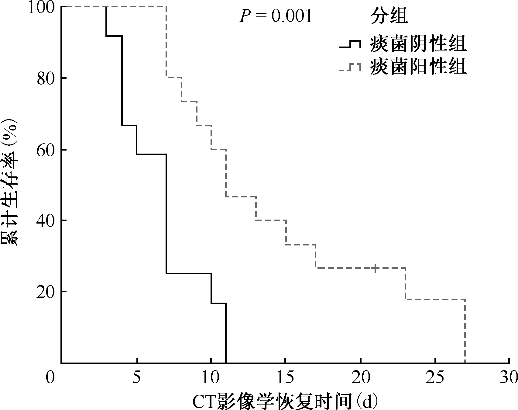
目的: 分析新型冠状病毒(简称“新冠病毒”)感染合并肺结核患者的临床表现、影像学特征及实验室检查指标情况,探讨合并肺结核对新冠病毒感染患者预后的影响。方法: 选取2022年12月至2023年1月于湖南省胸科医院住院并明确诊断为新冠病毒感染的患者作为研究对象,共140例。研究对象中单纯新冠病毒感染患者57例(新冠组);新冠病毒感染合并肺结核患者83例(新冠合并结核组),其中,合并耐药肺结核患者32例。收集研究对象的人口学特征、临床特征、实验室检查结果、预后情况。采用病例-对照设计,首先根据研究对象是否合并肺结核,将其分为新冠组和新冠合并结核组,比较两组研究对象的人口学特征、临床表现、实验室检查结果及预后情况;然后,将新冠合并结核组按照是否耐药、是否痰菌阳性、治疗分类进行分层分析。采用Kaplan-Meierr(KM)生存分析比较组间的生存差异,并进一步探讨影响合并肺结核对新冠病毒感染患者预后的危险因素。结果: 新冠合并结核组的预后更差,CT影像学恢复时间[中位数(四分位数)]为8.5(7.0,11.5)d,与新冠组相比 [7.0(5.0,10.0)d],差异有统计学意义(U=785.000,P=0.049)。新冠合并结核组患者首发症状不典型,发热的患者占38.6%(32/83),低于新冠组(56.1%,32/57),差异有统计学意义(χ2=4.311,P=0.040)。两组患者的影像学表现也有较大差异,新冠组最常出现的是典型磨玻璃影(52.6%,30/57),高于新冠合并结核组的21.7%(18/83),差异有统计学意义(χ2=14.362,P<0.001);新冠合并结核组的影像学表现复杂,最常见的为斑片影(91.6%,76/83)和条索影(91.6%,76/83),高于新冠组的56.1%(32/57)和28.1%(16/57),差异均有统计学意义(χ2值分别为24.052和60.471,P值均<0.001)。新冠合并结核组患者的淋巴细胞计数[中位数(四分位数)]为1.0(0.7,1.6)×109/L,明显低于新冠组的1.3(0.9,2.0)×109/L,差异有统计学意义(U=1736.000,P=0.015);白细胞计数[中位数(四分位数)]为5.0(3.8,7.1)×109/L,明显低于新冠组的6.0(4.8,7.9)×109/L,差异有统计学意义(U=1800.000,P=0.024);中性粒细胞计数[中位数(四分位数)]为3.3(2.2,4.7)×109/L,明显低于新冠组的3.6(3.0,5.2)×109/L,差异有统计学意义(U=1865.000,P=0.049);细胞毒性T细胞(CD3+CD8+)计数[中位数(四分位数)]为344.2(239.6,457.3)/μl,明显低于新冠组的567.6(437.8,618.6)/μl,差异有统计学意义(U=74.000,P=0.009)。生存分析显示,痰菌阳性是影响新冠合并结核患者预后的危险因素。结论: 新冠病毒感染合并肺结核患者住院时间及胸部影像学恢复时间延迟,合并痰菌阳性肺结核的新冠病毒感染患者预后更差。

关键词: 结核,肺, 冠状病毒感染, 共病现象, 疾病特征, 预后

Abstract:

Objective: This study aims to delineate the clinical presentations, radiographic features, and laboratory findings of patients co-infected with the novel coronavirus (COVID-19) and pulmonary tuberculosis, with a particular focus on assessing the prognostic implications of tuberculosis in the context of COVID-19 infection. Methods: The cohort comprised 140 patients admitted to Hunan Provincial Chest Hospital between December 2022 and January 2023 with confirmed COVID-19 infection. This group included 57 patients with isolated COVID-19 infection and 83 patients with concurrent pulmonary tuberculosis, of which 32 were diagnosed with drug-resistant tuberculosis. Comprehensive data on demographic characteristics, clinical manifestations, laboratory diagnostics, and outcomes were systematically gathered for analysis. A case-control study design was utilized. Initially, subjects were stratified into either the isolated COVID-19 group or the COVID-19 with tuberculosis co-infection group, facilitating a comparative analysis of demographic data, clinical manifestations, laboratory outcomes, and prognostic data. Subsequently, patients within the tuberculosis co-infection group were further categorized based on drug-resistance status, sputum test results, and treatment classification to refine the analytical depth. Kaplan-Meier survival analysis was employed to delineate the survival disparities between the groups. This analysis was instrumental in further identifying and evaluating the risk factors that may influence the prognosis of patients afflicted with both COVID-19 and pulmonary tuberculosis. Results: Patients co-infected with COVID-19 and tuberculosis exhibited a notably poorer prognosis, as evidenced by prolonged chest imaging recovery times (median (interquartile range, IQR)) of 8.5 (7.0, 11.5) days, compared to 7.0 (5.0, 10.0) days in the COVID-19-only group, achieving statistical significance (U=785.000, P=0.049). Furthermore, initial symptoms in the co-infected cohort were less typical, with fever presenting in only 38.6% (32/83) of cases, significantly lower than the 56.1% (32/57) observed in the COVID-19-only group (χ2=4.311, P=0.040). Radiographic findings between the groups revealed marked differences. In the isolated COVID-19 group, ground glass opacities were predominant, appearing in 52.6% (30/57) of cases, significantly exceeding the 21.7% (18/83) observed in the tuberculosis co-infected group (χ2=14.362, P<0.001). Conversely, the imaging profiles in the co-infected group were notably more complex, with patchy opacities and linear streaks being the most prevalent, identified in 91.6% (76/83) of cases—substantially higher than the 56.1% (32/57) and 28.1% (16/57) seen in the COVID-19-only group, with statistically significant differences (χ2=24.052 and 60.471, P<0.001 respectively). In patients with concurrent COVID-19 and tuberculosis, lymphocyte counts were significantly reduced at 1.0×10⁹/L (IQR: 0.7×10⁹/L to 1.6×10⁹/L), compared to 1.3×10⁹/L (IQR: 0.9×10⁹/L to 2.0×10⁹/L) in those with COVID-19 alone (U=1736.000, P=0.015). Similarly, white blood cell counts were lower in the co-infected group, recorded at 5.0×10⁹/L (IQR: 3.8×10⁹/L to 7.1×10⁹/L), versus 6.0×10⁹/L (IQR: 4.8×10⁹/L to 7.9×10⁹/L) in the COVID-19 only group, a statistically significant difference (U=1800.000, P=0.024). Neutrophil counts further supported these findings, being lower at 3.3×10⁹/L (IQR: 2.2×10⁹/L to 4.7×10⁹/L) compared to 3.6×10⁹/L (IQR: 3.0×10⁹/L to 5.2×10⁹/L) in patients with only COVID-19 (U=1865.000, P=0.049). Moreover, cytotoxic T cells (CD3+CD8+) counts were markedly reduced in the co-infected group at 344.2/μl (IQR: 239.6 to 457.3/μl), significantly lower than 567.6/μl (IQR: 437.8 to 618.6/μl) observed in the COVID-19 group (U=74.000, P=0.009). Survival analysis identified sputum positivity as a significant prognostic risk factor in these patients. Conclusion: Patients co-infected with COVID-19 and pulmonary tuberculosis experienced prolonged hospitalization and delayed recovery in chest imaging. Notably, those with sputum-positive pulmonary tuberculosis had significantly poorer prognostic outcomes.

Key words: Tuberculosis, pulmonary, Coronavirus infections, Comorbidity, Disease characteristics, Prognosis

中图分类号: