Email Alert | RSS    帮助

中国防痨杂志 ›› 2022, Vol. 44 ›› Issue (3): 258-263.doi: 10.19982/j.issn.1000-6621.20210711

• 论著 • 上一篇    下一篇

结核性脊膜炎MRI表现及治疗后动态变化研究

李多1, 吕岩2, 吕平欣3()   

  1. 1首都医科大学附属北京友谊医院放射科,北京 100050
    2首都医科大学附属北京胸科医院影像中心,北京 101149
    3北京老年医院放射科,北京 100095
  • 收稿日期:2021-12-20 出版日期:2022-03-10 发布日期:2022-03-08
  • 通信作者: 吕平欣 E-mail:lpx1209@163.com

Study of MRI characteristics of spinal tuberculous meningitis and the change after anti-tuberculosis treatment

LI Duo1, LYU Yan2, LYU Ping-xin3()   

  1. 1Department of Radiology, Beijing Friendship Hospital, Capital Medical University,Beijing 100050, China
    2Department of Radiology, Beijing Chest Hospital, Capital Medical University, Beijing 101149, China
    3Department of Radiology, Beijing Geriatric Hospital,Beijing 100095,China
  • Received:2021-12-20 Online:2022-03-10 Published:2022-03-08
  • Contact: LYU Ping-xin E-mail:lpx1209@163.com

摘要:

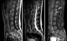
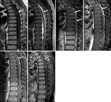
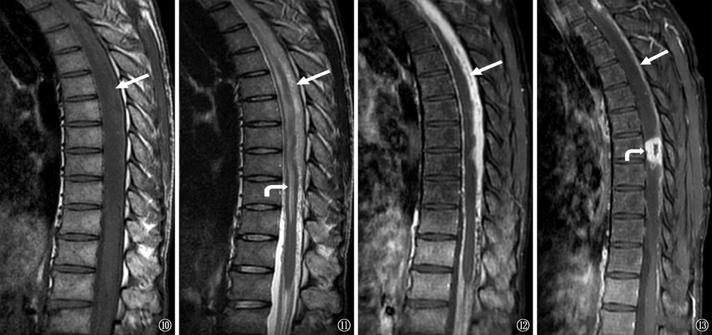
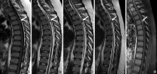
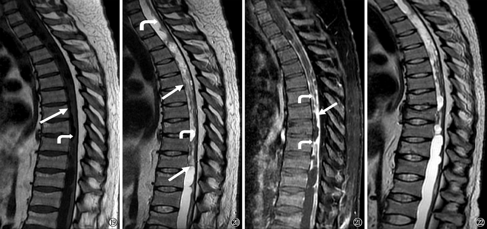
目的: 研究结核性脊膜炎MRI表现、治疗后演变及患者预后情况,以提高临床认识。方法: 通过《首都医科大学附属北京胸科医院病案电子化管理系统》和《图像获取及传输系统》(PACS),收集2012年1月1日至2016年12月31日确诊并有完整MRI资料的31例结核性脊膜炎患者,记录脊膜、蛛网膜下腔及脊髓改变。 结果: 96.8%(30/31)的患者有脊膜增厚和强化。48.4%(15/31)的患者蛛网膜下腔不规则或狭窄,12.9%(4/31)的患者蛛网膜下腔闭塞,6.5%(2/31)的患者有髓外硬膜内结核瘤。脊髓炎见于51.6%(16/31)的患者,而髓内结核瘤见于9.7%(3/31)的患者。58.3%(7/12)的患者在随访MRI中显示病情缓解,25.0%(3/12)的患者有进展,16.7%(2/12)患者的MRI表现无变化。71.0%(22/31)的结核性脊膜炎患者预后不良,包括13例死亡及9例残疾。结论: MRI增强扫描能发现增厚强化的脊膜、脊髓及蛛网膜下腔改变,并且能观察抗结核治疗后的疗效,可以作为结核性脊膜炎患者首选的影像检查方法。

关键词: 结核,脑膜, 软脑脊膜, 磁共振成像

Abstract:

Objective: To describe MRI characteristics of spinal tuberculous meningitis, the changes after anti-tuberculosis treatment and the prognosis of patients. Methods: Using the electronic medical record management system of Beijing Chest Hospital Affiliated to Capital Medical University and Picture Archiving and Communication System (PACS), 31 patients whose complete MRI data were maintained and diagnosed as tuberculous spinal meningitis from 2012 to 2016 were selected as study objects. Two radiologists reviewed MRI retrospectively and the changes of spinal meninges, subarachnoid space and spinal cord were recorded. Results: The MRI showed that 96.8% (30/31) patients had thickened and enhanced meninges, 48.4% (15/31) had irregular or narrow subarachnoid space, 12.9% (4/31) had obliteration of the subarachnoid space, and 6.5% (2/31) had intradural extramedullary tuberculomas. Myelitis was detected in 51.6% (16/31) patients, while intramedullary tuberculoma was detected in 9.7% (3/31) patients. Furthermore, 58.3% (7/12) patients showed remission in the follow-up MRI, 25.0% (3/12) patients had progression; and 16.7% (2/12) patients showed no change in MRI. 71.0% patients (22/31) had a poor prognosis, including 13 deaths and 9 disability cases. Conclusion: Enhanced MRI can show the thickened and enhanced meninges, change in the subarachnoid space and in the spinal cord. MRI can also reflect the changes after anti-tuberculosis treatment. Enhanced MRI should be the first choice of imaging examination for patients with tuberculous spinal meningitis.

Key words: Tuberculosis,meningeal, Pia mater, Magnetic resonance imaging

中图分类号: