Email Alert | RSS    帮助

中国防痨杂志 ›› 2020, Vol. 42 ›› Issue (6): 558-562.doi: 10.3969/j.issn.1000-6621.2020.06.005

• 论著 • 上一篇    下一篇

耐多药肺结核临床治愈患者CT随访转归征象的研究

张培泽, 付亮, 谭洁, 王玉香, 陈涛, 邓国防()   

  1. 518112 国家感染性疾病临床医学研究中心 深圳市第三人民医院肺二科
  • 收稿日期:2020-04-28 出版日期:2020-06-10 发布日期:2020-06-11
  • 通信作者: 邓国防 E-mail:jxxk1035@yeah.net
  • 基金资助:
    “十三五”国家科技重大专项(2018ZX10715004-002-007)

CT imaging characteristics of cured multidrug-resistant pulmonary tuberculosis

ZHANG Pei-ze, FU Liang, TAN Jie, WANG Yu-xiang, CHEN Tao, DENG Guo-fang()   

  1. National Clinical Research Center for Infectious Disease/Second Department of Pulmonary Diseases, Third People’s Hospital of Shenzhen, Shenzhen 518112, China
  • Received:2020-04-28 Online:2020-06-10 Published:2020-06-11
  • Contact: DENG Guo-fang E-mail:jxxk1035@yeah.net

摘要:

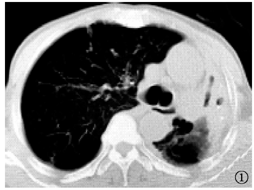
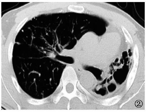
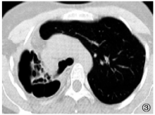
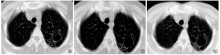
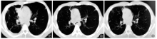
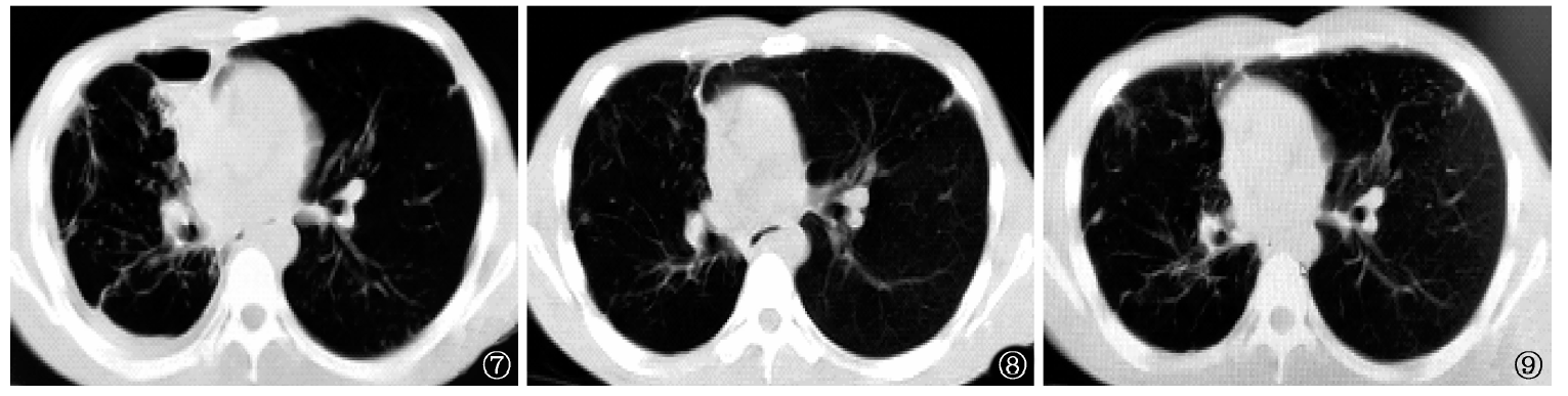
目的 评价耐多药肺结核(MDR-PTB)患者临床治愈后CT随访转归征象的情况。方法 回顾性分析2012年3月至2018年3月深圳市第三人民医院通过细菌学确诊的MDR-PTB临床治愈并随访2年无复发的患者,共计42例。其中初治患者18例(初治组),复治患者24例(复治组),分别对停药时、停药12个月时、停药24个月时患者的肺部CT征象转归情况进行分析。结果 42例患者在停药时、停药12个月时和停药24个月时,肺结核活动性征象的检出率分别为57.1%(24/42)、42.9%(18/42)和31.0%(13/42),稳定性征象的检出率分别为69.0%(29/42)、81.0%(34/42)和83.3%(35/42),不确定性征象的检出率均为40.5%(17/42)。在停药时,初治组与复治组活动性征象检出率分别为33.3%(6/18)和75.0%(18/24),稳定性征象检出率分别为50.0%(9/18)和83.3%(20/24),差异均有统计学意义(χ 2=7.292,P=0.007;χ 2=5.347,P=0.021);停药12个月时,两组患者活动性征象检出率分别为27.8%(5/18)和54.2%(13/24),稳定性征象检出率分别为83.3%(15/18)和79.2%(19/24),差异均无统计学意义(χ 2=2.925,P=0.087;χ 2=0.116,P=1.000);停药24个月时,两组患者活动性征象检出率分别为22.2%(4/18)和37.5%(9/24),稳定性征象检出率分别为88.9%(16/18)和79.2%(19/24),差异均无统计学意义(χ 2=1.123,P=0.289;χ 2=0.700,P=0.679)。在停药时、停药12个月时和停药24个月时,不确定性征象在初治组的检出率均为16.7%(3/18),在复治组的检出率均为58.3%(14/24),两组比较差异均有统计学意义(χ 2值均为7.412,P值均为0.006)。 结论 MDR-PTB患者达到临床治愈停药标准后,CT表现以损伤延迟修复为主要表现;与初治MDR-PTB患者比较,复治MDR-PTB患者肺部CT扫描显示出更多不确定征象。

关键词: 结核, 肺, 结核, 抗多种药物性, 临床对照试验, 体层摄影术, X线计算机, 回顾性研究

Abstract:

Objective To evaluate the characteristics of lung CT image in patients with cured multidrug-resistant pulmonary tuberculosis (MDR-PTB). Methods Forty-two patients who were diagnosed with MDR-TB from March 2012 to March 2018 in the Third People’s Hospital of Shenzhen and cured and no relapse occurred in two years were enrolled. Eighteen patients were divided into newly treated group and 24 patients were retreated groups. Consecutive lung CT images of 42 cured MDR-PTB patients were retrieved. The dynamic CT sign change at the time point of cure, 12 and 24 months after cure were analyzed. Results Respectively, 57.1% (24/42), 42.9% (18/42), and 31.0% (13/42) patients had active signs of PTB at the time point of cure, 12 months after cure, and 24 months after cure. Stable signs were observed in 69.0% (29/42), 81.0% (34/42), and 83.3% (35/42) patients at these time points. Uncertain signs were found in 40.5% (17/42) patients. At the time of cure, active signs were observed in 33.3% (6/18) newly treated group, and 75.0% (18/24) retreated group, and stable signs in 50.0% (9/18) newly treated and 83.3% (20/24) in retreated. The differences between two groups were statistically significant (χ 2=7.292, P=0.007; χ 2=5.347, P=0.021). At 12 months after cure, active signs were observed in 27.8% (5/18) newly treated, and 54.2% (13/24) in retreated, and stable signs in 83.3% (15/18) newly treated and in 79.2% (19/24) retreated. The differences between two groups were not statistically significant (χ 2=2.925, P=0.087; χ 2=0.116, P=1.000). At 24 months after cure, active signs were found in 22.2% (4/18) newly treated, and 37.5% (9/24) retreated, and stable signs in 88.9% (16/18) newly treated, and 79.2% (19/24) in retreated, and no statistical differences were found (χ 2=1.123, P=0.289; χ 2=0.700, P=0.679). At these different time points, uncertain signs were observed in 16.7% (3/18) newly treated, and 58.3% (14/24) retreated, and there was a statistical significance between the two groups (χ 2=7.412, P=0.006; χ 2=7.412, P=0.006; χ 2=7.412, P=0.006). Conclusion CT images showed signs of delayed repairment of pulmonary damage in most MDR-PTB patients after cure. Retreated MDR-PTB patients had more uncertain signs than newly treated MDR-PTB patients.

Key words: Tuberculosis, pulmonary, Tuberculosis, multidrug-resistant, Controlled clinical trial, Tomography, X-ray computed, Retrospective studies