Email Alert | RSS    帮助

中国防痨杂志 ›› 2019, Vol. 41 ›› Issue (4): 414-420.doi: 10.3969/j.issn.1000-6621.2019.04.009

• 论著 • 上一篇    下一篇

磁共振体素内不相干运动扩散加权成像在诊断脊柱结核中的价值

陈鹰,郑欢露,姚娟,李白艳,郭辉()   

  1. 830054 乌鲁木齐,新疆医科大学第一附属医院放射科
  • 收稿日期:2018-10-27 出版日期:2019-04-10 发布日期:2019-04-08
  • 通信作者: 郭辉 E-mail:guohui9804@126.com
  • 基金资助:
    新疆维吾尔自治区自然科学基金面上项目(2017D01C300)

Value of intravoxel incoherent motion diffusion weighted magnetic resonance imaging in diagnosis of spinal tuberculosis

Ying CHEN,Huan-lu ZHENG,Juan YAO,Bai-yan LI,Hui GUO()   

  1. Department of Radiology, First Affiliated Hospital of Xinjiang Medical University, Urumqi 830054, China
  • Received:2018-10-27 Online:2019-04-10 Published:2019-04-08
  • Contact: Hui GUO E-mail:guohui9804@126.com

摘要:

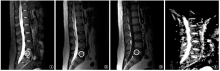
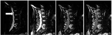
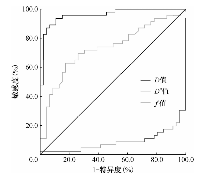
目的 探讨磁共振体素内不相干运动(intravoxel incoherent motion,IVIM)扩散加权成像(diffusion-weighted imaging,DWI)在脊柱结核患者中灌注分数(perfusion fraction,f)、单纯扩散系数(pure diffusion coefficient,D)、假性扩散系数(pseudodiffusion coefficient,D *)的参数值范围及其诊断价值。方法 选择2018年1—9月在新疆医科大学第一附属医院经临床活检、手术组织标本病理检查证实的18例脊柱结核患者作为结核组,男13例,女5例;年龄20~79岁,平均(40.39±14.84)岁;结核病变累及椎体46个,其中5例胸椎结核病变共累及23个椎体,13例腰椎结核病变共累及23个椎体。同期在就诊患者或人群中选取与结核组患者性别构成比相同、平均年龄相差不超过5岁、与结核组受累椎体节段一致且椎体正常的18名正常志愿者作为对照组。36例(名)受检者均行Signa 3.0 T MR常规和IVIM-DWI序列扫描(包括自旋回波矢状面T1WI、T2WI、压脂序列扫描,冠状面轴面T2WI序列),采用双指数模型后处理软件,得到脊柱结核组(病变椎体、跳跃2椎体、椎间盘)和与之相对应的正常对照组(正常椎体、椎间盘)的IVIM定量,应用IVIM模型计算两组fDD *等参数指标并进行比较,采用SPSS 16.0软件进行统计学分析,fDD *均为非正态分布资料,采用秩和检验,以P<0.05为差异有统计学意义。采用受试者工作曲线(ROC曲线)分析结核病变椎体fDD *参数值的曲线下最大面积、敏感度、特异度、最佳诊断阈值。结果 结核组病变椎体f值[12.91(8.15,22.73)%]低于对照组正常椎体[37.16(30.45,47.07)%](Z=6.841,P<0.001);D值[0.88(0.73,1.40)×10 -3mm 2/s]、D *值[39.99(20.15,66.35)×10 -3mm 2/s]明显高于正常椎体[分别为0.07(-0.12,0.28)×10 -3mm 2/s,20.37(12.26,29.97)×10 -3mm 2/s](Z值分别为7.598、3.842,P值均<0.001)。结核组病变椎体与跳跃2个椎体的正常椎体f值比较,前者低于后者 [40.51(33.75,46.28)%](Z=3.421,P=0.001);D值前者明显高于后者 [0.05(-0.20,0.15)×10 -3mm 2/s(Z=3.743,P<0.001)。结核组病变椎间盘f值[6.72(4.36,11.53)%]、D值[2.06(1.92,2.26)×10 -3mm 2/s]均高于对照组正常椎间盘[分别为5.72(3.00,7.85)%、1.88(1.79,1.85)×10 -3mm 2/s](Z=2.276,P=0.023;Z=3.919,P<0.001)。ROC曲线获得D值的曲线下最大面积为0.960,敏感度为95.74%,特异度为87.56%,最佳诊断阈值为0.63×10 -3mm 2/s。结论 IVIM-DWI可定量评估病变水分子扩散和微血管灌注特性,通过IVIM中fDD *各定量参数值范围分析,D值对脊柱结核诊断敏感度、特异度最高,为临床诊断脊柱结核可提供有效的参考。

关键词: 结核, 脊柱, 磁共振成像, 弥散, 回波平面成像, 分子成像, 诊断, 鉴别, 评价研究

Abstract:

Objective To explore the following parameter range of intravoxel incoherent motion-diffusion-weighted magnetic resonance imaging: perfusion fraction (f), pure diffusion coefficient (D) and pseudo-diffusion coefficient (D *), as well as its diagnostic value in diagnosis of patients with spinal tuberculosis (TB). Methods Eighteen patients with spinal tuberculosis, who were confirmed by pathological examination with clinical biopsy and surgical tissue specimens at the First Affiliated Hospital of Xinjiang Medical University from January to September 2018, were recruited as TB group, including 13 males and 5 females and the age ranged from 20 to 79 years old with an average of (40.39±14.84) years old. Among those patients, tuberculosis had involved 46 vertebral bodies in total, including 23 vertebral bodies in 5 cases with thoracic TB and 23 vertebral bodies in 13 cases with lumbar TB. In the same period, 18 volunteers with normal vertebral bodies were recruited as control group, which had the same gender composition ratio with the TB group, and the difference of its average age was within 5 years old compared to the TB group. All 36 cases in both TB group and control group received Signa 3.0 T MR routine scan and IVIM-DWI sequence scan (including spin echo sagittal T1WI, T2WI, lipostatic sequence scan, coronal axial T2WI sequence). A double exponential model post-processing software was used to obtain the IVIM quantifications of the cases in TB group (the diseased vertebral body, the jumping vertebral body, the intervertebral disc) and in control group (normal vertebral body, intervertebral disc). The IVIM model was used to calculate the parametric indicators (f, D, D*, etc.) of the cases in TB group and control group respectively and the results were compared by using SPSS 16.0 software. The non-normal distribution data were analyzed by rank-sum test. P<0.05 was regarded as statistically significant difference. The receiver operating curve (ROC curve) was used to analyze the maximum area under the curve, sensitivity, specificity and optimal diagnostic threshold of the parameters f, D and D * of the vertebral body. Results The f-value of the vertebral body in the tuberculosis group (12.91 (8.15, 22.73)%) was lower than that in the control group (37.16 (30.45, 47.07)%) (Z=6.841, P<0.001); the D value (0.88 (0.73, 1.40)×10 -3mm 2/s) and D * value (39.99 (20.15, 66.35)×10 -3mm 2/s) in the TB group was significantly higher than that in the control group respectively (0.07 (-0.12, 0.28)×10 -3mm 2)/s, 20.37 (12.26, 29.97)×10 -3mm 2/s) (Z=7.598, 3.842, Ps<0.001). The f value of TB vertebral body in the TB group was lower than that of two vertebral bodies in the control group (40.51 (33.75, 46.28)%) (Z=3.421, P=0.001); the D values were significantly higher than the latter (0.05 (-0.20, 0.15)×10 -3mm 2/s (Z=3.743, P<0.001; Z=1.730, P=0.042). The f value (6.72 (4.36, 11.53)%) and the D value (2.06 (1.92, 2.26)×10 -3mm 2/s) of the intervertebral disc in the tuberculosis group were higher than those in the control group respectively (5.72 (3.00, 7.85)%, 1.88 (1.79, 1.85)×10 -3 mm 2/s) (Z=2.276, P=0.023; Z=3.919, P<0.001). The maximum area under the curve of D value which obtained from the ROC curve was 0.960, the sensitivity was 95.74%, the specificity was 87.56%, and the optimal diagnostic threshold was 0.63×10 -3 mm 2/s. Conclusion IVIM-DWI can quantitatively evaluate the diffusion and microvascular perfusion characteristics of diseased water molecules. Through analysis of the range of quantitative parameter (f, D and D *) value in IVIM, D value has the highest sensitivity and specificity in the diagnosis of spinal tuberculosis, which provides a valuable reference for clinical diagnosis of spinal tuberculosis.

Key words: Tuberculosis, spinal, Diffusion magnetic resonance imaging, Echo-planar imaging, Molecu-lar imaging, Diagnosis, differential, Evaluation studies