Email Alert | RSS    帮助

中国防痨杂志 ›› 2019, Vol. 41 ›› Issue (3): 288-293.doi: 10.3969/j.issn.1000-6621.2019.03.009

• 论著 • 上一篇    下一篇

特征金字塔网络在胸部X线摄影图像上筛检肺结核的价值

曹盼1,王斐1,刘哲1,刘锦程2,梁矿立2,袁吉欣2,池峰3,黄烨东3,杨健1()   

  1. 1 710061 西安交通大学第一附属医院医学放射科
    2 陕西省结核病防治院
    3 西安盈谷网络科技有限公司
  • 收稿日期:2019-01-03 出版日期:2019-03-10 发布日期:2019-03-15
  • 通信作者: 杨健 E-mail:yj1118@mail.xjtu.edu.cn
  • 基金资助:
    国家重点研发计划(2016YFC0100300);国家自然科学基金面上项目(81471631);国家自然科学基金面上项目(81771810);国家自然科学基金面上项目(51706178);2011年教育部“新世纪优秀人才支持计划”(NCET-11-0438)

Value of FPN in pulmonary tuberculosis screening on the thoracic radiography images

Pan CAO1,Fei WANG1,Zhe LIU1,Jin-cheng LIU2,Kuang-li LIANG2,Ji-xin YUAN2,Feng CHI3,Ye-dong HUANG3,Jian YANG1()   

  1. 1 Department of Diagnostic Radiology, the First Affiliated Hospital of Xi’an Jiao tong University, Xi’an 710061,China
  • Received:2019-01-03 Online:2019-03-10 Published:2019-03-15
  • Contact: Jian YANG E-mail:yj1118@mail.xjtu.edu.cn

摘要:

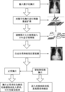
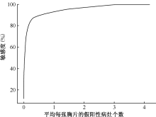
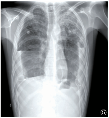
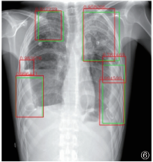
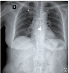
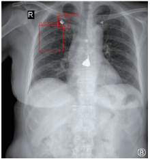
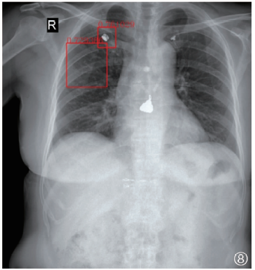
目的 评估特征金字塔网络(FPN)在胸部X线摄影图像(以下简称“胸片”)上对肺结核进行筛检的价值。方法 本研究采用回顾性分析,收集2016年1月至2017年12月陕西省结核病防治院住院的490例肺结核患者胸片和100名门诊健康体检者胸片,另纳入美国国立卫生研究院公开数据集中国深圳和美国马里兰州蒙哥马利县分别收集的332例和58例肺结核患者胸片。采用FPN对胸片和病灶分别进行分类和定位,由2名结核病院影像科医师对以上数据中的肺结核胸片进行审查和图像标注,将标注好的肺结核胸片经数据调整、扩增后送入FPN,对FPN进行训练,得到最终检测模型,然后使用独立的数据集来测试FPN的性能和泛化能力,以痰涂片阳性和有丰富经验的结核病专科医院影像科医生评估为标准,分析FPN区分肺结核患者胸片和健康人胸片的敏感度、特异度、准确度,以人工标记的病灶为标准评价FPN定位肺结核病灶的敏感度和假阳性率。图像中病变检测定位使用了自由响应受试者工作特性曲线(FROC)得分来评价FPN的性能。结果 在测试集上FPN诊断肺结核的敏感度、特异度和准确度分别为96.0%(96/100)、76.0%(76/100)、86.0%(172/200)。在100张测试集阳性胸片上共标记226处病灶,FPN共检出242处病灶,敏感度和假阳性率分别为87.6%(198/226)和14.0%(34/242),自由响应曲线FROC定位得分最高达88.0%。结论 FPN可对肺结核患者胸片和健康人胸片进行有效分类,并且实现对病灶位置的定位,为实现基于深度学习网络进行肺结核分类和病灶定位提供了参考依据。

关键词: 结核, 肺, 放射摄影术, 胸部, 人工智能, 诊断, 鉴别, 深度学习, 自动筛查

Abstract:

Objective To evaluate the value of feature pyramid network (FPN) for screening pulmonary tuberculosis on the thoracic radiography images (hereinafter referred to as “chest images”).Methods In this retros-pective study, chest images of 490 pulmonary tuberculosis patients who were hospitalized in the Tuberculosis Hospital of Shaanxi Province from January 2016 to December 2017 were collected, as well as chest images of 100 healthy outpatient controls. In addition, chest images of 332 and 58 pulmonary tuberculosis patients separately from Shenzhen, China and Montgomery, Maryland, USA in the NIH public dataset were also included. FPN was employed to classify and localize the radiographs and lesions. Two radiologists from tuberculosis hospitals examined and labeled the chest images of pulmonary tuberculosis patients in the above data. After data alignment and augmentation, the annotated radiographs were sent to the FPN, and the FPN was trained to obtain the final detection model. Then, the performance and generalization ability of FPN were tested with independent dataset, and the sensitivity, specificity and accuracy of FPN in distinguishing chest images of pulmonary tuberculosis patients from healthy controls were analyzed according to the criteria of positive sputum smear and evaluation by experienced radiologists. Meanwhile, the sensitivity and false positive rate of FPN in localizing tuberculosis lesions were evaluated based on manually labeled lesions. For the lesion detection and localization in the images, the free response receiver operating characteristic (FROC) score was used to evaluate the performance of FPN.Results The sensitivity, specificity and accuracy of FPN in diagnosing pulmonary tuberculosis on the test sets were 96.0% (96/100), 76.0% (76/100) and 86.0% (172/200), respectively. A total of 226 lesions were labeled on 100 positive chest images, while 242 lesions were detected in FPN, with sensitivity and false positive rate of 87.6% (198/226) and 14.0% (34/242), respectively. The FROC localization score could be up to 88.0%. Conclusion FPN can effectively differentiate chest images of pulmonary tuberculosis patients from healthy controls and implement the localization of lesions, which provide a reference for the deep learning-based classification and lesion localization of tuberculosis.

Key words: Tuberculosis, pulmonary, Radiography thoracic, Chest X-ray, Artificial intelligence, Diagnosis, differential, Deep learning, Automatic screening