
Chinese Journal of Antituberculosis ›› 2025, Vol. 47 ›› Issue (4): 444-453.doi: 10.19982/j.issn.1000-6621.20240579
• Original Articles • Previous Articles Next Articles
Hu Yifan, Du Boping, Wu Yadong, Zhu Chuanzhi, Zhang Lanyue, Jia Hongyan, Sun Qi, Pan Liping, Zhang Zongde, Li Zihui( )
)
Received:2024-12-20
Online:2025-04-10
Published:2025-04-02
Contact:
Li Zihui, Email: Supported by:CLC Number:
Hu Yifan, Du Boping, Wu Yadong, Zhu Chuanzhi, Zhang Lanyue, Jia Hongyan, Sun Qi, Pan Liping, Zhang Zongde, Li Zihui. Experimental study on the role of Mce4C in the uptake and utilization of cholesterol by Mycobacterium tuberculosis[J]. Chinese Journal of Antituberculosis, 2025, 47(4): 444-453. doi: 10.19982/j.issn.1000-6621.20240579
Add to citation manager EndNote|Ris|BibTeX
URL: https://www.zgflzz.cn/EN/10.19982/j.issn.1000-6621.20240579
| 引物名称 | 引物序列(5'~3') |
|---|---|
| sigA-F | GTCTGGGATGAAGACGAGT |
| sigA-R | CGATCTGTTTGAGGTAGGC |
| mce4C-F | CGCCGAACAGGTCAACAA |
| mce4C-R | GCAACATCGTCGATCCCAGA |
| 组别 | mce4C相对表达量 | q12值 | P值 | q13值 | P值 | q23值 | P值 |
|---|---|---|---|---|---|---|---|
| 苏通培养基添加胆固醇培养1周 | 1.936 | 0.413 | 10.010 | 0.001 | 8.076 | 0.003 | |
| 组1:无胆固醇组(对照) | 1.000±0.588 | ||||||
| 组2:0.01%胆固醇 | 1.390±0.162 | ||||||
| 组3:0.1%胆固醇 | 3.622±1.031 | ||||||
| F值 | 28.200 | ||||||
| P值 | 0.001 | ||||||
| 苏通培养基添加胆固醇培养4周 | 2.777 | 0.202 | 6.225 | 0.011 | 3.447 | 0.111 | |
| 组1:无胆固醇组(对照) | 1.006±0.030 | ||||||
| 组2:0.01%胆固醇 | 1.234±0.163 | ||||||
| 组3:0.1%胆固醇 | 1.518±0.182 | ||||||
| F值 | 9.724 | ||||||
| P值 | 0.013 | ||||||
| 含6%甘油苏通培养基添加胆固醇培养1周 | 0.734 | 0.865 | 6.302 | 0.010 | 5.569 | 0.018 | |
| 组1:无胆固醇组(对照) | 1.000±0.294 | ||||||
| 组2:0.01%胆固醇 | 1.103±0.241 | ||||||
| 组3:0.1%胆固醇 | 1.888±0.183 | ||||||
| F值 | 11.878 | ||||||
| P值 | 0.008 | ||||||
| 含6%甘油苏通培养基添加胆固醇培养4周 | 3.127 | 0.148 | 5.677 | 0.017 | 2.550 | 0.247 | |
| 组1:无胆固醇组(对照) | 1.000±0.107 | ||||||
| 组2:0.01%胆固醇 | 1.312±0.185 | ||||||
| 组3:0.1%胆固醇 | 1.568±0.210 | ||||||
| F值 | 8.084 | ||||||
| P值 | 0.020 |


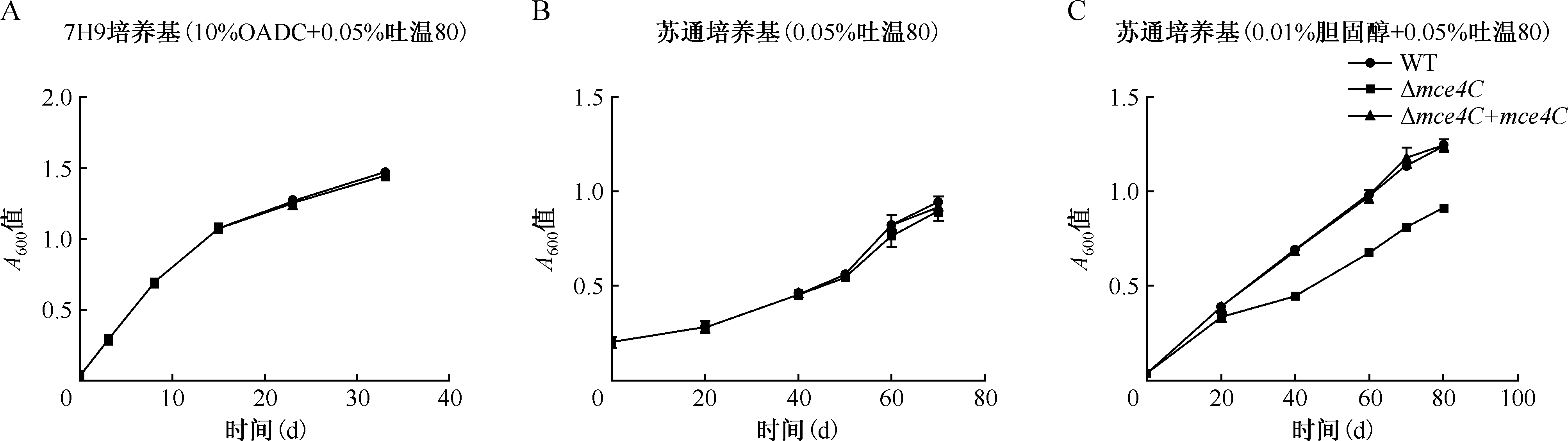
| 培养基 | A600值 (WT) | A600值 (Δmce4C) | A600值 (Δmce4C+mce4C) | t值a | P值 | t值b | P值 | t值c | P值 |
|---|---|---|---|---|---|---|---|---|---|
| 7H9培养基 | |||||||||
| 第0天 | 0.040±0.001 | 0.040±0.001 | 0.040±0.001 | -0.316 | 0.768 | 0.316 | 0.768 | 0.000 | 1.000 |
| 第3天 | 0.290±0.002 | 0.290±0.009 | 0.291±0.010 | -0.063 | 0.953 | 0.116 | 0.913 | -0.211 | 0.844 |
| 第8天 | 0.694±0.013 | 0.691±0.020 | 0.693±0.001 | -0.190 | 0.858 | 0.094 | 0.930 | 0.205 | 0.847 |
| 第15天 | 1.075±0.001 | 1.077±0.001 | 1.073±0.004 | -1.753 | 0.154 | 1.314 | 0.280 | 0.593 | 0.585 |
| 第23天 | 1.264±0.021 | 1.263±0.001 | 1.267±0.021 | 0.055 | 0.959 | 0.305 | 0.776 | 0.810 | 0.477 |
| 第33天 | 1.443±0.055 | 1.447±0.003 | 1.457±0.020 | -0.098 | 0.926 | 0.837 | 0.450 | 2.950 | 0.060 |
| 苏通培养基 | |||||||||
| 第0天 | 0.203±0.033 | 0.207±0.009 | 0.202±0.031 | -0.222 | 0.835 | -0.233 | 0.827 | 0.000 | 1.000 |
| 第20天 | 0.281±0.020 | 0.285±0.027 | 0.283±0.019 | -1.860 | 0.861 | -0.071 | 0.947 | -0.145 | 0.892 |
| 第40天 | 0.457±0.012 | 0.453±0.019 | 0.457±0.001 | 0.347 | 0.746 | 0.432 | 0.688 | -0.041 | 0.970 |
| 第50天 | 0.561±0.000 | 0.546±0.190 | 0.560±0.020 | 1.339 | 0.252 | 0.869 | 0.434 | 0.091 | 0.932 |
| 第60天 | 0.825±0.045 | 0.766±0.060 | 0.822±0.056 | 1.346 | 0.249 | 1.166 | 0.309 | 0.074 | 0.944 |
| 第70天 | 0.944±0.029 | 0.894±0.049 | 0.916±0.036 | 1.537 | 0.199 | 0.643 | 0.555 | 1.045 | 0.355 |
| 苏通培养基+胆固醇 | |||||||||
| 第0天 | 0.040±0.002 | 0.040±0.001 | 0.040±0.001 | 0.316 | 0.768 | 0.000 | 1.000 | 0.316 | 0.768 |
| 第20天 | 0.391±0.019 | 0.336±0.017 | 0.391±0.002 | 3.836 | 0.019 | 5.776 | 0.004 | 0.000 | 1.000 |
| 第40天 | 0.697±0.020 | 0.447±0.002 | 0.687±0.006 | 20.643 | <0.001 | 61.514 | <0.001 | 0.742 | 0.499 |
| 第60天 | 0.989±0.001 | 0.677±0.002 | 0.977±0.032 | 295.357 | <0.001 | 16.093 | <0.001 | 0.664 | 0.543 |
| 第70天 | 1.138±0.006 | 0.811±0.020 | 1.178±0.053 | 26.896 | <0.001 | 11.198 | <0.001 | -1.305 | 0.262 |
| 第80天 | 1.245±0.011 | 0.913±0.017 | 1.246±0.029 | 28.182 | <0.001 | 17.140 | <0.001 | -0.067 | 0.950 |
| 组别 | WT | Δmce4C | Δmce4C+mce4C | t值a | P值 | t值b | P值 | t值c | P值 |
|---|---|---|---|---|---|---|---|---|---|
| 菌体总胆固醇含量(μg/ml) | |||||||||
| 第1天 | 0.448±0.003 | 0.361±0.025 | 0.420±0.019 | 4.613 | 0.019 | 2.782 | 0.069 | 2.069 | 0.174 |
| 第7天 | 0.551±0.016 | 0.384±0.013 | 0.521±0.034 | 11.543 | 0.007 | 5.325 | 0.034 | 1.148 | 0.370 |
| 第14天 | 0.733±0.014 | 0.544±0.012 | 0.731±0.039 | 14.427 | 0.005 | 6.455 | 0.023 | 0.066 | 0.953 |
| 第21天 | 1.347±0.087 | 1.058±0.012 | 1.505±0.021 | 4.621 | 0.044 | 25.679 | 0.002 | -2.492 | 0.130 |
| 培养上清总胆固醇含量(μg/ml) | |||||||||
| 第1天 | 27.045±0.293 | 27.911±0.308 | 27.497±0.581 | -2.879 | 0.102 | -0.890 | 0.467 | -0.982 | 0.430 |
| 第7天 | 20.807±0.591 | 23.921±0.644 | 19.651±0.923 | -5.040 | 0.037 | -5.363 | 0.033 | 1.491 | 0.274 |
| 第14天 | 14.909±0.770 | 19.952±0.688 | 13.551±0.703 | -6.907 | 0.020 | 9.209 | 0.012 | 1.843 | 0.207 |
| 第21天 | 7.740±0.422 | 16.371±0.753 | 7.274±0.131 | -14.135 | 0.005 | -16.827 | 0.004 | 1.489 | 0.275 |
| 菌体荧光强度值 | |||||||||
| 第1天 | 0.544±0.001 | 0.478±0.012 | 0.533±0.023 | 7.501 | 0.017 | 2.973 | 0.097 | 0.644 | 0.585 |
| 第7天 | 0.809±0.028 | 0.510±0.012 | 0.794±0.050 | 13.883 | 0.005 | 7.867 | 0.016 | 0.380 | 0.741 |
| 第14天 | 0.889±0.017 | 0.743±0.014 | 0.875±0.019 | 9.990 | 0.002 | 7.816 | 0.016 | 0.882 | 0.443 |
| 第21天 | 1.630±0.027 | 0.848±0.002 | 1.840±0.044 | 40.166 | 0.001 | 31.735 | 0.001 | -5.706 | 0.059 |
| 培养上清荧光强度值 | |||||||||
| 第1天 | 13.974±0.214 | 16.980±0.345 | 13.122±0.849 | -10.481 | 0.009 | -7.502 | 0.017 | 5.100 | 0.306 |
| 第7天 | 4.588±0.030 | 5.179±0.054 | 4.376±0.054 | -13.479 | 0.005 | -14.878 | 0.004 | 4.846 | 0.404 |
| 第14天 | 2.590±0.066 | 4.048±0.018 | 2.600±0.012 | -30.184 | 0.001 | -94.905 | <0.001 | -0.205 | 0.857 |
| 第21天 | 1.316±0.044 | 2.482±0.039 | 1.360±0.012 | -28.352 | 0.001 | -30.878 | 0.001 | -8.896 | 0.052 |
| 感染时间 (h) | WT感染细胞 裂解液胆固醇 含量(μg/ml) | Δmce4C感染细胞 裂解液胆固醇 含量(μg/ml) | Δmce4C+mce4C感染 细胞裂解液胆固醇 含量(μg/ml) | t值a | P值 | t值b | P值 | t值c | P值 |
|---|---|---|---|---|---|---|---|---|---|
| 0 | 6.630±0.075 | 6.843±0.224 | 6.501±0.601 | -1.806 | 0.121 | -1.070 | 0.326 | 0.429 | 0.683 |
| 4 | 7.180±0.173 | 7.749±0.017 | 6.725±0.288 | -6.556 | 0.001 | -7.106 | <0.001 | 2.711 | 0.305 |
| 24 | 9.547±0.273 | 10.582±0.286 | 9.466±0.220 | -5.244 | 0.002 | -6.197 | 0.001 | 0.464 | 0.659 |
| 48 | 9.518±0.240 | 10.620±0.332 | 9.482±0.186 | -5.386 | 0.002 | -5.982 | 0.001 | 0.235 | 0.822 |
| 72 | 7.341±0.330 | 10.045±0.553 | 7.655±0.546 | -8.400 | <0.001 | 6.151 | 0.001 | 0.986 | 0.362 |


| [1] | World Health Organization. Global tuberculosis report 2024. Geneva: World Health Organization, 2024. |
| [2] |
Russell DG, Barry CE 3rd, Flynn JL. Tuberculosis: what we don’t know can, and does, hurt us. Science, 2010, 328(5980):852-856. doi:10.1126/science.1184784.
pmid: 20466922 |
| [3] |
Muñoz-Elías EJ, McKinney JD. Mycobacterium tuberculosis isocitrate lyases 1 and 2 are jointly required for in vivo growth and virulence. Nat Med, 2005, 11(6):638-644. doi:10.1038/nm1252.
pmid: 15895072 |
| [4] | Pandey AK, Sassetti CM. Mycobacterial persistence requires the utilization of host cholesterol. Proc Natl Acad Sci U S A, 2008, 105(11):4376-4380. doi:10.1073/pnas.0711159105. |
| [5] | Yang X, Nesbitt NM, Dubnau E, et al. Cholesterol metabolism increases the metabolic pool of propionate in Mycobacterium tuberculosis. Biochemistry, 2009, 48(18):3819-3821. doi:10.1021/bi9005418. |
| [6] |
Martens GW, Arikan MC, Lee J, et al. Hypercholesterolemia impairs immunity to tuberculosis. Infect Immun, 2008, 76(8): 3464-3472. doi:10.1128/iai.00037-08.
pmid: 18505807 |
| [7] | Kim MJ, Wainwright HC, Locketz M, et al. Caseation of human tuberculosis granulomas correlates with elevated host lipid metabolism. EMBO Mol Med, 2010, 2(7):258-274. doi:10.1002/emmm.201000079. |
| [8] | Schäfer G, Guler R, Murray G, et al. The role of scavenger receptor B 1 in infection with Mycobacterium tuberculosis in a murine model. PLoS One, 2009, 4(12): e8448. doi:10.1371/journal.pone.0008448. |
| [9] | Khan S, Islam A, Hassan MI, et al. Purification and structural characterization of Mce4A from Mycobacterium tuberculosis. Int J Biol Macromol, 2016, 93(Pt A): 235-241. doi:10.1016/j.ijbiomac.2016.06.059. |
| [10] | Bashiri G. Lipid transport across the mycobacterial cell envelope. IUCr J, 2021, 8(Pt 5): 711-712. doi:10.1107/S2052252521008885. |
| [11] |
Rathor N, Garima K, Sharma NK, et al. Expression profile of mce 4 operon of Mycobacterium tuberculosis following environmental stress. Int J Mycobacteriol, 2016, 5(3): 328-332. doi:10.1016/j.ijmyco.2016.08.004.
pmid: 27847019 |
| [12] | 张宗德, 李自慧, 杜博平, 等. 结核杆菌体内诱导基因的筛选及初步分析. 中华医学杂志, 2008, 88(3): 189-193. doi:10.3321/j.issn:0376-2491.2008.03.013. |
| [13] | Chandra P, Coullon H, Agarwal M, et al. Macrophage global metabolomics identifies cholestenone as host/pathogen cometabolite present in human Mycobacterium tuberculosis infection. J Clin Invest, 2022, 132(3):e152509. doi:10.1172/JCI152509. |
| [14] |
Freeman NE, Rusinol AE, Linton M, et al. Acyl-coenzyme A:cholesterol acyltransferase promotes oxidized LDL/oxysterol-induced apoptosis in macrophages. J Lipid Res, 2005, 46(9):1933-1943. doi:10.1194/jlr.M500101-JLR200.
pmid: 15995174 |
| [15] | Roca FJ, Whitworth LJ, Prag HA, et al. Tumor necrosis factor induces pathogenic mitochondrial ROS in tuberculosis through reverse electron transport. Science, 2022, 376(6600):eabh2841. doi:10.1126/science.abh2841. |
| [16] | Zhu Y, Choi D, Somanath PR, et al. Lipid-Laden Macrophages in Pulmonary Diseases. Cells, 2024, 13(11):889. doi:10.3390/cells13110889. |
| [17] | Sarathy JP, Dartois V. Caseum: a Niche for Mycobacterium tuberculosis Drug-Tolerant Persisters. Clin Microbiol Rev, 2020, 33(3):e00159-19. doi:10.1128/cmr.00159-19. |
| [18] | Rank L, Herring LE, Braunstein M. Evidence for the Mycobacterial Mce4 Transporter Being a Multiprotein Complex. J Bacteriol, 2021, 203(10):e00685-20. doi:10.1128/jb.00685-20. |
| [19] |
Perkowski EF, Miller BK, McCann JR, et al. An orphaned Mce-associated membrane protein of Mycobacterium tuberculosis is a virulence factor that stabilizes Mce transporters. Mol Microbiol, 2016, 100(1): 90-107. doi:10.1111/mmi.13303.
pmid: 26712165 |
| [20] | Klepp LI, Sabio Y, Garcia J, et al. Mycobacterial MCE proteins as transporters that control lipid homeostasis of the cell wall. Tuberculosis (Edinb), 2022, 132: 102162. doi:10.1016/j.tube.2021.102162. |
| [21] | Fieweger RA, Wilburn KM, Montague CR, et al. MceG stabilizes the Mce1 and Mce4 transporters in Mycobacterium tuberculosis. J Biol Chem, 2023, 299(3): 102910. doi:10.1016/j.jbc.2023.102910. |
| [22] | Asthana P, Singh D, Pedersen JS, et al. Structural insights into the substrate-binding proteins Mce1A and Mce4A from Mycobacterium tuberculosis. IUCr J, 2021, 8(Pt 5): 757-774. doi:10.1107/s2052252521006199. |
| [23] | Chen Z, Kong X, Ma Q, et al. The impact of Mycobacterium tuberculosis on the macrophage cholesterol metabolism pathway. Front Immunol, 2024, 15: 1402024. doi:10.3389/fimmu.2024.1402024. |
| [24] |
Pawełczyk J, Brzostek A, Minias A, et al. Cholesterol-dependent transcriptome remodeling reveals new insight into the contribution of cholesterol to Mycobacterium tuberculosis pathogenesis. Sci Rep, 2021, 11(1): 12396. doi:10.1038/s41598-021-91812-0.
pmid: 34117327 |
| [25] | Wilburn KM, Fieweger RA, VanderVen BC. Cholesterol and fatty acids grease the wheels of Mycobacterium tuberculosis pathogenesis. Pathog Dis, 2018, 76(2):fty021. doi:10.1093/femspd/fty021. |
| [26] | Kumar A, Bose M, Brahmachari V. Analysis of expression profile of mammalian cell entry (mce) operons of Mycobacterium tuberculosis. Infect Immun, 2003, 71(10): 6083-6087. doi:10.1128/iai.71.10.6083-6087.2003. |
| [27] | Mohn WW, van der Geize R, Stewart GR, et al. The actinobacterial mce 4 locus encodes a steroid transporter. J Biol Chem, 2008, 283(51): 35368-35374. doi:10.1074/jbc.M805496200. |
| [28] |
García-Fernández J, Papavinasasundaram K, Galán B, et al. Molecular and functional analysis of the mce 4 operon in Mycobacterium smegmatis. Environ Microbiol, 2017, 19(9):3689-3699. doi:10.1111/1462-2920.13869.
pmid: 28752922 |
| [29] | Nazarova EV, Montague CR, La T, et al. Rv3723/LucA coordinates fatty acid and cholesterol uptake in Mycobacterium tuberculosis. Elife, 2017, 6:e26969. doi:10.7554/eLife.26969. |
| [30] |
Ramón-García S, Stewart GR, Hui ZK, et al. The mycobacterial P 55 efflux pump is required for optimal growth on cholesterol. Virulence, 2015, 6(5):444-448. doi:10.1080/21505594.2015.1044195.
pmid: 26155739 |
| [1] | Sheng Jie, Hong Kaifeng, Mierzhati Aisha, Tang Wei, Dilixiati Abulizi. Study on the mechanism of IL-22 and p38 MAPK signaling pathways in inhibiting bone destruction in bone and joint tuberculosis [J]. Chinese Journal of Antituberculosis, 2025, 47(4): 454-459. |
| [2] | Hao Mingxiao, Mi Jie, Xu Zongyi. Effectiveness of a continuity of care model in patients with tuberculous meningitis [J]. Chinese Journal of Antituberculosis, 2025, 47(4): 477-481. |
| [3] | Huang Misun, Wu Yaning, Li Guilian, Liu Haican. Research advances of Mycobacterium tuberculosis enrichment technology [J]. Chinese Journal of Antituberculosis, 2025, 47(3): 369-373. |
| [4] | Zhang Chao, Yu Xia, Huang Hairong, Liu Wei, Liu Tao. Evaluation of the in vitro antimicrobial effects of sevoflurane on Mycobacterium tuberculosis [J]. Chinese Journal of Antituberculosis, 2025, 47(2): 158-163. |
| [5] | Liu Ruihua, Sarina , Wang Furong. Interaction between lung cancer and tuberculosis in disease development and progression [J]. Chinese Journal of Antituberculosis, 2025, 47(1): 102-111. |
| [6] | Chen Jifei, Huang Lihua, Luo Lanbo, Sui Wenxian, Pang Yu, Liu Aimei. Evaluation the efficacy of tongue swab-based PCR fluorescence probe method for pulmonary tuberculosis [J]. Chinese Journal of Antituberculosis, 2025, 47(1): 51-60. |
| [7] | Lu Hailin, Wang Wenfei, Tao Wenhui, Lin Peicong, Chen Xinchun, Deng Guofang, Xie Shuixiang. Oleic acid upregulates the expression of perilipin 2 enhancing macrophage clearance of Mycobacterium tuberculosis [J]. Chinese Journal of Antituberculosis, 2025, 47(1): 72-76. |
| [8] | Wang Yilin, Wu Xiao, Pang Yu, Li Shanshan. Immunomodulatory effect of orelabrutinib in host macrophages infected with mycobacterium [J]. Chinese Journal of Antituberculosis, 2024, 46(9): 1063-1068. |
| [9] | Palidanguli Abudureheman, Wang Senlu, Gulina Badeerhan, Wang Le, Zulikatiayi Abudula, Wang Xinqi, Maiwulajiang Yimamu, Wang Xijiang. Distribution of Mycobacterium tuberculosis genotypes in Kashgar region and their association with clinical characteristics of pulmonary tuberculosis patients [J]. Chinese Journal of Antituberculosis, 2024, 46(9): 1077-1082. |
| [10] | Xu Chunhua, Zhu Shiyu, Hu Yi, Yi Kehua, Song Canlei, Wang Zichun, Wu Yong, Wang Qing, Yang Qianru, Shen Xin. Analysis of screening effect of recombinant Mycobacterium tuberculosis fusion protein in screening Mycobacterium tuberculosis infection in close contacts of pulmonary tuberculosis patients [J]. Chinese Journal of Antituberculosis, 2024, 46(8): 897-902. |
| [11] | Zhang Lanyue, Wang Yingchao, Liu Weiyi, Shang Xuetian, Jia Hongyan, Zhu Chuanzhi, Zhang Zongde, Pan Liping. Study on the effect of thiol acetyltransferase mshD on the growth and stress response of Mycobacterium tuberculosis [J]. Chinese Journal of Antituberculosis, 2024, 46(8): 935-941. |
| [12] | Yu Lan, Chen Shuangshuang, Wang Nenhan, Tian Lili, Zhao Yanfeng, Fan Ruifang, Liu Haican, Li Chuanyou, Dai Xiaowei. Consistency between phenotypic resistance to fluoroquinolones and genetic mutations in rifampicin resistant Mycobacterium tuberculosis strains [J]. Chinese Journal of Antituberculosis, 2024, 46(8): 942-950. |
| [13] | He Xiangrong, Chen Hua, Chen Pinru, Liang Feng, Ren Huili, Zhu Jialou, Hu Jinxing, Tan Yaoju. A case report and literature review of Mycobacterium asiaticum pulmonary disease [J]. Chinese Journal of Antituberculosis, 2024, 46(7): 763-769. |
| [14] | Zhang Muli, Sun Zhaogang, Cao Tingming, Xie Zhongyao. The value of three proteins in diagnosing Mycobacterium tuberculosis infection [J]. Chinese Journal of Antituberculosis, 2024, 46(7): 808-814. |
| [15] | Fu Keyan, Zhu Bangzheng, Ye Jian. Research progress on interstitial lung disease combined with Mycobacterium tuberculosis infection [J]. Chinese Journal of Antituberculosis, 2024, 46(7): 823-829. |
| Viewed | ||||||
|
Full text |
|
|||||
|
Abstract |
|
|||||
 京公网安备11010202007215号
Total visitors: Visitors of today: Now online:
京公网安备11010202007215号
Total visitors: Visitors of today: Now online:
 This work is licensed under Creative Commons Attribution 3.0 License.