
Chinese Journal of Antituberculosis ›› 2024, Vol. 46 ›› Issue (4): 449-460.doi: 10.19982/j.issn.1000-6621.20240005
• Original Articles • Previous Articles Next Articles
Shang Xuetian1, Dong Jing1, Huang Mailing2, Sun Qi1, Jia Hongyan1, Zhang Lanyue1, Liu Qiuyue3, Yao Mingxu1, Wang Yingchao1, Ji Xiuxiu1, Du Boping1, Xing Aiying1, Pan Liping1( )
)
Received:2024-01-03
Online:2024-04-10
Published:2024-04-01
Contact:
Pan Liping
E-mail:panliping2006@163.com
Supported by:CLC Number:
Shang Xuetian, Dong Jing, Huang Mailing, Sun Qi, Jia Hongyan, Zhang Lanyue, Liu Qiuyue, Yao Mingxu, Wang Yingchao, Ji Xiuxiu, Du Boping, Xing Aiying, Pan Liping. Transcriptome study on peripheral blood mononuclear cells of latent tuberculosis infection individuals[J]. Chinese Journal of Antituberculosis, 2024, 46(4): 449-460. doi: 10.19982/j.issn.1000-6621.20240005
Add to citation manager EndNote|Ris|BibTeX
URL: http://www.zgflzz.cn/EN/10.19982/j.issn.1000-6621.20240005
| 分类/人口学特征 | HC组 | LTBI组 | 统计检验值 | P值 |
|---|---|---|---|---|
| 微阵列芯片集 | ||||
| 性别(名/例) | - | - | ||
| 男性 | 0 | 0 | ||
| 女性 | 4 | 4 | ||
| 年龄(岁) | ||||
| 范围 | 33~44 | 32~42 | - | - |
| 均数±标准差 | 36.5±4.4 | 38.0±4.5 | t=0.554 | 0.600 |
| qPCR验证集 | ||||
| 性别(名/例) | χ2=0.220 | 0.500 | ||
| 男性 | 7 | 6 | ||
| 女性 | 3 | 4 | ||
| 年龄(岁) | ||||
| 范围 | 21~58 | 26~68 | - | - |
| 均数±标准差 | 37.1±12.3 | 42.9±12.6 | t=1.045 | 0.310 |

| 基因 | 功能 | 芯片数据 | qPCR验证 | ||||||
|---|---|---|---|---|---|---|---|---|---|
| 差异倍数 | P值 | 表达趋势 | 差异倍数 | t值 | P值 | 表达趋势 | |||
| CAMK1G | 调节钙离子/钙调蛋白依赖性蛋白激酶活性 | 12.50 | 0.003 | 上调 | 4.36 | 1.296 | 0.212 | 上调 | |
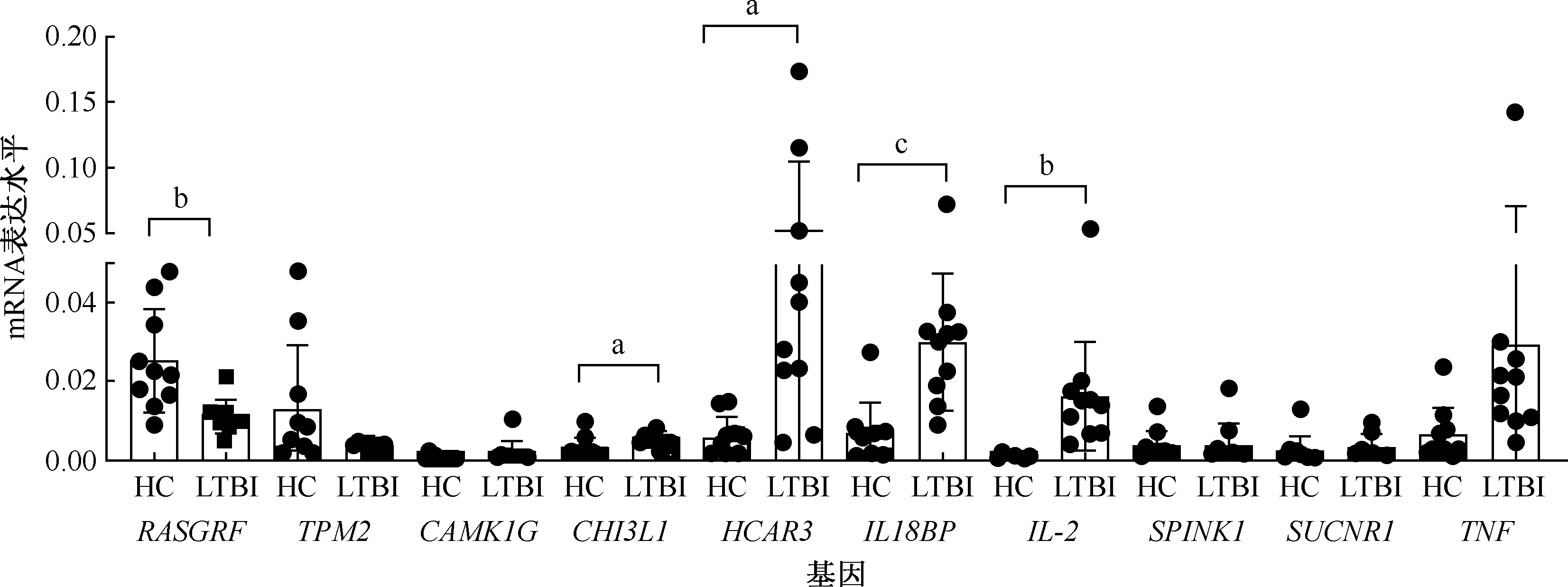
| IL2 | Th1型细胞因子,参与免疫反应 | 5.79 | 0.005 | 上调 | 23.46 | 3.539 | 0.002 | 上调 | |
| SPINK1 | 调节胞质钙离子浓度 | 5.37 | 0.007 | 上调 | 1.15 | 0.237 | 0.816 | 上调 | |
| SUCNR1 | 参与G蛋白偶联受体信号通路 | 4.56 | 0.001 | 上调 | 1.46 | 0.634 | 0.530 | 上调 | |
| HCAR3 | 参与G蛋白偶联受体信号通路 | 4.11 | <0.001 | 上调 | 8.76 | 2.678 | 0.015 | 上调 | |
| IL18BP | 参与Th1型免疫反应 | 4.10 | 0.002 | 上调 | 4.52 | 3.902 | 0.001 | 上调 | |
| CHI3L1 | 参与几丁质分解代谢 | 4.05 | <0.001 | 上调 | 1.87 | 2.338 | 0.031 | 上调 | |
| TNF | 促炎细胞因子 | 3.65 | <0.001 | 上调 | 3.49 | 1.782 | 0.092 | 上调 | |
| SASH1 | 参与蛋白质多泛素化 | 3.56 | 0.003 | 上调 | |||||
| CD274 | 参与适应性免疫反应 | 3.52 | 0.001 | 上调 | |||||
| ABCA7 | 参与脂质运输 | 0.49 | 0.008 | 下调 | |||||
| C1orf186 | 参与红细胞生成 | 0.49 | 0.002 | 下调 | |||||
| IKZF2 | 参与RNA聚合酶Ⅱ启动子转录调控 | 0.49 | 0.001 | 下调 | |||||
| ACAP1 | 参与蛋白质运输 | 0.47 | 0.006 | 下调 | |||||
| AQP3 | 正向调节免疫系统过程 | 0.46 | 0.003 | 下调 | |||||
| LY9 | 参与细胞黏附 | 0.46 | 0.007 | 下调 | |||||
| RGS14 | 参与G蛋白偶联受体信号通路 | 0.42 | 0.002 | 下调 | |||||
| MYLIP | 参与泛素依赖的蛋白质分解代谢过程 | 0.40 | 0.001 | 下调 | |||||
| RASGRP2 | 调节细胞生长 | 0.37 | 0.007 | 下调 | 0.44 | 3.279 | 0.004 | 下调 | |
| TPM2 | 调节ATP酶活性 | 0.37 | 0.002 | 下调 | 0.40 | 1.867 | 0.078 | 下调 | |


| 模块/聚类分析 | 条目 | 富集倍数 | P值 | 基因个数 |
|---|---|---|---|---|
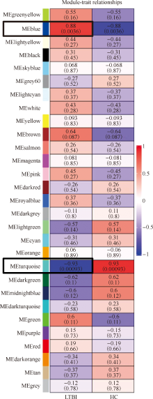
| 蓝色模块 | ||||
| BP | ||||
| 炎症应答 | 2.46 | <0.001 | 48 | |
| 溶酶体腔酸化 | 10.14 | <0.001 | 10 | |
| 信号转导 | 1.67 | <0.001 | 98 | |
| 细胞因子介导的信号通路 | 3.21 | <0.001 | 23 | |
| 突触囊泡腔酸化 | 11.16 | <0.001 | 8 | |
| 液泡酸化 | 8.73 | <0.001 | 9 | |
| 肌动蛋白细胞骨架组织 | 2.68 | <0.001 | 28 | |
| 白细胞介素-1β产生的正调节 | 4.33 | <0.001 | 13 | |
| MF | ||||
| 蛋白结合 | 1.12 | <0.001 | 664 | |
| 质子转运ATP酶活性,旋转机制 | 7.68 | <0.001 | 9 | |
| 相同蛋白结合 | 1.41 | <0.001 | 115 | |
| PDZ结构域结合 | 3.36 | <0.001 | 14 | |
| 受体结合 | 1.81 | 0.001 | 35 | |
| 细胞因子受体活性 | 3.74 | 0.001 | 10 | |
| 锰离子跨膜转运体活性 | 12.20 | 0.003 | 4 | |
| 信号受体活性 | 2.01 | 0.003 | 22 | |
| CC | ||||
| 质膜 | 1.46 | <0.001 | 347 | |
| 溶酶体膜 | 2.94 | <0.001 | 52 | |
| 细胞外外泌体 | 1.65 | <0.001 | 161 | |
| 内体膜 | 2.94 | <0.001 | 35 | |
| 细胞表面 | 2.09 | <0.001 | 60 | |
| 溶酶体 | 2.62 | <0.001 | 36 | |
| 胞液 | 1.27 | <0.001 | 312 | |
| 高尔基体 | 1.74 | <0.001 | 87 | |
| 蓝绿色模块 | ||||
| BP | ||||
| 转录正调控,DNA模板 | 1.82 | <0.001 | 63 | |
| RNA聚合酶Ⅱ启动子转录的正向调节 | 1.59 | <0.001 | 93 | |
| 染色质组织 | 2.22 | <0.001 | 34 | |
| 炎症应答 | 1.94 | <0.001 | 40 | |
| 参与细胞凋亡过程的半胱氨酸型内肽酶活性的激活 | 3.60 | <0.001 | 14 | |
| RNA聚合酶Ⅱ启动子转录的调控 | 1.41 | <0.001 | 116 | |
| 细胞周期 | 2.00 | <0.001 | 35 | |
| 神经管闭合 | 3.39 | <0.001 | 13 | |
| MF | ||||
| 蛋白结合 | 1.15 | <0.001 | 724 | |
| 磷脂酰肌醇结合 | 3.10 | <0.001 | 17 | |
| 磷脂酰肌醇-3,4-二磷酸结合 | 5.35 | 0.001 | 8 | |
| 染色质结合 | 1.68 | 0.001 | 42 | |
| 肌动蛋白丝结合 | 2.07 | 0.001 | 24 | |
| RNA聚合酶Ⅱ核心启动子近端区域序列特异性DNA结合 | 1.38 | 0.002 | 84 | |
| 转录辅阻遏物活性 | 2.07 | 0.003 | 21 | |
| 脂质结合 | 2.02 | 0.005 | 20 | |
| CC | ||||
| 胞液 | 1.41 | <0.001 | 367 | |
| 细胞质 | 1.30 | <0.001 | 349 | |
| 细胞核 | 1.25 | <0.001 | 353 | |
| 细胞膜 | 1.31 | <0.001 | 224 | |
| 染色质 | 1.59 | <0.001 | 82 | |
| 核浆 | 1.24 | <0.001 | 234 | |
| 质膜的细胞质侧 | 2.89 | 0.001 | 15 | |
| 中心体 | 1.69 | 0.001 | 45 |
| 模块/聚类分析 | 条目 | 富集倍数 | P值 | 基因个数 |
|---|---|---|---|---|
| 蓝色模块 | ||||
| KEGG | ||||
| 溶酶体 | 3.93 | <0.001 | 29 | |
| 吞噬体 | 3.18 | <0.001 | 27 | |
| 肺结核 | 2.78 | <0.001 | 28 | |
| 类风湿性关节炎 | 3.65 | <0.001 | 19 | |
| 癌症通路 | 1.82 | <0.001 | 54 | |
| 肿瘤中PD-L1的表达和PD-1检查点通路 | 3.42 | <0.001 | 17 | |
| 人乳头瘤病毒感染 | 2.05 | <0.001 | 38 | |
| 铁死亡 | 4.80 | <0.001 | 11 | |
| 蓝绿色模块 | ||||
| KEGG | ||||
| 破骨细胞分化 | 2.95 | <0.001 | 21 | |
| 甲状腺激素信号通路 | 2.98 | <0.001 | 19 | |
| FcγR介导的吞噬作用 | 3.13 | <0.001 | 16 | |
| B细胞受体信号通路 | 3.17 | 0.001 | 14 | |
| 糖尿病并发症中的AGE-RAGE信号通路 | 2.85 | 0.001 | 15 | |
| 脂质与动脉粥样硬化 | 2.12 | 0.001 | 24 | |
| 小细胞肺癌 | 2.89 | 0.001 | 14 | |
| C型凝集素受体信号通路 | 2.74 | 0.001 | 15 |


| [1] | Getahun H, Matteelli A, Chaisson RE, et al. Latent Mycobacterium tuberculosis infection. N Engl J Med, 2015, 372(22): 2127-2135. doi:10.1056/NEJMra1405427. |
| [2] | 中国防痨协会. 高危人群结核分枝杆菌潜伏感染检测及预防性治疗专家共识. 中国防痨杂志, 2021, 43(9): 874-878. doi:10.3969/j.issn.1000-6621.2021.09.004. |
| [3] | World Health Organization. Latent tuberculosis infection: updated and consolidated guidelines for programmatic management. Geneva: World Health Organization, 2018. |
| [4] | Bagcchi S. WHO’s Global Tuberculosis Report 2022. Lancet Microbe, 2023, 4(1): e20. doi:10.1016/S2666-5247(22)00359-7. |
| [5] |
Zellweger JP, Sotgiu G, Corradi M, et al. The diagnosis of latent tuberculosis infection (LTBI): currently available tests, future developments, and perspectives to eliminate tuberculosis (TB). Med Lav, 2020, 111(3):170-183. doi:10.23749/mdl.v111i3.9983.
pmid: 32624559 |
| [6] | Qin H, Wang Y, Huang L, et al. Efficacy and Risk Factors of Interferon-Gamma Release Assays among HIV-Positive Individuals. Int J Environ Res Public Health, 2023, 20(5):4556. doi:10.3390/ijerph20054556. |
| [7] | Shen BJ, Lin HH. Time-dependent association between cancer and risk of tuberculosis: A population-based cohort study. Int J Infect Dis, 2021, 108: 340-346. doi:10.1016/j.ijid.2021.05.037. |
| [8] | Cadena J, Rathinavelu S, Lopez-Alvarenga JC, et al. The re-emerging association between tuberculosis and diabetes: Lessons from past centuries. Tuberculosis (Edinb), 2019, 116 S: S89-S97. doi:10.1016/j.tube.2019.04.015. |
| [9] |
Singhania A, Verma R, Graham CM, et al. A modular trans-criptional signature identifies phenotypic heterogeneity of human tuberculosis infection. Nat Commun, 2018, 9(1):2308. doi:10.1038/s41467-018-04579-w.
pmid: 29921861 |
| [10] | Tabone O, Verma R, Singhania A, et al. Blood transcriptomics reveal the evolution and resolution of the immune response in tuberculosis. J Exp Med, 2021, 218(10): e20210915. doi:10.1084/jem.20210915. |
| [11] | Chen C, Wu Y, Li J, et al. TBtools-Ⅱ: A “one for all, all for one” bioinformatics platform for biological big-data mining. Mol Plant, 2023, 16(11):1733-1742. doi:10.1016/j.molp.2023.09.010. |
| [12] | Tang D, Chen M, Huang X, et al. SRplot: A free online platform for data visualization and graphing. PLoS One, 2023, 18(11): e294236. doi:10.1371/journal.pone.0294236. |
| [13] |
Carlson MR, Zhang B, Fang Z, et al. Gene connectivity, function, and sequence conservation: predictions from modular yeast co-expression networks. BMC Genomics, 2006, 7: 40. doi:10.1186/1471-2164-7-40.
pmid: 16515682 |
| [14] | Verma A, Ghoshal A, Dwivedi VP, et al. Tuberculosis: The success tale of less explored dormant Mycobacterium tuberculosis. Front Cell Infect Microbiol, 2022, 12:1079569. doi:10.3389/fcimb.2022.1079569. |
| [15] | Khabibullina NF, Kutuzova DM, Burmistrova IA, et al. The Biological and Clinical Aspects of a Latent Tuberculosis Infection. Trop Med Infect Dis, 2022, 7(3):48. doi:10.3390/tropicalmed7030048. |
| [16] | Cohen GM. Caspases: the executioners of apoptosis. Biochem J, 1997, 326 (Pt 1): 1-16. doi:10.1042/bj3260001. |
| [17] |
Blomgran R, Desvignes L, Briken V, et al. Mycobacterium tuberculosis inhibits neutrophil apoptosis, leading to delayed activation of naive CD 4 T cells. Cell Host Microbe, 2012, 11(1):81-90. doi:10.1016/j.chom.2011.11.012.
pmid: 22264515 |
| [18] | Nisa A, Kipper FC, Panigrahy D, et al. Different modalities of host cell death and their impact on Mycobacterium tuberculosis infection. Am J Physiol Cell Physiol, 2022, 323(5):C1444-C1474. doi:10.1152/ajpcell.00246.2022. |
| [19] |
Keane J, Remold HG, Kornfeld H. Virulent Mycobacterium tuberculosis strains evade apoptosis of infected alveolar macrophages. J Immunol, 2000, 164(4):2016-2020. doi:10.4049/jimmunol.164.4.2016.
pmid: 10657653 |
| [20] |
Zhang W, Ellingson L, Frascoli F, et al. An investigation of tuberculosis progression revealing the role of macrophages apoptosis via sensitivity and bifurcation analysis. J Math Biol, 2021, 83(3):31. doi:10.1007/s00285-021-01655-6.
pmid: 34436682 |
| [21] | Kim CH. Chemokine-chemokine receptor network in immune cell trafficking. Curr Drug Targets Immune Endocr Metabol Disord, 2004, 4(4):343-361. doi:10.2174/1568008043339712. |
| [22] | Slight SR, Khader SA. Chemokines shape the immune responses to tuberculosis. Cytokine Growth Factor Rev, 2013, 24(2):105-113. doi:10.1016/j.cytogfr.2012.10.002. |
| [23] | Barclay AM, Ninaber DK, van Veen S, et al. Airway epithelial cells mount an early response to mycobacterial infection. Front Cell Infect Microbiol, 2023, 13:1253037. doi:10.3389/fcimb.2023.1253037. |
| [24] | Jang S, Uzelac A, Salgame P. Distinct chemokine and cytokine gene expression pattern of murine dendritic cells and macrophages in response to Mycobacterium tuberculosis infection. J Leukoc Biol, 2008, 84(5):1264-1270. doi:10.1189/jlb.1107742. |
| [25] | Guler R, Ozturk M, Sabeel S, et al. Targeting Molecular Inflammatory Pathways in Granuloma as Host-Directed Therapies for Tuberculosis. Front Immunol, 2021, 12: 733853. doi:10.3389/fimmu.2021.733853. |
| [26] | Algood HM, Chan J, Flynn JL. Chemokines and tuberculosis. Cytokine Growth Factor Rev, 2003, 14(6):467-477. doi:10.1016/s1359-6101(03)00054-6. |
| [27] | Scott HM, Flynn JL. Mycobacterium tuberculosis in chemokine receptor 2-deficient mice: influence of dose on disease progression. Infect Immun, 2002, 70(11):5946-5954. doi:10.1128/IAI.70.11.5946-5954.2002. |
| [28] |
Mack U, Migliori GB, Sester M, et al. LTBI: latent tuberculosis infection or lasting immune responses to M.tuberculosis? A TBNET consensus statement. Eur Respir J, 2009, 33(5):956-973. doi:10.1183/09031936.00120908.
pmid: 19407047 |
| [29] |
Orme IM, Robinson RT, Cooper AM. The balance between protective and pathogenic immune responses in the TB-infected lung. Nat Immunol, 2015, 16(1):57-63. doi:10.1038/ni.3048.
pmid: 25521685 |
| [30] | Simon HU, Yousefi S, Germic N, et al. The Cellular Functions of Eosinophils: Collegium Internationale Allergologicum (CIA) Update 2020. Int Arch Allergy Immunol, 2020, 181(1):11-23. doi:10.1159/000504847. |
| [31] | Bohrer AC, Castro E, Hu Z, et al. Eosinophils are part of the granulocyte response in tuberculosis and promote host resis-tance in mice. J Exp Med, 2021, 218(10): e20210469. doi:10.1084/jem.20210469. |
| [32] | Kumar R, Singh P, Kolloli A, et al. Immunometabolism of Phagocytes During Mycobacterium tuberculosis Infection. Front Mol Biosci, 2019, 6: 105. doi:10.3389/fmolb.2019.00105. |
| [33] |
Italiani P, Boraschi D. From Monocytes to M1/M2 Macrophages: Phenotypical vs. Functional Differentiation. Front Immunol, 2014, 5: 514. doi:10.3389/fimmu.2014.00514.
pmid: 25368618 |
| [34] | Verreck FA, de Boer T, Langenberg DM, et al. Human IL-23-producing type 1 macrophages promote but IL-10-producing type 2 macrophages subvert immunity to (myco)bacteria. Proc Natl Acad Sci U S A, 2004, 101(13):4560-4565. doi:10.1073/pnas.0400983101. |
| [35] | Mily A, Kalsum S, Loreti MG, et al. Polarization of M1 and M2 Human Monocyte-Derived Cells and Analysis with Flow Cytometry upon Mycobacterium tuberculosis Infection. J Vis Exp, 2020(163). doi:10.3791/61807. |
| [1] | Chen Ruiqi, Zhang Mingwu, Wang Wei, Chen Songhua, Liu Zhengwei, Chen Bin. Prevalence and influencing factors of latent tuberculosis infection among elderly rural residents in Changshan County, Zhejiang Province [J]. Chinese Journal of Antituberculosis, 2024, 46(4): 383-389. |
| [2] | Li Xueqiu, Kuang Haobin, Wang Ting, Min Fei, Tao Lan, Cao Xihui, Li Xiang, Liu Guobiao. Analysis of self-paid medical expenses and influencing factors for multidrug-resistant tuberculosis patients in Guangzhou City [J]. Chinese Journal of Antituberculosis, 2024, 46(4): 411-417. |
| [3] | Wen Shufang, Wei Rongrong, Li Haoran, Liu Yi. The role of CD4+ and CD8+T cells in the immune response to tuberculosis [J]. Chinese Journal of Antituberculosis, 2024, 46(4): 479-484. |
| [4] | Pei Shaojun, Ou Xichao. Interpretation of the World Health Organization’s Catalogue of mutations in Mycobacterium tuberculosis complex and their association with drug resistance (2nd Edition) [J]. Chinese Journal of Antituberculosis, 2024, 46(3): 260-266. |
| [5] | Yao Yangyang, Liang Changhua, Han Dongming, Cui Junwei, Pan Ben, Wang Huihui, Wei Zhengqi, Zhen Siyu, Wei Hanyu. Differentiation of pulmonary tuberculosis and nontuberculous mycobacterial pulmonary disease based on computed tomography radiomics combined with clinical features [J]. Chinese Journal of Antituberculosis, 2024, 46(3): 302-310. |
| [6] | Zhang Jing, Fu Ruonan, Wang Senlu, Feng Jianyu, Zhang Ling, Gulina Badeerhan, Zulikatiayi Abudula, Wang Xinqi. Analysis of willingness of receiving preventive treatment in patients with latent tuberculosis infection among close contacts of pulmonary tuberculosis in high-burden areas [J]. Chinese Journal of Antituberculosis, 2024, 46(2): 165-172. |
| [7] | Shang Xuetian, Pan Liping. Role of tissue kallikrein family in pathogenesis of microorganism infection [J]. Chinese Journal of Antituberculosis, 2024, 46(2): 239-244. |
| [8] | He Yijun, Cheng Jun, Gao Lei. Epidemiological investigation of latent tuberculosis infection should be carried out systematically [J]. Chinese Journal of Antituberculosis, 2024, 46(1): 1-7. |
| [9] | Zhang Lifan, Ma Yanan, Zou Xiaoqing, Zhang Yueqiu, Zhang Fengchun, Zeng Xiaofeng, Zhao Yan, Liu Shengyun, Zuo Xiaoxia, Wu Huaxiang, Wu Lijun, Li Hongbin, Zhang Zhiyi, Chen Sheng, Zhu Ping, Zhang Miaojia, Qi Wencheng, Liu Yi, Liu Huaxiang, Shi Xiaochun, Liu Xiaoqing, the Epidemiological Study and Therapeutic Evaluation of Rheumatic Patients with Tuberculosis Study Team. Latent tuberculosis infection rate and risk factors in patients with rheumatic diseases: a multi-center, cross-sectional study [J]. Chinese Journal of Antituberculosis, 2024, 46(1): 29-39. |
| [10] | Zhang Guoqin, Wei Wenliang, Zhang Zhi, Zhang Yuhua, Zhong Da, Zhang Fan. Factors for discrepancy between two methods in testing latent tuberculosis infection and effect of preventive anti-tuberculosis treatment among prisoners [J]. Chinese Journal of Antituberculosis, 2024, 46(1): 45-53. |
| [11] | Liu Mei, Wu Xia, Gu Xu, Li Nana, Zhang Wanmin, Zhang Xiaoke, Lan Yuanbo. Evaluation of the performance of InnowaveDX MTB/RIF in detecting Mycobacterium tuberculosis complex and rifampicin resistance [J]. Chinese Journal of Antituberculosis, 2024, 46(1): 70-74. |
| [12] | Qin Huifang, Liang Xiaoyan, Zhou Lingyun, Lan Yumei, Lin Mei, Zhang Zhitong, Huang Yan, Wei Xiaolin, Liang Dabin. Analysis on the effect of GeneXpert MTB/RIF single test and mixed test on sputum samples for actively screening tuberculosis in key population of Guangxi [J]. Chinese Journal of Antituberculosis, 2023, 45(9): 880-884. |
| [13] | Du Yu, Zhang Haipeng, Wang Peng. Research status and application progress of mycobacteria phages [J]. Chinese Journal of Antituberculosis, 2023, 45(9): 897-903. |
| [14] | Bi Xiuli, Geng Hong, Jin Jin. The role of myeloid system and CD4+T cells in Mycobacterium tuberculosis infection and immunopathology [J]. Chinese Journal of Antituberculosis, 2023, 45(9): 904-912. |
| [15] | Guo Tonglei, Xin Henan, Gao Lei. Interpretation of WHO consolidated guidelines on tuberculosis: Module 1: prevention: tuberculosis preventive treatment [J]. Chinese Journal of Antituberculosis, 2023, 45(8): 723-727. |
| Viewed | ||||||
|
Full text |
|
|||||
|
Abstract |
|
|||||
 京公网安备11010202007215号
Total visitors: Visitors of today: Now online:
京公网安备11010202007215号
Total visitors: Visitors of today: Now online:
 This work is licensed under Creative Commons Attribution 3.0 License.