Email Alert | RSS    帮助

中国防痨杂志 ›› 2022, Vol. 44 ›› Issue (11): 1180-1186.doi: 10.19982/j.issn.1000-6621.20220190

• 论著 • 上一篇    下一篇

双层探测器光谱CT多参数图像诊断咯血患者中体-肺循环分流的价值

渠慧芳1, 王武章1, 汪立明1, 邓薇薇2, 刘晓敏2, 杨济生1, 郭晓雯1, 张运曾3, 鲁忆南1, 金锋3,4()   

  1. 1山东省公共卫生临床中心影像中心,济南 250013
    2飞利浦医疗科技公司临床科学部,上海 200072
    3山东省公共卫生临床中心胸部外科中心,济南 250013
    4山东省传染性呼吸疾病重点实验室,济南 250013
  • 收稿日期:2022-05-19 出版日期:2022-11-10 发布日期:2022-11-03
  • 通信作者: 金锋 E-mail:2547875348@qq.com
  • 基金资助:
    山东省医药卫生科技发展计划项目(2019WS530)

Diagnostic value of spectral images derived from dual-layer spectral detector CT in bronchial-pulmonary shunts in patients with hemoptysis

Qu Huifang1, Wang Wuzhang1, Wang Liming1, Deng Weiwei2, Liu Xiaomin2, Yang Jisheng1, Guo Xiaowen1, Zhang Yunzeng3, Lu Yinan1, Jin Feng3,4()   

  1. 1Center for Medicine Imaging, Shandong Public Health Clinical Center, Ji’nan 250013, China
    2Clinical Science Department of Philips Medical Technology, Shanghai 200072, China
    3Center for Thoracic Surgery, Shandong Public Health Clinical Center, Ji’nan 250013, China
    4Key Laboratory for Infectious Respiratory Disease of Shandong Province, Ji’nan 250013, China
  • Received:2022-05-19 Online:2022-11-10 Published:2022-11-03
  • Contact: Jin Feng E-mail:2547875348@qq.com
  • Supported by:
    Projects of Medical and Health Technology Development Program in Shandong Province(2019WS530)

摘要:

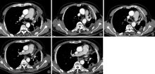
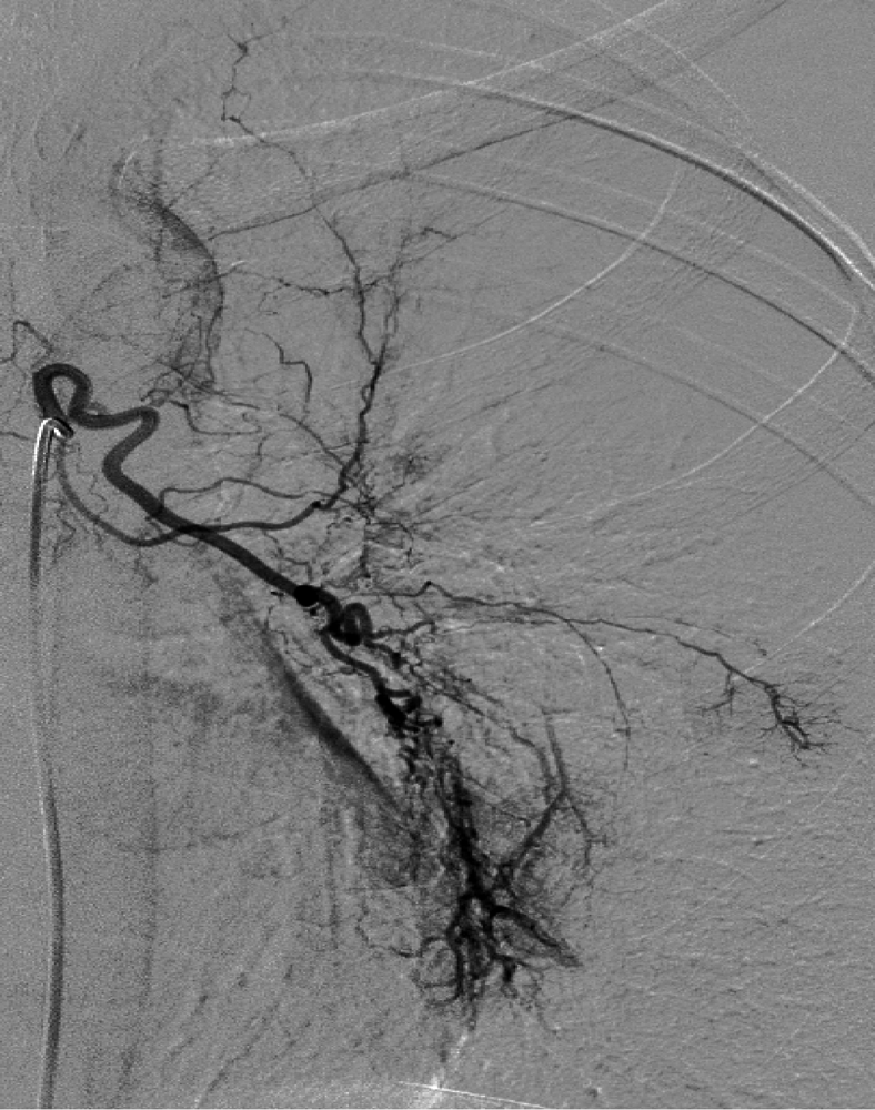
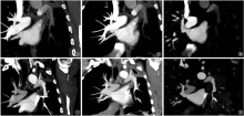
目的: 探索双层探测器光谱CT体动脉期光谱多参数图像诊断体-肺循环分流(BPS)的价值。方法: 回顾性分析2019年12月至2020年12月山东省胸科医院28 例经数字减影-血管造影(DSA)确诊BPS的咯血患者的光谱CT体动脉期重建的常规图像和光谱图像(单能级图像和碘密度图),以超选择性支气管相关体动脉造影为标准,比较光谱单能级图像和常规图像对BPS诊断的阳性率;同时,分别测量三组图像中主肺动脉干(ROI1)、BPS区域内异常显影的肺动脉管腔内(ROI2)及其叶根部肺动脉强化最明显区域(ROI3)的CT衰减值或碘密度,分析其变化趋势。同时,分析对侧正常肺动脉内与病变侧ROI2和ROI3分别对应的同级别同位置的肺动脉管腔(ROI4)及叶根部管腔(ROI5),分析其测量值及其与对侧变化的差异。结果: DSA图像共确诊39处BPS阳性肺动脉,其中光谱CT数据中体动脉期重建的40keV单能级图像(40keV virtual monoenergetic images,VMI40keV)对肺动脉分支错期显影显示,BPS阳性率分别为76.9%(30/39)和69.2%(27/39)。病变侧三组图像中ROI3的测量值[(539.00±152.09)HU]明显高于ROI1[(398.10±102.31)HU]和RO2[(441.40±115.52)HU]的测量值,差异有统计学意义(F=9.990,P<0.001)。对侧正常肺组织内的肺动脉比较显示,ROI1的测量值 [(393.95±120.03)HU]略大于ROI4[(396.40±146.01)HU]和ROI5[(361.00±135.40)HU],差异无统计学意义(F=0.290,P=0.753)。与对侧正常肺组织内的肺动脉比较,三组图像中在肺动脉沿血流分布的强化程度均呈现一定的变化趋势:BPS阳性侧肺动脉管腔内造影剂浓度沿血流方向逐渐增高,平均值由(398.10±102.31)HU上升至(539.00±152.09)HU;而正常肺血管的造影剂浓度变化较小[由(361.00±135.40)HU上升至(393.95±120.03)HU]。 结论: 光谱CT体动脉期光谱图像能够较为准确地诊断咯血患者中的BPS责任血管,有助于介入术前对BPS异常血管的预判。

关键词: 咯血, 肺循环, 体层摄影术, X线计算机, 血管造影术

Abstract:

Objective: To explore the diagnostic value of spectral images derived from dual-layer spectral detector CT (DLCT) in bronchial-pulmonary shunts (BPS). Methods: Conventional and spectral images (monoenergetic images and iodine density images) of spectral CT body arterial phase reconstruction of 28 patients with hemoptysis diagnosed as BPS by digital subtraction angiography (DSA) in Shandong Provincial Chest Hospital from December 2019 to December 2020 were retrospectively analyzed. Using DSA as the standard, the positive rates of spectral single level image and conventional image in the diagnosis of BPS were compared. The CT attenuation value or iodine density of the main pulmonary artery trunk (ROI1), the abnormally developed pulmonary artery lumen (ROI2) and the most obvious pulmonary artery enhancement area (ROI3) at the root of the lobe in the three groups of images were measured respectively, and the change trend was analyzed. Besides, of the same level and location of ROI2 and ROI3, the pulmonary artery lumen (ROI4) and lobar root lumen (ROI5) in the contralateral normal and diseased pulmonary artery were examined, and the measured values and differences with the contralateral changes were analyzed. Results: A total of 39 BPS positive pulmonary arteries were diagnosed by DSA images. Staggered development of pulmonary artery branches by 40 keV virtual monoenergetic images (VMI 40 keV) showed that the positive rates of BPS were 76.9% (30/39) and 69.2% (27/39), respectively. In the three groups of images on the abnormal side, the measured value of ROI3 ((539.00±152.09) HU) was significantly higher than those of ROI1 ((398.10±102.31) HU) and RO2 ((441.40±115.52) HU) (F=9.990, P <0.001). Compared with the pulmonary artery in the contralateral normal lung tissue, the measured value of ROI1 ((393.95±120.03) HU) was slightly greater than those of ROI4 ((396.40±146.01) HU) and ROI5 ((361.00±135.40) HU). There was no statistically significant difference (F=0.290, P=0.753). Compared with the pulmonary artery in the contralateral normal pulmonary tissue, the enhancement degree of pulmonary artery distribution along the blood flow in the three images showed a certain trend: the contrast agent concentration in the pulmonary artery lumen of the BPS positive side increased gradually along the blood flow direction, with the average value rising from (398.10±102.31) HU to (539.00±152.09) HU; however, the contrast agent concentration of normal pulmonary vessels changes little, increased from (361.00±135.40) HU to (393.95±120.03) HU. Conclusion: Spectral images based on DLCT could improve the diagnosis of BPS responsible vessels in patients with hemoptysis, which is helpful to predict the abnormal blood vessels of BPS before DSA.

Key words: Hemoptysis, Pulmonary circulation, Tomography, X-ray computed, Angiography

中图分类号: