| [1] |
Bloom BR. A half-century of research on tuberculosis: Successes and challenges. J Exp Med, 2023, 220(9): e20230859. doi:10.1084/jem.20230859.
|
| [2] |
中华医学会结核病学分会, 结核病病理学诊断专家共识编写组. 中国结核病病理学诊断专家共识. 中华结核和呼吸杂志, 2017, 40(6): 419-425. doi:10.3760/cma.j.issn.1001-0939.2017.06.005.
|
| [3] |
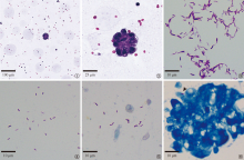
赵艳丽, 车南颖. 《结核病病理学诊断规范》团体标准解读. 中国防痨杂志, 2024, 46(4): 371-374. doi:10.19982/j.issn.1000-6621.20240074.
|
| [4] |
中国医疗保健国际交流促进会临床微生物与感染分会, 中华医学会检验医学分会临床微生物学组, 中华医学会微生物学和免疫学分会临床微生物学组. 综合医院结核分枝杆菌感染实验室检查共识. 中华检验医学杂志, 2022, 45(4): 343-353. doi:10.3760/cma.j.cn114452-20211118-00722.
|
| [5] |
World Health Organization. Practical manual on tuberculosis laboratory strengthening, 2022 update. Geneva: World Health Organization, 2022.
|
| [6] |
Wu RI, Mark EJ, Hunt JL. Staining for acid-fast bacilli in surgical pathology: practice patterns and variations. Hum Pathol, 2012, 43(11): 1845-1851. doi:10.1016/j.humpath.2012.01.006.
pmid: 22542129
|
| [7] |
叶恩如, 陈伟飞, 翁寿向, 等. 新型阳性对照在抗酸杆菌染色中的应用. 中华病理学杂志, 2020, 49(5): 485-486. doi:10.3760/cma.j.cn112151-20190909-00489.
|
| [8] |
余光银, 陶丽丽, 黄楚强, 等. 一种抗酸染色阳性质控结核菌蜡块的制作. 中华病理学杂志, 2023, 52(3): 291-292. doi:10.3760/cma.j.cn112151-20220712-00597.
|
| [9] |
中华人民共和国国家卫生和计划生育委员会. WS 288—2017 肺结核诊断. 结核与肺部疾病杂志, 2024, 5(4): 376-378. doi:10.19983/j.issn.2096-8493.2024022.
|
| [10] |
方伟建, 龚伟, 朱忆凌, 等. 结核病诊断中抗酸染色阳性对照芯片的制作及其应用. 中国防痨杂志, 2015, 37(5): 541-542. doi:10.3969/j.issn.1000-6621.2015.05.017.
|
| [11] |
Schewe C, Goldmann T, Grosser M, et al. Inter-laboratory validation of PCR-based detection of Mycobacterium tuberculosis in formalin-fixed, paraffin-embedded tissues. Virchows Arch, 2005, 447(3): 573-585. doi:10.1007/s00428-005-1233-3.
|
| [12] |
Cabras AD, Kremer M, Schulz S, et al. Quality assessment in diagnostic molecular pathology:: experience from a German-Austrian-Swiss multicenter trial. Virchows Arch, 2000, 437(1): 46-51. doi:10.1007/s004280000212.
pmid: 10963379
|
| [13] |
陈昕, 王晨, 陈琦, 等. 一种新型抗酸染色阳性对照在病理诊断中的应用. 临床与实验病理学杂志, 2022, 38(1): 117-118. doi:10.13315/j.cnki.cjcep.2022.01.030.
|
| [14] |
杨润, 程书亚, 刘路录, 等. 一种抗酸染色阳性对照品的制作及其应用. 诊断病理学杂志, 2024, 31(2): 167,171. doi:10.3969/j.issn.1007-8096.2024.02.022.
|
| [15] |
Barletta F, Vandelannoote K, Collantes J, et al. Standardization of a TaqMan-Based Real-Time PCR for the Detection of Mycobacterium tuberculosis-Complex in Human Sputum. Am J Trop Med Hyg, 2014, 91(4): 709-714. doi:10.4269/ajtmh.13-0603.
|
| [16] |
Noordhoek GT, Mulder S, Wallace P, et al. Multicentre quality control study for detection of Mycobacterium tuberculosis in clinical samples by nucleic amplification methods. Clin Microbiol Infect, 2004, 10(4): 295-301. doi:10.1111/j.1198-743X.2004.00825.x.
|
| [17] |
Ferrer I, Armstrong J, Capellari S, et al. Effects of formalin fixation, paraffin embedding, and time of storage on DNA preservation in brain tissue: a BrainNet Europe study. Brain Pathol, 2007, 17(3): 297-303. doi:10.1111/j.1750-3639.2007.00073.x.
pmid: 17465988
|

 ), Che Nanying1(
 京公网安备11010202007215号
Total visitors: Visitors of today: Now online:
京公网安备11010202007215号
Total visitors: Visitors of today: Now online:
 This work is licensed under Creative Commons Attribution 3.0 License.