
Chinese Journal of Antituberculosis ›› 2020, Vol. 42 ›› Issue (11): 1142-1152.doi: 10.3969/j.issn.1000-6621.2020.11.002
• Original Articles • Previous Articles Next Articles
LIU Fang-chao, YANG Xin-ting, JIANG Hui, DUAN Hong-fei, LIANG Qing-tao, LI Hua, YANG Yang, GUO Chao, ZHANG Yun, SHAO Ling-ling, CHEN Xiao-you( )
)
Online:2020-11-10
Published:2020-11-13
Contact:
CHEN Xiao-you
E-mail:chenxy1998@hotmail.com
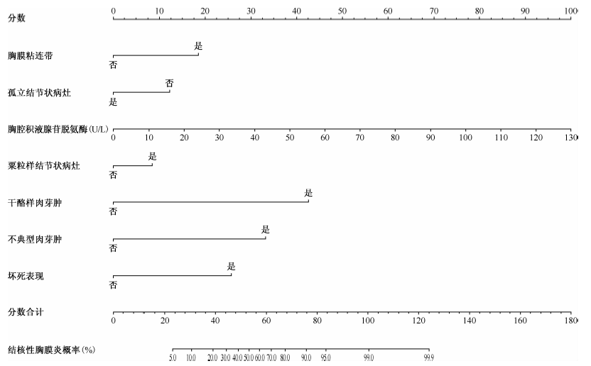
LIU Fang-chao, YANG Xin-ting, JIANG Hui, DUAN Hong-fei, LIANG Qing-tao, LI Hua, YANG Yang, GUO Chao, ZHANG Yun, SHAO Ling-ling, CHEN Xiao-you. Evaluation of the diagnosis value of nomogram based on the clinical and internal thoracoscopic features in pleural effusion patients complicated with tuberculous pleurisy[J]. Chinese Journal of Antituberculosis, 2020, 42(11): 1142-1152. doi: 10.3969/j.issn.1000-6621.2020.11.002
Add to citation manager EndNote|Ris|BibTeX
URL: https://www.zgflzz.cn/EN/10.3969/j.issn.1000-6621.2020.11.002
| 指标 | 全部患者 (206例) | 其他原因胸腔积液组 (77例) | 结核性胸膜炎组 (129例) | 统计检验值 | P值 | ||||
|---|---|---|---|---|---|---|---|---|---|
| 一般资料 | |||||||||
| 性别 | χ2=2.484 | 0.115 | |||||||
| 男 | 147(71.4) | 50(64.9) | 97(75.2) | ||||||
| 女 | 59(28.6) | 27(35.1) | 32(24.8) | ||||||
| 年龄(岁) | χ2=6.609 | 0.010 | |||||||
| ≥60 | 66(32.0) | 33(42.9) | 33(25.6) | ||||||
| <60 | 140(68.0) | 44(57.1) | 96(74.4) | ||||||
| 吸烟 | χ2=1.409 | 0.235 | |||||||
| 否 | 131(63.6) | 45(58.4) | 86(66.7) | ||||||
| 是 | 75(36.4) | 32(41.6) | 43(33.3) | ||||||
| 胸腔积液部位 | χ2=0.734 | 0.693 | |||||||
| 单侧 | 192(93.2) | 71(92.2) | 121(93.8) | ||||||
| 双侧 | 14(6.8) | 6(7.8) | 8(6.2) | ||||||
| 指标 | 全部患者 (206例) | 其他原因胸腔积液组 (77例) | 结核性胸膜炎组 (129例) | 统计检验值 | P值 | ||||
| 胸腔积液量 | χ2=2.510 | 0.285 | |||||||
| 低于肺门 | 134(65.0) | 49(63.6) | 85(65.9) | ||||||
| 高于肺门 | 72(35.0) | 28(36.4) | 44(34.1) | ||||||
| 胸腔镜下形态学特征 | |||||||||
| 胸膜充血 | χ2=3.816 | 0.050 | |||||||
| 否 | 66(32.0) | 31(40.3) | 35(27.1) | ||||||
| 是 | 140(68.0) | 46(59.7) | 94(72.9) | ||||||
| 粟粒样结节状病灶 | -a | <0.001a | |||||||
| 否 | 148(71.8) | 72(93.5) | 76(58.9) | ||||||
| 是 | 58(28.2) | 5(6.5) | 53(41.1) | ||||||
| 纤维素沉积 | χ2=13.760 | <0.001 | |||||||
| 否 | 142(68.9) | 65(84.4) | 77(59.7) | ||||||
| 是 | 64(31.1) | 12(15.6) | 52(40.3) | ||||||
| 胸膜粘连带 | χ2=28.413 | <0.001 | |||||||
| 否 | 90(43.7) | 52(67.5) | 38(29.5) | ||||||
| 是 | 116(56.3) | 25(32.5) | 91(70.5) | ||||||
| 胸膜增厚 | χ2=0.504 | 0.477 | |||||||
| 否 | 166(80.6) | 64(83.1) | 102(79.1) | ||||||
| 是 | 40(19.4) | 13(16.9) | 27(20.9) | ||||||
| 孤立结节状病灶 | χ2=0.420 | 0.516 | |||||||
| 否 | 125(60.7) | 33(42.9) | 92(71.3) | ||||||
| 是 | 81(39.3) | 44(57.1) | 37(28.7) | ||||||
| 病理形态学特征 | |||||||||
| 干酪样肉芽肿 | -a | <0.001a | |||||||
| 否 | 153(74.3) | 75(97.4) | 76(58.9) | ||||||
| 是 | 53(25.7) | 2(2.6) | 53(41.1) | ||||||
| 不典型肉芽肿 | -a | <0.001a | |||||||
| 否 | 155(75.2) | 74(96.1) | 81(62.8) | ||||||
| 是 | 51(24.8) | 3(3.9) | 48(37.2) | ||||||
| 坏死表现 | -a | 0.330a | |||||||
| 否 | 192(93.2) | 74(96.1) | 118(91.5) | ||||||
| 是 | 14(6.8) | 3(3.9) | 11(8.5) | ||||||
| 非特异性炎性反应 | χ2=0.145 | 0.702 | |||||||
| 否 | 166(80.6) | 61(79.2) | 105(81.4) | ||||||
| 是 | 40(19.4) | 16(20.8) | 24(18.6) | ||||||
| 胸腔积液实验室检查指标 | |||||||||
| 白细胞计数(个/μl) | 2321.0(1014.0,3879.0) | 1280.0(659.5,2612.0) | 2702.0(1620.0,4272.0) | Z=-4.577 | <0.001 | ||||
| 单核细胞百分比 | 0.93(0.81,0.97) | 0.85(0.65,0.93) | 0.95(0.87,0.98) | Z=-5.281 | 0.005 | ||||
| 腺苷脱氨酶(U/L) | 34.3(15.4,50.8) | 14.0(10.0,18.3) | 46.0(35.0,59.0) | Z=-9.332 | <0.001 | ||||
| 总蛋白(g/L) | 48.9(45.1,52.6) | 49.2(42.7,51.1) | 48.9(46.2,52.9) | Z=-1.101 | 0.270 | ||||
| 乳酸脱氢酶(U/L) | 333.0(254.0,506.0) | 294.0(193.0,343.0) | 377.0(274.0,540.0) | Z=-2.839 | 0.005 | ||||
| 胸膜组织检查 | 检测例数 | 阳性例数 | 阳性率(%) |
|---|---|---|---|
| 病理切片抗酸染色 | 106 | 42 | 39.6 |
| 病理切片核酸检测 | 69 | 35 | 50.7 |
| 胸膜组织抗酸染色 | 83 | 2 | 2.4 |
| 胸膜组织GeneXpert MTB/RIF | 103 | 33 | 32.0 |
| 胸膜组织结核分枝杆菌培养 | 110 | 32 | 29.1 |
| 以上方法联合评价a | 129 | 75 | 58.1 |
| 指标 | 训练集合计 (162例) | 其他原因胸腔积液组 (58例) | 结核性胸膜炎组 (104例) | 统计检验值 | P值 | ||||
|---|---|---|---|---|---|---|---|---|---|
| 一般资料 | |||||||||
| 性别 | χ2=2.798 | 0.094 | |||||||
| 男 | 111(68.5) | 35(60.3) | 76(73.1) | ||||||
| 女 | 51(31.5) | 23(39.7) | 28(26.9) | ||||||
| 年龄组(岁) | χ2=6.021 | 0.014 | |||||||
| ≥60 | 53(32.7) | 26(44.8) | 27(26.0) | ||||||
| <60 | 109(67.3) | 32(55.2) | 77(74.0) | ||||||
| 单侧胸腔积液 | - | 0.525a | |||||||
| 是 | 11(6.8) | 5(8.6) | 6(5.8) | ||||||
| 否 | 151(93.2) | 53(91.4) | 98(94.2) | ||||||
| 大量胸腔积液 | χ2=4.770 | 0.092 | |||||||
| 低于肺门 | 109(67.3) | 42(72.4) | 67(64.4) | ||||||
| 超过肺门 | 53(32.7) | 16(27.6) | 37(35.6) | ||||||
| 吸烟 | χ2=1.853 | 0.173 | |||||||
| 否 | 106(65.4) | 34(58.6) | 72(69.2) | ||||||
| 是 | 56(34.6) | 24(41.4) | 32(30.8) | ||||||
| 胸腔镜下形态学特征 | |||||||||
| 胸膜充血 | χ2=3.272 | 0.071 | |||||||
| 否 | 50(30.9) | 23(39.7) | 27(26.0) | ||||||
| 是 | 112(69.1) | 35(60.3) | 77(74.0) | ||||||
| 指标 | 训练集合计 (162例) | 其他原因胸腔积液组 (58例) | 结核性胸膜炎组 (104例) | 统计检验值 | P值 | ||||
| 粟粒样结节状病灶 | χ2=29.701 | <0.001 | |||||||
| 否 | 114(70.4) | 56(96.6) | 58(55.8) | ||||||
| 是 | 48(29.6) | 2(3.4) | 46(44.2) | ||||||
| 纤维素沉积 | χ2=11.397 | 0.001 | |||||||
| 否 | 110(67.9) | 49(84.5) | 61(58.7) | ||||||
| 是 | 52(32.1) | 9(15.5) | 43(41.3) | ||||||
| 胸膜粘连带 | χ2=21.742 | <0.001 | |||||||
| 否 | 67(41.4) | 38(65.5) | 29(27.9) | ||||||
| 是 | 95(58.6) | 20(34.5) | 75(72.1) | ||||||
| 胸膜增厚 | χ2=0.539 | 0.462 | |||||||
| 否 | 132(81.5) | 49(84.5) | 83(79.8) | ||||||
| 是 | 30(18.5) | 9(15.5) | 21(20.2) | ||||||
| 孤立结节状病灶 | χ2=13.266 | <0.001 | |||||||
| 否 | 100(61.7) | 25(43.1) | 75(72.1) | ||||||
| 是 | 62(38.3) | 33(56.9) | 29(27.9) | ||||||
| 病理形态学特征 | |||||||||
| 干酪样肉芽肿 | - | <0.001a | |||||||
| 否 | 120(74.1) | 57(98.3) | 63(60.6) | ||||||
| 是 | 42(25.9) | 1(1.7) | 41(39.4) | ||||||
| 不典型肉芽肿 | - | <0.001a | |||||||
| 否 | 118(72.8) | 55(94.8) | 63(60.6) | ||||||
| 是 | 44(27.2) | 3(5.2) | 41(39.4) | ||||||
| 坏死表现 | - | 0.330a | |||||||
| 否 | 151(93.2) | 56(96.6) | 95(91.3) | ||||||
| 是 | 11(6.8) | 2(3.4) | 9(8.7) | ||||||
| 非特异性炎性反应 | χ2=0.233 | 0.630 | |||||||
| 否 | 129(79.6) | 45(77.6) | 84(80.8) | ||||||
| 是 | 33(20.4) | 13(22.4) | 20(19.2) | ||||||
| 胸腔积液实验室检查指标 | |||||||||
| 白细胞计数(个/μl)b | 2344.0(1010.0,3879.0) | 1330.5(635.0,2615.0) | 2692.0(1617.5,4278.5) | Z=-4.035 | <0.001 | ||||
| 单核细胞百分比b | 0.91(0.82,0.97) | 0.88(0.78,0.94) | 0.95(0.84,0.98) | Z=-3.080 | 0.002 | ||||
| 腺苷脱氨酶 (U/L)b | 35.0(16.0,51.0) | 14.0(11.0,19.0) | 46.4(34.4,55.6) | Z=-8.960 | <0.001 | ||||
| 总蛋白(g/L)b | 48.1(46.0,50.9) | 46.6(45.3,48.8) | 49.2(46.8,51.8) | Z=-3.510 | 0.274 | ||||
| 乳酸脱氢酶(U/L)b | 362.0(216.0,577.0) | 216.0(84.0,376.0) | 419.0(303.5,600.5) | Z=-4.975 | <0.001 | ||||
| 指标 | β值 | s | Wald χ2值 | P值 | OR值 | 95%CI值 |
|---|---|---|---|---|---|---|
| 腺苷脱氨酶 | 0.075 | 0.022 | 11.980 | <0.001 | 0.928 | 0.042~1.145 |
| 粟粒样结节状病灶 | 0.405 | 0.529 | 0.587 | 0.444 | 2.248 | 0.283~17.851 |
| 胸膜粘连带 | 0.903 | 0.348 | 6.717 | 0.009 | 6.083 | 1.553~23.828 |
| 孤立结节状病灶 | -0.595 | 0.353 | 2.839 | 0.092 | 0.305 | 0.076~1.214 |
| 干酪样肉芽肿 | 2.072 | 0.590 | 12.330 | <0.001 | 63.063 | 14.240~238.301 |
| 不典型肉芽肿 | 1.617 | 0.441 | 13.472 | <0.001 | 25.377 | 8.513~72.694 |
| 坏死表现 | 1.253 | 0.552 | 5.166 | 0.023 | 12.264 | 2.412~53.527 |

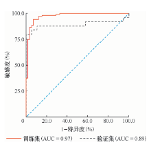
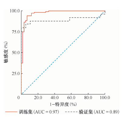
| 评价指标 | 训练集 | 验证集 |
|---|---|---|
| AUC | 0.97(0.95~0.99) | 0.89(0.78~0.98) |
| 敏感度(%) | 92.31(85.12~96.37) | 80.44(58.65~92.42) |
| 特异度(%) | 93.42(81.51~97.66) | 100.00(79.65~100.00) |
| 正确指数 | 0.85 | 0.80 |
| 阳性预测值(%) | 96.31(89.49~98.03) | 100.00(80.00~100.00) |
| 阴性预测值(%) | 87.14(76.39~94.14) | 79.45(57.31~92.25) |
| 阳性似然比 | 13.38(5.19~34.51) | - |
| 阴性似然比 | 0.08(0.04~0.16) | 0.20(0.09~0.44) |

| [1] |
Willendrup F, Bodtger U, Colella S, et al. Diagnostic accuracy and safety of semirigid thoracoscopy in exudative pleural effusions in Denmark. J Bronchology Interv Pulmonol, 2014,21(3):215-219. doi: 10.1097/LBR.0000000000000088.
doi: 10.1097/LBR.0000000000000088 URL pmid: 24992129 |
| [2] | Na MJ. Diagnostic tools of pleural effusion. Tuberc Respir Dis (Seoul), 2014,76(5):199-210. doi: 10.4046/trd.2014.76.5.199. |
| [3] |
Rodrigues LV, Samouco G, Gomes R, et al. Effectiveness and safety of local anesthetic, semi-flexible pleuroscopy-experience from a peripheral hospital. Pulmonology, 2019,25(1):9-14. doi: 10.1016/j.pulmoe.2018.05.003.
doi: 10.1016/j.pulmoe.2018.05.003 URL pmid: 29898873 |
| [4] |
Wang Z, Xu LL, Wu YB, et al. Diagnostic value and safety of medical thoracoscopy in tuberculous pleural effusion. Respir Med, 2015,109(9):1188-1192. doi: 10.1016/j.rmed.2015.06.008.
doi: 10.1016/j.rmed.2015.06.008 URL pmid: 26166016 |
| [5] |
Sivagnaname Y, Radhakrishnan P, Maria Selvam A. Thoracoscopic pleural brushing-an innovative method of pleural sampling in diagnostic medical thoracoscopy. Adv Respir Med, 2019,87(5):257-260. doi: 10.5603/ARM.2019.0046.
doi: 10.5603/ARM.2019.0046 URL pmid: 31680224 |
| [6] |
Kong XL, Zeng HH, Chen Y, et al. The visual diagnosis of tuberculous pleuritis under medical thoracoscopy: a retrospective series of 91 cases. Eur Rev Med Pharmacol Sci, 2014,18(10):1487-1495.
URL pmid: 24899607 |
| [7] |
Haralsingh A, Rawlins R. The role of thoracoscopic biopsies in the diagnosis of pleural tuberculosis. Respir Med Case Rep, 2019,27:100846. doi: 10.1016/j.rmcr.2019.100846.
doi: 10.1016/j.rmcr.2019.100846 URL pmid: 31061789 |
| [8] | 陈正贤. 内科胸腔镜. 北京: 人民卫生出版社, 2008: 16. |
| [9] |
Ruan SY, Chuang YC, Wang JY, et al. Revisiting tuberculous pleurisy: pleural fluid characteristics and diagnostic yield of mycobacterial culture in an endemic area. Thorax, 2012,67(9):822-827. doi: 10.1136/thoraxjnl-2011-201363.
doi: 10.1136/thoraxjnl-2011-201363 URL pmid: 22436167 |
| [10] |
Zhou X, Jiang P, Huan X, et al. Ultrasound-Guided versus Thoracoscopic Pleural Biopsy for Diagnosing Tuberculous Pleurisy Following Inconclusive Thoracentesis: A Rando-mized, Controlled Trial. Med Sci Monit, 2018,24:7238-7248. doi: 10.12659/MSM.912506.
doi: 10.12659/MSM.912506 URL pmid: 30303950 |
| [11] |
Kho SS, Chan SK, Yong MC, et al. Drainage of multilocula-ted tuberculous pleural effusion by medical thoracoscopy: When and why should it be considered? Med J Malaysia, 2018,73(1):49-50.
URL pmid: 29531204 |
| [12] |
Wang XJ, Yang Y, Wang Z, et al. Efficacy and safety of diagnostic thoracoscopy in undiagnosed pleural effusions. Respiration, 2015,90(3):251-255. doi: 10.1159/000435962.
doi: 10.1159/000435962 URL pmid: 26279455 |
| [13] |
Chen RL, Zhang YQ, Wang J, et al. Diagnostic value of medi-cal thoracoscopy for undiagnosed pleural effusions. Exp Ther Med, 2018,16(6):4590-4594. doi: 10.3892/etm.2018.6742.
doi: 10.3892/etm.2018.6742 URL pmid: 30542409 |
| [14] |
Christopher DJ, Dinakaran S, Gupta R, et al. Thoracoscopic pleural biopsy improves yield of Xpert MTB/RIF for diagnosis of pleural tuberculosis. Respirology, 2018,23(7):714-717. doi: 10.1111/resp.13275.
doi: 10.1111/resp.13275 URL pmid: 29486527 |
| [15] |
He Y, Zhang W, Huang T, et al. Evaluation of a diagnostic flow chart applying medical thoracoscopy, adenosine deaminase and T-SPOT.TB in diagnosis of tuberculous pleural effusion. Eur Rev Med Pharmacol Sci, 2015,19(19):3563-3568.
URL pmid: 26502844 |
| [16] |
Huo Z, Yang M, Chen J, et al. Improved early diagnosis of difficult cases of tuberculous pleural effusion by combination of thoracoscopy with immunological tests. Int J Infect Dis, 2019,81:38-42. doi: 10.1016/j.ijid.2019.01.045.
doi: 10.1016/j.ijid.2019.01.045 URL pmid: 30710790 |
| [17] |
Yin Y, Eberhardt R, Wang XB, et al. Semi-Rigid Thoracoscopic Punch Biopsy Using a Hybrid Knife with a High-Pressure Water Jet for the Diagnosis of Pleural Effusions. Respiration, 2016,92(3):192-196. doi: 10.1159/000448556.
doi: 10.1159/000448556 URL pmid: 27577029 |
| [18] |
Lei Z, Li J, Wu D, et al. Nomogram for Preoperative Estimation of Microvascular Invasion Risk in Hepatitis B Virus-Related Hepatocellular Carcinoma Within the Milan Criteria. JAMA Surg, 2016,151(4):356-363. doi: 10.1001/jamasurg.2015.4257.
doi: 10.1001/jamasurg.2015.4257 URL pmid: 26579636 |
| [1] | Zhu Mingzhi, Shao Yanqin, Fan Dapeng, Liu Libin, Mei Bin, Dai Lingshan, Cai Long. Diagnostic value of urine lipoarabinomannan antigen detection in extrapulmonary tuberculosis [J]. Chinese Journal of Antituberculosis, 2025, 47(4): 471-476. |
| [2] | Senior Department of Tuberculosis, the 8th Medical Center of Chinese PLA General Hospital , Editorial Board of Chinese Journal of Antituberculosis , Basic and Clinical Speciality Committees of Tuberculosis Control Branch of China International Exchange , Promotive Association for Medical and Health Care . Expert consensus on multidisciplinary diagnosis and treatment of tuberculous peritonitis [J]. Chinese Journal of Antituberculosis, 2025, 47(3): 243-257. |
| [3] | Duan Hongfei, Tao Yong. Interpretation of social organization standard of Diagnosis Specification of Intraocular Tuberculosis [J]. Chinese Journal of Antituberculosis, 2025, 47(3): 258-261. |
| [4] | Gong Sheng, Wang Ning, Li Dan, Li Gang, Liu Yu, Jiang Liangshuang, Yao Xiaojun. Comparative study of fluorescence staining method and inflation-deflation method for thoracoscopic pulmonary tuberculosis segmentectomy [J]. Chinese Journal of Antituberculosis, 2025, 47(3): 292-297. |
| [5] | Jia Hui, Jing Hui, Ling Xiaojie, Wang Yan, Li Xuezheng. The diagnostic value of GeneXpert MTB/RIF Ultra in detecting sputum samples for newly diagnosed pulmonary tuberculosis [J]. Chinese Journal of Antituberculosis, 2025, 47(3): 298-304. |
| [6] | Shi Yuru, Gu Dejian, Wu Jing, Liu Ting, Qin Linghan, Yue Li, Qi Yingjie. Diagnostic value of probe capture-based targeted next-generation sequencing and metagenomic next-generation sequencing for detecting Mycobacterium tuberculosis in bronchoalveolar lavage fluid [J]. Chinese Journal of Antituberculosis, 2025, 47(3): 305-311. |
| [7] | Yang Ziyi, Chen Suting. Research progress on bedaquiline resistance and drug resistance diagnosis [J]. Chinese Journal of Antituberculosis, 2025, 47(3): 374-379. |
| [8] | Qiu Yong, Quan Zhuo, Qu Rong, Tian Fajun, Li Meng, Wang Gengsheng, Wang Ya, Guo Mingcheng, Gao Qian. Evaluation of different tuberculosis diagnostic tools for detecting patients in a primary-level clinic in rural China: a real-world retrospective study [J]. Chinese Journal of Antituberculosis, 2025, 47(2): 181-188. |
| [9] | Zhao Yue, Wang Haoran, Cheng Meijin, Wang Wei, Liang Ruixia, Huang Hairong. The evaluation of the smear-positive and Xpert-negative outcome as an early indicator of nontuberculous mycobacteria existence in clinical specimen [J]. Chinese Journal of Antituberculosis, 2025, 47(1): 61-65. |
| [10] | Wang Xiaomin, Chen Jinyun, Zeng Yuqin, Ma Quan, Kong Xingxing, Meng Jianzhou, Lu Shuihua. Interpretation of the third edition of WHO consolidated guidelines on tuberculosis: module 3: diagnosis: rapid diagnostics for tuberculosis detection [J]. Chinese Journal of Antituberculosis, 2024, 46(9): 1006-1022. |
| [11] | Mei Chunlin, Yang Chengqing, Du Ronghui, Cao Tanze, Feng Wei, Chen Shufang, Liu Xiuping, Ou Jiali. Diagnostic accuracy of GeneXpert MTB/RIF in detecting pulmonary tuberculosis with extremely low loads of MTB in bronchoalveolar lavage fluid in general hospitals [J]. Chinese Journal of Antituberculosis, 2024, 46(9): 1037-1041. |
| [12] | Zhong Lingshan, Wang Li, Zhang Shuo, Li Nan, Yang Qingyuan, Ding Wenlong, Chen Xingzhi, Huang Chencui, Xing Zhiheng. A machine learning model based on CT images combined with radiomics and semantic features for diagnosis of nontuberculous mycobacterium lung disease and pulmonary tuberculosis [J]. Chinese Journal of Antituberculosis, 2024, 46(9): 1042-1049. |
| [13] | Li Wenhan, Yang Jing, Li Chunhua. Research progress of artificial intelligence in pulmonary tuberculosis imaging diagnosis and drug resistance prediction [J]. Chinese Journal of Antituberculosis, 2024, 46(9): 1098-1103. |
| [14] | Duan Hongfei. Diagnosis and treatment of nontuberculous mycobacteria diseases in the past 60 years [J]. Chinese Journal of Antituberculosis, 2024, 46(8): 863-868. |
| [15] | Liu Ling, Zeng Yi, Wang Jin, Liu Xiaoling, Liu Yan, Lin Feishen, Guo Jing. Construction and evaluation of a model for predicting malnutrition in patients with pulmonary tuberculosis and diabetes mellitus [J]. Chinese Journal of Antituberculosis, 2024, 46(8): 903-909. |
| Viewed | ||||||
|
Full text |
|
|||||
|
Abstract |
|
|||||
 京公网安备11010202007215号
Total visitors: Visitors of today: Now online:
京公网安备11010202007215号
Total visitors: Visitors of today: Now online:
 This work is licensed under Creative Commons Attribution 3.0 License.