
Chinese Journal of Antituberculosis ›› 2019, Vol. 41 ›› Issue (2): 202-209.doi: 10.3969/j.issn.1000-6621.2019.02.015
• Original Articles • Previous Articles Next Articles
Duo LI,Kun FANG,Jue WANG,Zhen ZHOU,Ping-xin LYU( )
)
Received:2018-07-01
Online:2019-02-10
Published:2019-02-01
Contact:
Ping-xin LYU
E-mail:lpx1209@163.com
Duo LI,Kun FANG,Jue WANG,Zhen ZHOU,Ping-xin LYU. Analysis of CT image classification and clinical characteristics of nontuberculous mycobacterial pulmonary disease[J]. Chinese Journal of Antituberculosis, 2019, 41(2): 202-209. doi: 10.3969/j.issn.1000-6621.2019.02.015
Add to citation manager EndNote|Ris|BibTeX
URL: https://www.zgflzz.cn/EN/10.3969/j.issn.1000-6621.2019.02.015


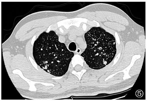

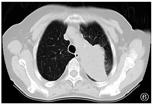
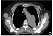
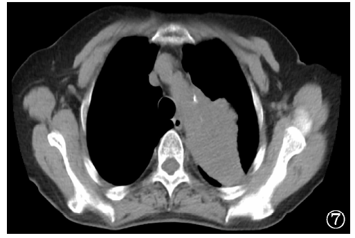


| CT征象 | 上叶空洞型 (36例) | 结节支气管 扩张型(61例) | 混合型 (15例) | 不易分类型 (20例) | χ2值 | P值 |
|---|---|---|---|---|---|---|
| 空洞 | 36(100.0) | 28(45.9) | 15(100.0) | 3(15.0) | 56.790a | <0.001 |
| 支气管扩张 | 19(52.8) | 61(100.0) | 13(86.7) | 8(40.0) | 51.464 | <0.001 |
| 肺气肿 | 24(66.7) | 8(13.1) | 8(53.3) | 5(25.0) | 32.155 | <0.001 |
| 间质纤维化 | 12(33.3) | 0(0.0) | 0(0.0) | 2(10.0) | 25.557a | <0.001 |
| 淋巴结肿大 | 7(19.4) | 10(16.4) | 4(26.7) | 4(20.0) | 1.135a | 0.789 |
| 淋巴结钙化或密度增高 | 12(33.3) | 22(36.1) | 5(33.3) | 7(35.0) | 0.092 | 0.993 |
| 胸膜增厚 | 22(61.1) | 14(23.0) | 10(66.7) | 6(30.0) | 19.432 | <0.001 |
| 胸腔积液 | 4(11.1) | 6(9.8) | 2(13.3) | 2(10.0) | 0.512a | 0.973 |
| 临床情况 | 上叶空洞型 (34例) | 结节支气管 扩张型(54例) | 混合型 (14例) | 不易分类型 (16例) | χ2值 | P值 |
|---|---|---|---|---|---|---|
| 咳嗽 | 31(91.2) | 50(92.6) | 13(92.9) | 12(75.0) | 3.861a | 0.226 |
| 咳痰 | 29(85.3) | 48(88.9) | 12(85.7) | 11(68.8) | 3.697a | 0.300 |
| 咯血 | 10(29.4) | 25(46.3) | 4(28.6) | 4(25.0) | 4.268 | 0.234 |
| 发热 | 21(61.8) | 26(48.1) | 8(57.1) | 8(50.0) | 6.184a | 0.402 |
| 痰涂片抗酸染色阳性 | 23(67.6) | 18(33.3) | 10(71.4) | 1(6.2) | 28.967 | 0.000 |
| 痰培养阳性 | 31(91.2) | 43(79.6) | 13(92.9) | 13(81.2) | 3.667 | 0.715 |
| 吸烟 | 22(64.7) | 7(13.0) | 3(21.4) | 5(31.2) | 26.697 | 0.000 |
| 并发糖尿病 | 3(8.8) | 1(1.9) | 4(28.6) | 1(6.2) | 9.197a | 0.012 |
| 菌种 | 上叶空洞型 (36例) | 结节支气管 扩张型(61例) | 混合型 (15例) | 不易分类型 (20例) | 合计 (132例) |
|---|---|---|---|---|---|
| 鸟-胞内分枝杆菌复合群 | 22 | 34 | 13 | 8 | 77 |
| 脓肿分枝杆菌 | 6 | 22 | 1 | 7 | 36 |
| 堪萨斯分枝杆菌 | 4 | 4 | 1 | 3 | 12 |
| 蟾蜍分枝杆菌 | 2 | 0 | 0 | 0 | 2 |
| 戈登分枝杆菌 | 1 | 0 | 0 | 1 | 2 |
| 偶然分枝杆菌 | 0 | 0 | 0 | 1 | 1 |
| 苏尔加分枝杆菌 | 1 | 0 | 0 | 0 | 1 |
| 龟分枝杆菌 | 0 | 1 | 0 | 0 | 1 |
| 合计 | 36 | 61 | 15 | 20 | 132 |
| CT征象 | MAC肺病(77例) | 脓肿分枝杆菌肺病(36例) | χ2值 | P值 |
|---|---|---|---|---|
| 空洞 | 51(66.2) | 19(52.8) | 1.884 | 0.170 |
| 支气管扩张 | 62(80.5) | 30(83.3) | 0.128 | 0.720 |
| 肺气肿 | 30(39.0) | 8(22.2) | 3.079 | 0.079 |
| 间质纤维化 | 10(13.0) | 2(5.6) | 0.753a | 0.386 |
| 纵隔淋巴结肿大 | 20(26.0) | 3(8.3) | 4.709a | 0.030 |
| 纵隔淋巴结钙化或密度增高 | 29(37.7) | 9(25.0) | 1.762 | 0.184 |
| 胸膜增厚 | 34(44.2) | 13(36.1) | 0.654 | 0.419 |
| 胸腔积液 | 5(6.5) | 8(22.2) | 4.516 | 0.034 |
| [1] |
Prevots DR, Shaw PA, Strickland D , et al. Nontuberculous mycobacterial lung disease prevalence at four integrated health care delivery systems. Am J Respir Crit Care Med, 2010,182(7):970-976.
doi: 10.1164/rccm.201002-0310OC URL |
| [2] |
Yu X, Liu P, Liu G , et al. The prevalence of non-tuberculous mycobacterial infections in mainland China: Systematic review and meta-analysis. J Infect, 2016,73(6):558-567.
doi: 10.1016/j.jinf.2016.08.020 URL |
| [3] | 吕平欣, 马大庆 . 常见非结核分枝杆菌肺病的CT表现. 中华放射学杂志, 2015,49(3):238-240. |
| [4] | 中华医学会结核病学分会. 非结核分枝杆菌病诊断与治疗专家共识. 中华结核和呼吸杂志, 2012,35(8):572-579. |
| [5] |
Okumura M, Iwai K, Ogata H , et al. Clinical factors on cavitary and nodular bronchiectatic types in pulmonary Mycobacterium avium complex disease. Internal Medicine, 2008,47(16):1465-1472.
doi: 10.2169/internalmedicine.47.1114 URL |
| [6] |
Chung MJ, Lee KS, Koh WJ , et al. Thin-section CT findings of nontuberculous mycobacterial pulmonary diseases: comparison between Mycobacterium avium-intracellulare complex and Mycobacterium abscessus infection. J Korean Med Sci, 2005,20(5):777-783.
doi: 10.3346/jkms.2005.20.5.777 URL |
| [7] |
Erasmus JJ , McAdams HP, Farrell MA, et al. Pulmonary nontuberculous mycobacterial infection: radiologic manifestations. Radiographics, 1999,19(6):1487-1505.
doi: 10.1148/radiographics.19.6.g99no101487 URL |
| [8] |
Wallace RJ Jr, Zhang Y, Brown BA , et al. Polyclonal Mycobacterium avium complex infections in patients with nodular bronchiectasis. Am J Respir Crit Care Med, 1998,158(4):1235-1244.
doi: 10.1164/ajrccm.158.4.9712098 URL |
| [9] |
Park TY, Chong S, Jung JW , et al. Natural course of the nodular bronchiectatic form of Mycobacterium avium complex lung disease: Long-term radiologic change without treatment. PLoS One, 2017,12(10):e0185774.
doi: 10.1371/journal.pone.0185774 URL |
| [10] |
Carrillo MC, Patsios D, Wagnetz U , et al. Comparison of the spectrum of radiologic and clinical manifestations of pulmonary disease caused by Mycobacterium avium complex and Mycobacterium xenopi. Can Assoc Radiol J, 2014,65(3):207-213.
doi: 10.1016/j.carj.2013.05.006 URL |
| [11] |
Huang LK, Wang HH, Lai YC , et al. The impact of glycemic status on radiological manifestations of pulmonary tuberculosis in diabetic patients. PLoS One, 2017,12(6):e0179750.
doi: 10.1371/journal.pone.0179750 URL |
| [12] |
Polverosi R, Guarise A, Balestro E , et al. High-resolution CT of nontuberculous mycobacteria pulmonary infection in immunocompetent, non-HIV-positive patients. Radiol Med, 2010,115(2):191-204.
doi: 10.1007/s11547-009-0479-2 URL |
| [13] |
Koh WJ, Lee KS, Kwon OJ , et al. Bilateral bronchiectasis and bronchiolitis at thin-section CT: diagnostic implications in nontuberculous mycobacterial pulmonary infection. Radiology, 2005,235(1):282-288.
doi: 10.1148/radiol.2351040371 URL |
| [14] |
Kwak N, Lee CH, Lee HJ , et al. Non-tuberculous mycobacterial lung disease: diagnosis based on computed tomography of the chest. Eur Radiol, 2016,26(12):4449-4456.
doi: 10.1007/s00330-016-4286-6 URL |
| [15] | Koh WJ, Yu CM, Suh GY , et al. Pulmonary TB and NTM lung disease: comparison of characteristics in patients with AFB smear-positive sputum. Int J Tuberc Lung Dis, 2006,10(9):1001-1007. |
| [16] |
Kendall BA, Varley CD, Choi D , et al. Distinguishing tuberculosis from nontuberculous mycobacteria lung disease, Oregon, USA. Emerg Infect Dis, 2011,17(3):506-509.
doi: 10.3201/eid1703.101164 URL |
| [17] |
Kim C, Park SH, Oh SY , et al. Comparison of chest CT fin-dings in nontuberculous mycobacterial diseases vs. Mycobacterium tuberculosis lung disease in HIV-negative patients with cavities. PLoS One, 2017,12(3):e0174240.
doi: 10.1371/journal.pone.0174240 URL |
| [18] | 李芳, 贺伟, 周新华 , 等. 非结核分枝杆菌肺病和活动性肺结核的高分辨率表现异同性分析. 中国防痨杂志, 2018,40(5):499-505. |
| [19] |
Hong SJ, Kim TJ, Lee JH , et al. Nontuberculous mycobacterial pulmonary disease mimicking lung cancer: Clinicoradiologic features and diagnostic implications. Medicine (Baltimore), 2016,95(26):e3978.
doi: 10.1097/MD.0000000000003978 URL |
| [20] |
Kim YK, Hahn S, Uh Y , et al. Comparable characteristics of tuberculous and non-tuberculous mycobacterial cavitary lung diseases. Int J Tuberc Lung Dis, 2014,18(6):725-729.
doi: 10.5588/ijtld.13.0871 URL |
| [21] |
段鸿飞, 王敬, 初乃惠 , 等. 鸟-胞内分枝杆菌复合菌组肺病与脓肿分枝杆菌肺病临床表现的对比观察. 中华结核和呼吸杂志, 2012,35(8):588-591.
doi: 10.3760/cma.j.issn.1001-0939.2012.08.008 |
| [22] |
贺伟, 宁锋刚, 李成海 , 等. 鸟-胞内分枝杆菌复合群肺病和脓肿分枝杆菌肺病的CT影像学比较. 中国防痨杂志, 2014,36(8):700-705.
doi: 10.3969/j.issn.1000-6621.2014.08.020 |
| [1] | Wang Yingchao, Liu Weiyi, Ji Xiuxiu, Shang Xuetian, Jia Hongyan, Zhang Lanyue, Sun Qi, Du Boping, Zhu Chuanzhi, Pan Liping, Zhang Zongde. Profile analysis of circRNA expression and identification of diagnostic markers in peripheral blood mononuclear cells of tuberculosis patients [J]. Chinese Journal of Antituberculosis, 2025, 47(4): 460-470. |
| [2] | Zhang Peize, Gao Qian, Deng Guofang. [18F]FDT-PET-CT technology that may bring revolutionary changes to tuberculosis clinical research [J]. Chinese Journal of Antituberculosis, 2025, 47(3): 262-265. |
| [3] | Song Feier, Mao Yanjun, Xia Qiuyue, Zhou Yang, Lin Huan. The prevalence and influencing factors of post-tuberculosis lung disease: A Meta-analysis [J]. Chinese Journal of Antituberculosis, 2025, 47(3): 322-330. |
| [4] | Zhao Yue, Wang Haoran, Cheng Meijin, Wang Wei, Liang Ruixia, Huang Hairong. The evaluation of the smear-positive and Xpert-negative outcome as an early indicator of nontuberculous mycobacteria existence in clinical specimen [J]. Chinese Journal of Antituberculosis, 2025, 47(1): 61-65. |
| [5] | Fan Jun, Wang Heng, Lan Tinglong, Dong Weijie, Tang Kai, Li Yuan, Yan Guangxuan, Xu Shangsheng, Kang Zhigang, Qin Shibing. Clinical characteristics and surgical outcomes of 12 cases of non-tuberculous mycobacterial spondylitis [J]. Chinese Journal of Antituberculosis, 2025, 47(1): 87-95. |
| [6] | Zhong Lingshan, Wang Li, Zhang Shuo, Li Nan, Yang Qingyuan, Ding Wenlong, Chen Xingzhi, Huang Chencui, Xing Zhiheng. A machine learning model based on CT images combined with radiomics and semantic features for diagnosis of nontuberculous mycobacterium lung disease and pulmonary tuberculosis [J]. Chinese Journal of Antituberculosis, 2024, 46(9): 1042-1049. |
| [7] | Chen Shuangshuang, Tian Lili, Wang Nenhan, Yang Xinyu, Zhao Yanfeng, Li Chuanyou, Dai Xiaowei. Analysis of in vitro antibacterial effects of 17 antibiotics against rapidly growing mycobacteria in the Beijing area [J]. Chinese Journal of Antituberculosis, 2024, 46(9): 1056-1062. |
| [8] | Wang Fei, Hua Duo, Guo Jianjian, Liu Chang, Han Lu, Ren Yi. Characteristic analysis of non-tuberculous mycobacterial pulmonary disease patients in Wuhan area from 2021 to 2023 [J]. Chinese Journal of Antituberculosis, 2024, 46(9): 1069-1076. |
| [9] | Li Wenhan, Yang Jing, Li Chunhua. Research progress of artificial intelligence in pulmonary tuberculosis imaging diagnosis and drug resistance prediction [J]. Chinese Journal of Antituberculosis, 2024, 46(9): 1098-1103. |
| [10] | Duan Hongfei. Diagnosis and treatment of nontuberculous mycobacteria diseases in the past 60 years [J]. Chinese Journal of Antituberculosis, 2024, 46(8): 863-868. |
| [11] | Xu Chunhua, Zhu Shiyu, Hu Yi, Yi Kehua, Song Canlei, Wang Zichun, Wu Yong, Wang Qing, Yang Qianru, Shen Xin. Analysis of screening effect of recombinant Mycobacterium tuberculosis fusion protein in screening Mycobacterium tuberculosis infection in close contacts of pulmonary tuberculosis patients [J]. Chinese Journal of Antituberculosis, 2024, 46(8): 897-902. |
| [12] | Tan Shouyong. Research progress on comprehensive treatment beyond antibiotic therapy for nontuberculous mycobacterium pulmonary disease [J]. Chinese Journal of Antituberculosis, 2024, 46(8): 967-970. |
| [13] | Chai Dongyu, Qin Shuyi, Zhang Ronghua, Zou Nannan, Wang Xin. Analysis of risk factors for viral pneumonia combined with invasive pulmonary mycosis [J]. Chinese Journal of Antituberculosis, 2024, 46(7): 750-755. |
| [14] | Zhang Hui, Ge Li, Zhang Yuhan, Feng Ruie. Clinicopathologic characteristics of 34 cases non-tuberculous mycobacterial disease [J]. Chinese Journal of Antituberculosis, 2024, 46(7): 756-762. |
| [15] | He Xiangrong, Chen Hua, Chen Pinru, Liang Feng, Ren Huili, Zhu Jialou, Hu Jinxing, Tan Yaoju. A case report and literature review of Mycobacterium asiaticum pulmonary disease [J]. Chinese Journal of Antituberculosis, 2024, 46(7): 763-769. |
| Viewed | ||||||
|
Full text |
|
|||||
|
Abstract |
|
|||||
 京公网安备11010202007215号
Total visitors: Visitors of today: Now online:
京公网安备11010202007215号
Total visitors: Visitors of today: Now online:
 This work is licensed under Creative Commons Attribution 3.0 License.