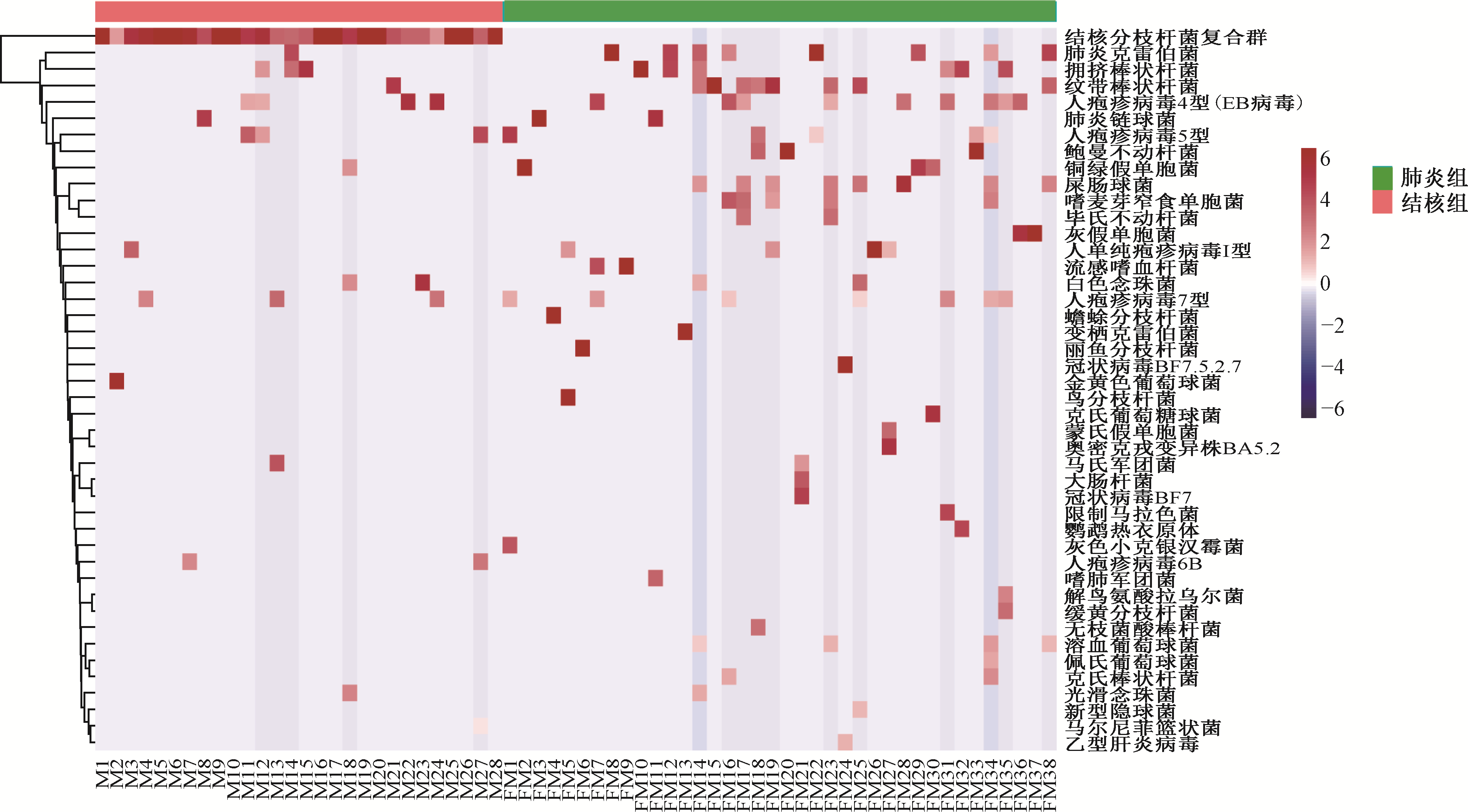
| [1] |
Hu Y, Cheng M, Liu B, et al. Metagenomic analysis of the lung microbiome in pulmonary tuberculosis-a pilot study. Emerg Microbes Infect, 2020, 9(1): 1444-1452. doi:10.1080/22221751.2020.1783188.
|
| [2] |
Hu Y, Kang Y, Liu X, et al. Distinct lung microbial community states in patients with pulmonary tuberculosis. Sci China Life Sci, 2020, 63(10): 1522-1533. doi:10.1007/s11427-019-1614-0.
|
| [3] |
Adami AJ, Cervantes JL. The microbiome at the pulmonary alveolar niche and its role in Mycobacterium tuberculosis infection. Tuberculosis (Edinb), 2015, 95(6): 651-658. doi:10.1016/j.tube.2015.07.004.
|
| [4] |
Hong BY, Maulén NP, Adami AJ, et al. Microbiome Changes during Tuberculosis and Antituberculous Therapy. Clin Microbiol Rev, 2016, 29(4): 915-926. doi:10.1128/CMR.00096-15.
|
| [5] |
Sulaiman I, Wu BG, Li Y, et al. Evaluation of the airway microbiome in nontuberculous mycobacteria disease. Eur Respir J, 2018, 52(4): 1800810. doi:10.1183/13993003.00810-2018.
|
| [6] |
张晨晨, 谭卫国, 郭卉欣, 等. 结核分枝杆菌感染对痰液菌群结构的影响. 中国防痨杂志, 2021, 43(4): 357-363. doi:10.3969/j.issn.1000-6621.2021.04.011.
|
| [7] |
谢锦慧, 喻容, 石国民, 等. 初治肺结核肠道菌群改变与免疫指标的相关性研究. 中华预防医学杂志, 2021, 55(12): 1486-1490. doi:10.3760/cma.j.cn112150-20210728-00721.
|
| [8] |
Tsay JJ, Wu BG, Sulaiman I, et al. Lower Airway Dysbiosis Affects Lung Cancer Progression. Cancer Discov, 2021, 11(2): 293-307. doi:10.1158/2159-8290.CD-20-0263.
|
| [9] |
Chao Y, Li J, Gong Z, et al. Rapid discrimination between tuberculosis and sarcoidosis using next-generation sequencing. Int J Infect Dis, 2021, 108: 129-136. doi:10.1016/j.ijid.2021.05.028.
pmid: 34004327
|
| [10] |
温立旻, 侯代伦. 肺结核合并肺癌影像学评估方法现状及进展. 中国防痨杂志, 2023, 45(6): 620-624. doi:10.19982/j.issn.1000-6621.20230045.
|
| [11] |
田丽丽, 陈双双, 樊瑞芳, 等. 四种方法联合检测对病原学阴性肺结核的诊断价值. 中国防痨杂志, 2023, 45(2): 144-150. doi:10.19982/j.issn.1000-6621.20220443.
|
| [12] |
Xu R, Lu R, Zhang T, et al. Temporal association between human upper respiratory and gut bacterial microbiomes during the course of COVID-19 in adults. Commun Biol, 2021, 4(1): 240. doi:10.1038/s42003-021-01796-w.
pmid: 33603076
|
| [13] |
Rueca M, Fontana A, Bartolini B, et al. Investigation of Nasal/Oropharyngeal Microbial Community of COVID-19 Patients by 16S rDNA Sequencing. Int J Environ Res Public Health, 2021, 18(4): 2174. doi:10.3390/ijerph18042174.
|
| [14] |
Millares L, Pascual S, Monton C, et al. Relationship between the respiratory microbiome and the severity of airflow limitation, history of exacerbations and circulating eosinophils in COPD patients. BMC Pulm Med, 2019, 19(1): 112. doi:10.1186/s12890-019-0867-x.
pmid: 31234826
|
| [15] |
Rosas-Salazar C, Kimura KS, Shilts MH, et al. SARS-CoV-2 infection and viral load are associated with the upper respiratory tract microbiome. J Allergy Clin Immunol, 2021, 147(4): 1226-1233.e2. doi:10.1016/j.jaci.2021.02.001.
pmid: 33577896
|
| [16] |
Lira-Lucio JA, Falfán-Valencia R, Ramírez-Venegas A, et al. Lung Microbiome Participation in Local Immune Response Regulation in Respiratory Diseases. Microorganisms, 2020, 8(7):1059. doi:10.3390/microorganisms8071059.
|
| [17] |
Natalini JG, Singh S, Segal LN. The dynamic lung microbiome in health and disease. Nat Rev Microbiol, 2023, 21(4):222-235. doi:10.1038/s41579-022-00821-x.
|
| [18] |
Pérez-Cobas AE, Ginevra C, Rusniok C, et al. The respiratory tract microbiome, the pathogen load, and clinical interventions define severity of bacterial pneumonia. Cell Rep Med, 2023, 4(9): 101167. doi:10.1016/j.xcrm.2023.101167.
|
| [19] |
Vogelzang A, Guerrini MM, Minato N, et al. Microbiota-an amplifier of autoimmunity. Curr Opin Immunol, 2018, 55: 15-21. doi:10.1016/j.coi.2018.09.003.
pmid: 30248521
|
| [20] |
Marsland BJ, Gollwitzer ES. Host-microorganism interactions in lung diseases. Nat Rev Immunol, 2014, 14(12): 827-835. doi:10.1038/nri3769.
pmid: 25421702
|

 ), He Yukun2, Zhou Dexun2, Zhang Pingji1
 京公网安备11010202007215号
Total visitors: Visitors of today: Now online:
京公网安备11010202007215号
Total visitors: Visitors of today: Now online:
 This work is licensed under Creative Commons Attribution 3.0 License.