| [1] |
ZHOU Lin, LIU Er-yong, MENG Qing-lin, CHEN Ming-ting, ZHOU Xin-hua, GAO Wei-wei, LIN Ming-gui, XIE Ru-ming.
Evaluation of the quality of pulmonary tuberculosis diagnosis after the implementation of the newly revised WS 288-2017 Diagnosis for pulmonary tuberculosis standards
[J]. Chinese Journal of Antituberculosis, 2020, 42(9): 910-915.
|
| [2] |
LIANG Rui-yun, FANG Wei-jun, REN Hui-li, LI Hui-ru, ZHANG Hui.
Study on CT manifestations of non-tuberculous mycobacterium pulmonary disease patients with and without diabetes mellitus
[J]. Chinese Journal of Antituberculosis, 2020, 42(9): 962-967.
|
| [3] |
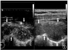
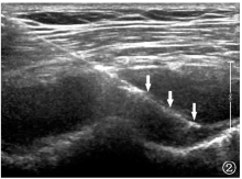
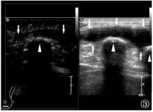
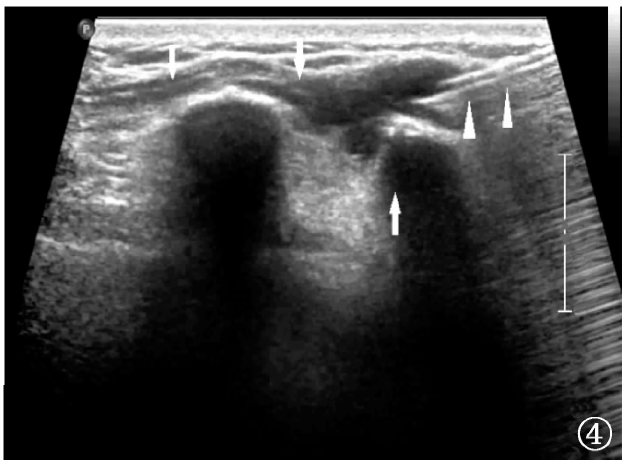
MA Ting-long, HAN Yi, CHENG Xu, LIU Zhi-dong.
Clinical observation on treatment effectiveness of transdermal ultrasound-mediated drug delivery combined with oral anti-tuberculosis drug in patients with chest wall tuberculosis
[J]. Chinese Journal of Antituberculosis, 2020, 42(9): 968-972.
|
| [4] |
ZHANG Li-juan, TAO Xiao, XIA Li-li, WEI Fen-fen, ZHENG Qi.
Value of clinical pathway table of enhanced recovery cluster nursing in patients with spinal tuberculosis during perioperative period
[J]. Chinese Journal of Antituberculosis, 2020, 42(9): 981-986.
|
| [5] |
LI Ai-fang, CUI Xiao-li, KANG Lei, LEI Jing, DANG Li-yun, YANG Han.
Value of fluorescence PCR probe melting curve method in detecting resistance of Mycobacterium tuberculosis
[J]. Chinese Journal of Antituberculosis, 2020, 42(9): 998-1001.
|
| [6] |
ZHAO Tie-niu, JIANG Shuang-shuang, HUANG Li, HU Xue-mei.
Comparative study of multislice spiral CT and color Doppler ultrasonography in the diagnosis of bone and joint tuberculosis
[J]. Chinese Journal of Antituberculosis, 2020, 42(8): 845-849.
|
| [7] |
BAO Rui, LIU Xiao-yang, REN Peng, ZHANG Feng, LIANG Hai-yan, WANG Ru, FU Ling, GAN Di-shou.
Analysis of surgical treatment characteristics in patients with spinal tuberculosis and HIV co-infection
[J]. Chinese Journal of Antituberculosis, 2020, 42(6): 645-648.
|
| [8] |
LI Bang-yin, PU Yu, HE Min, HE Lei, HUAN Ming-cang, CAI Yu-guo, LIU Lin, JIANG Xi.
Clinical characteristics of HIV-positive spinal tuberculosis patients and effect analysis of strengthening perioperative management
[J]. Chinese Journal of Antituberculosis, 2020, 42(5): 449-453.
|
| [9] |
ZHAO Ben-nan, LIU Da-feng, LIU Ya-ling, YANG Ming, LAN Li-juan, DU Qing.
Clinical analysis of hypothyroidism after anti-tuberculosis treatment in patients with multidrug-resistant tuberculosis
[J]. Chinese Journal of Antituberculosis, 2020, 42(5): 465-471.
|
| [10] |
ZHANG Jing, CHEN Xi, WANG Bin, FU Lei, LU Yu, CHEN Xiao-you.
Establishment of modified propidium monoazide (PMAxx)-quantitative PCR assay and its application for identification of antituberculosis drug activity
[J]. Chinese Journal of Antituberculosis, 2020, 42(5): 472-480.
|
| [11] |
ZHU Jian-kun, MENG Qian, JIN Feng.
Exploration of the expression level of lipoxin A4 and its influence mechanism in process of tuberculous pleural fibrosis
[J]. Chinese Journal of Antituberculosis, 2020, 42(5): 489-492.
|
| [12] |
YANG Han,YANG Jing-fen,WU Hao,CUI Xiao-li,DANG Li-yun.
Value of MicroDST test in detecting sensitivity of first-line anti-tuberculosis drugs
[J]. Chinese Journal of Antituberculosis, 2020, 42(4): 380-384.
|
| [13] |
LYU Yan,WANG Jue,LI Fang,HE Wei,ZHOU Zhen,MU Jing,ZHOU Xin-hua.
Analysis of CT imaging findings appearing as focal ground glass opacity in patients with pulmonary tuberculosis
[J]. Chinese Journal of Antituberculosis, 2020, 42(3): 204-209.
|
| [14] |
CHEN Qi-yi,LI Jing-jing,XU Yun-liang,LYU Zhi-bin,WEI Lian-gui,XU Dong-hai,XIE Ru-ming,CHEN Bu-dong.
Magnetic resonance neuroimaging differential diagnosis of tuberculous meningitis and toxoplasmosis encephalopathy in HIV infected individuals
[J]. Chinese Journal of Antituberculosis, 2020, 42(3): 222-226.
|
| [15] |
ZHANG Ming-hui,ZHANG Qiu-di,ZHANG Su-juan,SUN Yi-fang.
Study on chest CT findings of 55 patients with HIV-negative pulmonary cryptococcosis
[J]. Chinese Journal of Antituberculosis, 2020, 42(3): 233-239.
|

 ),Dong-ming SU,Jun MENG,Ning HE,Cai-fen WANG
 京公网安备11010202007215号
Total visitors: Visitors of today: Now online:
京公网安备11010202007215号
Total visitors: Visitors of today: Now online:
 This work is licensed under Creative Commons Attribution 3.0 License.