
Chinese Journal of Antituberculosis ›› 2022, Vol. 44 ›› Issue (12): 1227-1241.doi: 10.19982/j.issn.1000-6621.20220361
• Guideline·Standard·Consensus • Previous Articles Next Articles
Multidisciplinary Diagnosis and Treatment Committee of the Chinese Anti-tuberculosis Association, Editorial Board of Chinese Journal of Antituberculosis, Clinical Multidisciplinary Cooperation Working Group of the Radiology Branch of the Chinese Medical Association
Received:2022-09-22
Online:2022-12-10
Published:2022-12-02
Supported by:CLC Number:
Multidisciplinary Diagnosis and Treatment Committee of the Chinese Anti-tuberculosis Association, Editorial Board of Chinese Journal of Antituberculosis, Clinical Multidisciplinary Cooperation Working Group of the Radiology Branch of the Chinese Medical Association. Expert consensus on clinical differential diagnosis and treatment of intrathoracic sarcoidosis and pulmonary tuberculosis under the background of tuberculosis epidemic[J]. Chinese Journal of Antituberculosis, 2022, 44(12): 1227-1241. doi: 10.19982/j.issn.1000-6621.20220361
Add to citation manager EndNote|Ris|BibTeX
URL: http://www.zgflzz.cn/EN/10.19982/j.issn.1000-6621.20220361
| 序号 | 推荐意见 | 一致性评分 (分, | 变异系数 (%) |
|---|---|---|---|
| 1 | 结节病是一种可累及全身多器官的肉芽肿性疾病,当与结核病鉴别困难时,应积极寻找胸外器官受累证据,提高诊断效率 | 9.88±0.64 | 6.48 |
| 2 | 应采用高分辨率CT和增强CT对胸内病变进行系统评价,并结合动态CT随访,提出倾向性诊断 | 9.46±1.22 | 12.90 |
| 3 | 支气管镜检查应常规用于胸内结节病和菌阴肺结核鉴别,应重视支气管黏膜活检技术的普及和应用。根据病变特点选择经支气管镜肺活检和淋巴结针吸活检用于肺组织和纵隔/肺门淋巴结病变诊断 | 9.92±0.40 | 4.07 |
| 4 | 支气管肺泡灌洗液结核分枝杆菌分子生物学检测或培养应作为结节病与结核病鉴别的常规手段;支气管肺泡灌洗液中的CD4+/CD8+T细胞比值对两者鉴别可能具有价值,诊断尚需结合临床 | 9.54±0.85 | 8.90 |
| 5 | 病理科医师对于肉芽肿病变,在病理报告时应详细描述肉芽肿形态、分布特征及伴随病变,并提出倾向性意见 | 9.79±0.62 | 6.31 |
| 6 | 病理报告的肉芽肿病变,建议常规做特殊染色寻找结核分枝杆菌,对需要与结核病鉴别的活检及手术标本应进行分枝杆菌培养和菌种鉴定 | 9.87±0.64 | 6.48 |
| 7 | 结节病与结核病鉴别时,建议将分子生物学技术用于组织学标本结核分枝杆菌检测,但不能替代分枝杆菌培养作为诊断金标准,阳性结果需结合临床表现综合考虑 | 9.75±0.67 | 6.86 |
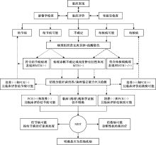
| 8 | 应采用结核科和呼吸科共同主导的多学科合作模式,开展结节病与结核病鉴别诊断、处置和随访工作 | 9.75±0.79 | 8.06 |
| 9 | 经多学科联合会诊或完善相关检查后仍难区分胸内结节病与肺结核时,经权衡利弊和知情同意后,建议选择诊断性抗结核治疗 | 9.00±1.24 | 13.75 |
| 10 | 应对结节病患者中的结核发病高风险人群进行结核分枝杆菌潜伏感染筛查,包括接受肿瘤坏死因子拮抗剂、长期接受糖皮质激素和(或)其他免疫抑制剂治疗者,肺结核密切接触者,HIV感染者,血液透析和器官移植的结节病患者 | 9.38±1.55 | 16.56 |
| 11 | 建议对结节病高风险人群中的结核分枝杆菌潜伏感染者采用异烟肼联合利福喷丁(利福平)的方案进行预防性抗结核治疗 | 9.13±1.30 | 14.23 |


| 不确定性征象 | 结节病可能 | 结核病可能 |
|---|---|---|
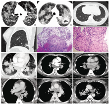
| 微结节簇集征 | 占9.9%,中上肺分布为主,团簇状,微结节排列不规则,伴中轴间质和间隔间质不规则增厚 | 占2.6%,中上肺分布为主,单发或多发团簇状,微结节排列规则,不伴或伴轻度间隔间质增厚 |
| 星系征 | 占7.3%~27%,结节聚集融合成大结节,边界不清,周围伴淋巴管周分布微结节,淋巴结肿大常见,空洞极少见 | 占1.9%~9.3%,结节融合形成局灶性实变,可坏死形成空洞,伴发树芽征或小叶中心结节,淋巴结肿大少见 |
| 反晕征 | 占5.3%,晕环呈结节表现,常伴肺内淋巴管周分布结节和(或)肺门淋巴结肿大 | 占3.6%~13.9%,晕环呈结节表现,常伴发树芽征或小叶中心结节,一般不伴淋巴结肿大 |
| 空洞 | 占2.2%~6.8%,大小不一,薄壁空洞多见,较大空洞见分隔带;厚壁空洞见于活动期,随着病情好转,洞壁变薄 | 占19.6%,空洞形态多样,常见引流支气管征,伴发树芽征 |
| 斑块与肿块 | 占12%~25%,多发或双侧分布,形态不规则,边界不清,伴发淋巴管周分布结节 | 占15.9%,球形病变,边界清,伴卫星结节,呈小叶中心分布 |
| 实变 | 占12%,多为结节融合所致,密度多不均匀,伴或不伴支气管充气征 | 占69.6%,渗出性肺实变,节段性分布,密度均匀,可进展成干酪性肺实变 |
| 淋巴结坏死或 强化不均匀 | 占5.9%,个别淋巴结局灶性坏死或强化不均,偶可见环形强化,淋巴结边界清晰,不融合 | 78.1%的淋巴结呈低密度或环形强化;淋巴结结外浸润、融合,边界不清,侵蚀支气管壁形成支气管淋巴瘘 |

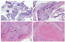
| 支持特征 | 不支持特征 |
|---|---|
| 沿淋巴道分布 | 缺乏淋巴道分布特征 |
| 显微镜下肉芽肿位于肺间质 | 显微镜下肺泡腔内肉芽肿 |
| 肉芽肿排列紧密,大小相对一致,融而不合 | 肺泡腔机化,肉芽肿松散,大小不一,融合 |
| 非坏死性(偶有点灶坏死) | 大片坏死 |
| 结节周边纤维组织增生 | 较多炎症细胞浸润 |
| 炎细胞少 | 淋巴滤泡形成 |
| 常见包涵体 | 抗酸染色阳性和(或)培养阳性 |
| 支持特征 | 不支持特征 |
|---|---|
| 沿气道分布或随机分布 | 沿淋巴道周围分布 |
| 显微镜下肉芽肿位于肺泡腔内及间质 | 显微镜下肉芽肿仅见于肺间质 |
| 肉芽肿排列紧密或松散、大小不一,融合 | |
| 干酪样坏死 | |
| 较多淋巴细胞浸润 | |
| 肺泡腔内浆液性、纤维素渗出 | |
| 抗酸染色阳性和(或)培养阳性 |


| [1] |
汪小鹏, 赵妍妍, 黎春艳, 等. 1303例肺结节病临床荟萃分析. 现代中西医结合杂志, 2013, 22(18):2009-2011. doi:10.3969/j.issn.1008-8849.2013.18.034.
doi: 10.3969/j.issn.1008-8849.2013.18.034 |
| [2] |
王洪武. 结节病概述及误诊原因分析. 临床误诊误治, 2002, 15(6):403-407. doi:10.3969/j.issn.1002-3429.2002.06.002.
doi: 10.3969/j.issn.1002-3429.2002.06.002 |
| [3] |
Madan K, Sryma PB, Pattnaik B, et al. Clinical Profile of 327 patients with Sarcoidosis in India: An Ambispective Cohort Study in a Tuberculosis (TB) Endemic Population. Lung India, 2022, 39(1):51-57. doi:10.4103/lungindia.lungindia_960_20.
doi: 10.4103/lungindia.lungindia_960_20 pmid: 34975053 |
| [4] | World Health Organization. Global tuberculosis report 2021. Geneva: World Health Organization, 2021. |
| [5] |
中国医学科学院病原生物学研究所, 中国疾病预防控制中心, 中国科学院地理科学与资源研究所. 全国结核分枝杆菌潜伏感染率估算专家共识. 中国防痨杂志, 2022, 44(1):4-8. doi:10.19982/j.issn.1000-6621.20210662.
doi: 10.19982/j.issn.1000-6621.20210662 |
| [6] | 中华人民共和国国家卫生和计划生育委员会. WS 288—2017肺结核诊断. 2017-11-09. |
| [7] |
中华医学会呼吸病学分会间质性肺疾病学组, 中国医师协会呼吸医师分会间质性肺疾病工作委员会. 中国肺结节病诊断和治疗专家共识. 中华结核和呼吸杂志, 2019, 42(9):685-693. doi:10.3760/cma.j.issn.1001-0939.2019.09.007.
doi: 10.3760/cma.j.issn.1001-0939.2019.09.007 |
| [8] |
Gupta D, Agarwal R, Aggarwal AN, et al. Molecular evidence for the role of mycobacteria in sarcoidosis: a meta-analysis. Eur Respir J, 2007, 30(3):508-516. doi:10.1183/09031936.00002607.
doi: 10.1183/09031936.00002607 pmid: 17537780 |
| [9] |
Atkins D, Best D, Briss PA, et al. Grading quality of evidence and strength of recommendations. BMJ, 2004, 328(7454):1490. doi:10.1136/bmj.328.7454.1490.
doi: 10.1136/bmj.328.7454.1490 URL |
| [10] |
Li CW, Tao RJ, Zou DF, et al. Pulmonary sarcoidosis with and without extrapulmonary involvement: a cross-sectional and observational study in China. BMJ Open, 2018, 8(2):e018865. doi:10.1136/bmjopen-2017-018865.
doi: 10.1136/bmjopen-2017-018865 |
| [11] |
Baughman RP, Teirstein AS, Judson MA, etal. Clinical characteristics of patients in a case control study of sarcoidosis. Am J Respir Crit Care Med, 2001, 164(<W>10 Pt 1):1885-1889. doi:10.1164/ajrccm.164.10.2104046.
doi: 10.1164/ajrccm.164.10.2104046 URL |
| [12] | Judson MA, Boan AD, Lackland DT. The clinical course of sarcoidosis: presentation, diagnosis, and treatment in a large white and black cohort in the United States. Sarcoidosis Vasc Diffuse Lung Dis, 2012, 29(2):119-127. |
| [13] |
Acharya NR, Browne EN, Rao N, et al. Distinguishing Features of Ocular Sarcoidosis in an International Cohort of Uveitis Patients. Ophthalmology, 2018, 125(1):119-126. doi:10.1016/j.ophtha.2017.07.006.
doi: S0161-6420(17)31125-9 pmid: 28823384 |
| [14] |
Laura D, Lee Y, Farhangi M, et al. Ocular Manifestations of Sarcoidosis in a South Florida Population. Clin Ophthalmol, 2020, 14:3741-3746. doi:10.2147/OPTH.S278373.
doi: 10.2147/OPTH.S278373 URL |
| [15] |
徐祖辉, 刘礼亲, 王巧智, 等. 病原学阳性住院肺结核患者并发肺外结核的流行特征及其影响因素研究. 中国防痨杂志, 2021, 43(11):1164-1170. doi:10.3969/j.issn.1000-6621.2021.11.011.
doi: 10.3969/j.issn.1000-6621.2021.11.011 |
| [16] |
Testi I, Agrawal R, Mehta S, et al. Ocular tuberculosis: Where are we today? Indian J Ophthalmol, 2020, 68(9):1808-1817. doi:10.4103/ijo.IJO_1451_20.
doi: 10.4103/ijo.IJO_1451_20 pmid: 32823397 |
| [17] |
Studdy P, Bird R, James DG. Serum angiotensin-converting enzyme (SACE) in sarcoidosis and other granulomatous disorders. Lancet, 1978, 2(8104-5):1331-1334. doi:10.1016/s0140-6736(78)91972-4.
doi: 10.1016/s0140-6736(78)91972-4 pmid: 82838 |
| [18] |
Brice EA, Friedlander W, Bateman ED, et al. Serum angiotensin-converting enzyme activity, concentration, and specific activity in granulomatous interstitiallung disease, tubercul osis, and COPD. Chest, 1995, 107(3):706-710. doi:10.1378/chest.107.3.706.
doi: 10.1378/chest.107.3.706 pmid: 7874941 |
| [19] | Abe Y, Mizuki M, Tsuda T, et al. Elevated serum angiotensin-converting enzyme in miliary tuberculosis. Kekkaku, 1985, 60(11):573-576. |
| [20] |
Crouser ED, Maier LA, Wilson KC, et al. Diagnosis and Detection of Sarcoidosis. An Official American Thoracic Society Clinical Practice Guideline. Am J Respir Crit Care Med, 2020, 201(8):e26-e51. doi:10.1164/rccm.202002-0251ST.
doi: 10.1164/rccm.202002-0251ST |
| [21] |
Chopra A, Foulke L, Judson MA. Sarcoidosis associated pleural effusion: Clinical aspects. Respir Med, 2022, 191:106723. doi:10.1016/j.rmed.2021.106723.
doi: 10.1016/j.rmed.2021.106723 |
| [22] |
Palmucci S, Torrisi SE, Caltabiano DC, et al. Clinical and radiological features of extra-pulmonary sarcoidosis: a pictorial essay. Insights Imaging, 2016, 7(4):571-587. doi:10.1007/s13244-016-0495-4.
doi: 10.1007/s13244-016-0495-4 URL |
| [23] |
Bhalla AS, Das A, Naranje P, et al. Dilemma of diagnosing thoracic sarcoidosis in tuberculosis endemic regions: An imaging-based approach. Part 1. Indian J RadiolImaging, 2017, 27(4):369-379. doi:10.4103/ijri.IJRI_200_17.
doi: 10.4103/ijri.IJRI_200_17 |
| [24] |
Zeng Y, Zhai XL, Wáng YXJ, et al. Illustration of a number of atypical computed tomography manifestations of active pulmonary tuberculosis. Quant Imaging Med Surg, 2021, 11(4):1651-1667. doi:10.21037/qims-20-1323.
doi: 10.21037/qims-20-1323 URL |
| [25] |
Zhan X, Zhang L, Wang Z, et al. Reversed Halo Sign: Presents in Different Pulmonary Diseases. PLoS One, 2015, 10(6):e0128153. doi:10.1371/journal.pone.0128153.
doi: 10.1371/journal.pone.0128153 |
| [26] |
Hours S, Nunes H, Kambouchner M, et al. Pulmonary cavitary sarcoidosis:clinico-radiologic characteristics and natural history of a rare form of sarcoidosis. Medicine (Baltimore), 2008, 87(3):142-151. doi:10.1097/MD.0b013e3181775a73.
doi: 10.1097/MD.0b013e3181775a73 URL |
| [27] |
路希伟, 伍建林, 张国庆, 等. 涂阴、涂阳活动性肺结核CT征象的对照研究. 中国医学影像技术, 2007, 23(9):1337-1341. doi:10.3321/j.issn:1003-3289.2007.09.021.
doi: 10.3321/j.issn:1003-3289.2007.09.021 |
| [28] |
Hong JH, Yoon SH, Goo JM, et al. Clustered micronodules as predominant manifestation on CT: A sign of active but indolently evolving pulmonary tuberculosis. PLoS One, 2020, 15(4):e0231537. doi:10.1371/journal.pone.0231537.
doi: 10.1371/journal.pone.0231537 |
| [29] |
Martini K, Loubet A, Bankier A, et al. Nodular reverse halo sign in active pulmonary tuberculosis: A rare CT feature? DiagnInterv Imaging, 2020, 101(5):281-287. doi:10.1016/j.diii.2020.01.013.
doi: 10.1016/j.diii.2020.01.013 |
| [30] |
Nakatsu M, Hatabu H, Morikawa K, et al. Large coalescent parenchymal nodules in pulmonary sarcoidosis: “sarcoid galaxy” sign. AJR Am J Roentgenol, 2002, 178(6):1389-1393. doi:10.2214/ajr.178.6.1781389.
doi: 10.2214/ajr.178.6.1781389 URL |
| [31] |
吕岩, 李成海, 谢汝明, 等. 初治活动性继发性肺结核的HRCT影像研究. 中华实验和临床感染病杂志(电子版), 2016, 9(5):643-648. doi:10.3877/cma.j.issn.1674-1358.2015.05.011.
doi: 10.3877/cma.j.issn.1674-1358.2015.05.011 |
| [32] | Avital M, Hadas-Halpern I, Deeb M, et al. Radiological findings in sarcoidosis. Isr Med Assoc J, 2008, 10(8/9):572-574. |
| [33] |
王峰, 童朝晖, 王臻, 等. 结节病胸膜病变合并胸腔积液六例报告及文献复习. 中华结核和呼吸杂志, 2015, 38(2):99-104. doi:10.3760/cma.j.issn.1001-0939.2015.02.008.
doi: 10.3760/cma.j.issn.1001-0939.2015.02.008 |
| [34] |
Ko JM, Park HJ, Kim CH. Clinicoradiologic evidence of pulmonary lymphatic spread in adult patients with tuberculosis. AJR Am J Roentgenol, 2015, 204(1):38-43. doi:10.2214/AJR.14.12908.
doi: 10.2214/AJR.14.12908 URL |
| [35] |
Heo JN, Choi YW, Jeon SC, et al. Pulmonary tuberculosis: another disease showing clusters of small nodules. AJR Am J Roentgenol, 2005, 184(2):639-642. doi:10.2214/ajr.184.2.01840639.
doi: 10.2214/ajr.184.2.01840639 URL |
| [36] | Koide T, Saraya T, Tsukahara Y, et al. Clinical significance of the “galaxy sign” in patients with pulmonary sarcoidosis in a Japanese single-center cohort. Sarcoidosis Vasc Diffuse Lung Dis, 2016, 33(3):247-252. |
| [37] |
Herráez Ortega I, Alonso Orcajo N, López González L. et al. The “sarcoid clustersign”. A new sign in high resolution chest CT. Radiologia, 2009, 51(5):495-499. doi:10.1016/j.rx.2009.05.003.
doi: 10.1016/j.rx.2009.05.003 |
| [38] |
Shorr AF, Torrington KG, Hnatiuk OW. Endobronchial biopsy for sarcoidosis: a prospective study. Chest, 2001, 120(1):109-114. doi:10.1378/chest.120.1.109.
doi: 10.1378/chest.120.1.109 pmid: 11451824 |
| [39] |
Polychronopoulos VS, Prakash UBS. Airway involvement in sarcoidosis. Chest, 2009, 136(5):1371-1380. doi:10.1378/chest.08-2569.
doi: S0012-3692(09)60695-4 pmid: 19892676 |
| [40] |
佟冰, 徐燕, 钟巍, 等. 支气管镜检查对肺结节病的诊断价值. 中华结核和呼吸杂志, 2015, 38(11):839-843. doi:10.3760/cma.j.issn.1001-0939.2015.11.009.
doi: 10.3760/cma.j.issn.1001-0939.2015.11.009 |
| [41] |
Göktalay T, Çelik P, Alpaydın AÖ, et al. The Role of Endobronchial Biopsy in the Diagnosis of Pulmonary Sarcoidosis. Turk Thorac J, 2016, 17(1):22-27. doi:10.5578/ttj.17.1.004.
doi: 10.5578/ttj.17.1.004 pmid: 29404117 |
| [42] |
中华医学会结核病学分会, 《中华结核和呼吸杂志》编辑委员会. 气管支气管结核诊断和治疗指南(试行). 中华结核和呼吸杂志, 2012, 35(8):581-587. doi:10.3760/cma.j.issn.1001-0939.2012.08.007.
doi: 10.3760/cma.j.issn.1001-0939.2012.08.007 |
| [43] |
Gilman MJ. Transbronchial biopsy in sarcoidosis. Chest, 1983, 83(1):159. doi:10.1378/chest.83.1.159a.
doi: 10.1378/chest.83.1.159a pmid: 6848328 |
| [44] |
Mok Y, Tan TY, Tay TR, et al. Do we need transbronchial lung biopsy if we have bronchoalveolar lavage Xpert MTB/RIF? Int J Tuberc Lung Dis, 2016, 20(5):619-624. doi:10.5588/ijtld.15.0463.
doi: 10.5588/ijtld.15.0463 pmid: 27084815 |
| [45] |
Mondoni M, Repossi A, Carlucci P, et al. Bronchoscopic techniques in the management of patients with tuberculosis. Int J Infect Dis, 2017, 64:27-37. doi:10.1016/j.ijid.2017.08.008.
doi: S1201-9712(17)30213-8 pmid: 28864395 |
| [46] |
Trisolini R, Lazzari Agli L, Cancellieri A, et al. The value of flexible transbronchial needle aspiration in the diagnosis of stage Ⅰ sarcoidosis. Chest, 2003, 124(6):2126-2130. doi:10.1378/chest.124.6.2126.
doi: 10.1378/chest.124.6.2126 pmid: 14665490 |
| [47] |
Cetinkaya E, Yildiz P, Altin S, et al. Diagnostic value of transbronchial needle aspiration by Wang 22-gauge cytology needle in intrathoracic lymphadenopathy. Chest, 2004, 125(2):527-531. doi:10.1378/chest.125.2.527.
doi: 10.1378/chest.125.2.527 pmid: 14769734 |
| [48] |
Sun J, Teng J, Yang H, et al. Endobronchial ultrasound-guided transbronchial needle aspiration in diagnosing intrathoracic tuberculosis. Ann Thorac Surg, 2013, 96(6):2021-2027. doi:10.1016/j.athoracsur.2013.07.005.
doi: 10.1016/j.athoracsur.2013.07.005 pmid: 24035300 |
| [49] |
Ye W, Zhang R, Xu X, et al. Diagnostic Efficacy and Safety of Endobronchial Ultrasound-Guided Transbronchial Needle Aspiration in Intrathoracic Tuberculosis: A Meta-analysis. J Ultrasound Med, 2015, 34(9):1645-1650. doi:10.7863/ultra.15.14.06017.
doi: 10.7863/ultra.15.14.06017 pmid: 26269299 |
| [50] |
Liu X, Hou XF, Gao L, et al. Indicators for prediction of Mycobacterium tuberculosis positivity detected with bronchoalveolar lavage fluid. Infect Dis Poverty, 2018, 7(1):22. doi:10.1186/s40249-018-0403-x.
doi: 10.1186/s40249-018-0403-x pmid: 29580276 |
| [51] |
Liu X, Chen Y, Ouyang H, et al. Tuberculosis Diagnosis by Metagenomic Next-generation Sequencing on Bronchoalveolar Lavage Fluid: a cross-sectional analysis. Int J Infect Dis, 2021, 104:50-57. doi:10.1016/j.ijid.2020.12.063.
doi: 10.1016/j.ijid.2020.12.063 URL |
| [52] | Yin Y, Qin J, Dai Y, et al. The CD4+/CD8+ Ratio in Pulmonary Tuberculosis: Systematic and Meta-Analysis Article. Iran J Public Health, 2015, 44(2):185-193. |
| [53] |
Tanriverdi H, Erboy F, Altinsoy B, et al. Bronchoalveolar Lavage Fluid Characteristics of Patients With Sarcoidosis and Nonsarcoidosis Interstitial Lung Diseases: Ten-Year Experience of a Single Center in Turkey. Iran Red Crescent Med J, 2015, 17(10):e31103. doi:10.5812/ircmj.31103.
doi: 10.5812/ircmj.31103 |
| [54] |
Greco S, Marruchella A, Massari M, et al. Predictive value of BAL cellular analysis in differentiating pulmonary tuberculosis and sarcoidosis. Eur Respir J, 2005, 26(2):360-361. doi:10.1183/09031936.05.00042905.
doi: 10.1183/09031936.05.00042905 pmid: 16055888 |
| [55] |
Rosen Y. Pathology of Granulomatous Pulmonary Diseases. Arch Pathol Lab Med, 2022, 146(2):233-251. doi:10.5858/arpa.2020-0543-RA.
doi: 10.5858/arpa.2020-0543-RA URL |
| [56] |
Jain D, Ghosh S, Teixeira L, et al. Pathology of pulmonary tuberculosis and non-tuberculous mycobacterial lung disease: Facts, misconceptions, and practical tips for pathologists. Semin DiagnPathol, 2017, 34(6):518-529. doi:10.1053/j.semdp.2017.06.003.
doi: 10.1053/j.semdp.2017.06.003 |
| [57] |
Rosen Y. Four decades of necrotizing sarcoid granulomatosis: what do we know now? Arch Pathol Lab Med, 2015, 139(2):252-262. doi:10.5858/arpa.2014-0051-RA.
doi: 10.5858/arpa.2014-0051-RA pmid: 25611109 |
| [58] |
方木通, 杨倩婷, 王仲元, 等. 病理组织中的病原学检查对结核病的诊断价值. 中华传染病杂志, 2021, 39(2):92-96. doi:10.3760/cma.j.cn311365-20191124-00391.
doi: 10.3760/cma.j.cn311365-20191124-00391 |
| [59] |
Nazarullah A, Nilson R, Maselli DJ, et al. Incidence and aetiologies of pulmonary granulomatous inflammation: a decade of experience. Respirology, 2015, 20(1):115-121. doi:10.1111/resp.12410.
doi: 10.1111/resp.12410 pmid: 25351289 |
| [60] | Carrol KC, Pfaller MA. 临床微生物学手册.12版.王辉, 马筱玲, 钱渊,等,译. 北京: 中华医学电子音像出版社, 2021. |
| [61] | World Health Organization. Automated Real-Time Nucleic Acid Amplification Technology for Rapid and Simultaneous Detection of Tuberculosis and Rifampicin Resistance: Xpert MTB/RIF Assay for the Diagnosis of Pulmonary and Extrapulmonary TB in Adults and Children: Policy Update. Geneva: World Health Organization, 2013. |
| [62] | Dhooria S, Gupta N, Bal A, et al. Role of Xpert MTB/RIF in differentiatingtuberculosis from sarcoidosis in patients with mediastinal lymphadenopathy undergoing EBUS-TBNA: a study of 147 patients. Sarcoidosis Vasc Diffuse Lung Dis, 2016, 33(3):258-266. |
| [63] |
周瑛, 李惠萍, 李秋红, 等. 实时定量聚合酶链反应在鉴别诊断结节病和结核病中的应用. 中华结核和呼吸杂志, 2009, 32(4):311-312. doi:10.3760/cma.j.issn.1001-0939.2009.04.024.
doi: 10.3760/cma.j.issn.1001-0939.2009.04.024 |
| [64] |
Fang C, Huang H, Xu Z. Immunological Evidence for the Role of Mycobacteria in Sarcoidosis: A Meta-Analysis. PLoS One, 2016, 11(8):e0154716. doi:10.1371/journal.pone.0154716.
doi: 10.1371/journal.pone.0154716 |
| [65] |
Hernández-Pando R, Jeyanathan M, Mengistu G, et al. Persistence of DNA from Mycobacterium tuberculosis in superficially normal lung tissue during latent infection. Lancet, 2000, 356(9248):2133-2138. doi:10.1016/s0140-6736(00)03493-0.
doi: 10.1016/s0140-6736(00)03493-0 pmid: 11191539 |
| [66] |
Li QH, Zhang Y, Zhao MM, et al. Simultaneous amplification and testing method for Mycobacterium tuberculosis rRNA to differentiate sputum-negative tuberculosis fromsarcoidosis. Am J Physiol Lung Cell Mol Physiol, 2019, 316(3):L519-L524. doi:10.1152/ajplung.00172.2018.
doi: 10.1152/ajplung.00172.2018 URL |
| [67] |
Chao Y, Li J, Gong Z, et al. Rapid discrimination between tuberculosis and sarcoidosis using next-generation sequencing. Int J Infect Dis, 2021, 108:129-136. doi:10.1016/j.ijid.2021.05.028.
doi: 10.1016/j.ijid.2021.05.028 pmid: 34004327 |
| [68] |
Tubach F, Salmon D, Ravaud P, et al. Risk of tuberculosis is higher with anti-tumor necrosis factor monoclonal antibody therapy than with soluble tumor necrosis factor receptor therapy: The three-year prospective French Research Axed on Tolerance of Biotherapies registry. Arthritis Rheum, 2009, 60(7):1884-1894. doi:10.1002/art.24632.
doi: 10.1002/art.24632 URL |
| [69] |
Zhang Z, Fan W, Yang G, et al. Risk of tuberculosis in patients treated with TNF-α antagonists: a systematic review and meta-analysis of randomised controlled trials. BMJ Open, 2017, 7(3):e012567. doi:10.1136/bmjopen-2016-012567.
doi: 10.1136/bmjopen-2016-012567 |
| [70] |
Jick SS, Lieberman ES, Rahman MU, et al. Glucocorticoid use, other associated factors, and the risk of tuberculosis. Arthritis Rheum, 2006, 55(1):19-26. doi:10.1002/art.21705.
doi: 10.1002/art.21705 URL |
| [71] |
Bass JB Jr, Farer LS, Hopewell PC, et al. Treatment of tuberculosis and tuberculosis infection in adults and children. American Thoracic Society and The Centers for Disease Control and Prevention. Am J Respir Crit Care Med, 1994, 149(5):1359-1374. doi:10.1164/ajrccm.149.5.8173779.
doi: 10.1164/ajrccm.149.5.8173779 URL |
| [72] |
Jamilloux Y, Valeyre D, Lortholary O, et al. The spectrum of opportunistic diseases complicating sarcoidosis. Autoimmun Rev, 2015, 14(1):64-74. doi:10.1016/j.autrev.2014.10.006.
doi: 10.1016/j.autrev.2014.10.006 pmid: 25305373 |
| [73] |
Duréault A, Chapelon C, Biard L, et al. Severe infections in sarcoidosis: Incidence, predictors and long-term outcome in a cohort of 585 patients. Medicine (Baltimore), 2017, 96(49):e8846. doi:10.1097/MD.0000000000008846.
doi: 10.1097/MD.0000000000008846 URL |
| [74] | World Health Organization. Latent tuberculosis infection: updated and consolidated guidelines for programmatic management. Geneva: World Health Organization, 2018. |
| [75] | Amicosante M. IGRAs for tuberculosis in sarcoidosis patients: is the immune response to mycobacteria helpful in the differential diagnosis or still a confounding factor? Sarcoidosis Vasc Diffuse Lung Dis, 2011, 28(2):85-86. |
| [76] |
Milman N, Søborg B, Svendsen CB, et al. Quantiferon test for tuberculosis screening in sarcoidosis patients. Scand J Infect Dis, 2011, 43(9):728-735. doi:10.3109/00365548.2011.582141.
doi: 10.3109/00365548.2011.582141 pmid: 21619424 |
| [77] | Gupta D, Kumar S, Aggarwal AN, et al. Interferon gamma release assay (QuantiFERON-TB Gold In Tube) in patients of sarcoidosis from a population with high prevalence of tuberculosis infection. Sarcoidosis Vasc Diffuse Lung Dis, 2011, 28(2):95-101. |
| [78] |
Piotrowski WJ, Adam B, Gwadera Ł, et al. QuantiFERON-TB-GOLD In-Tube in patients with sarcoidosis. Adv Respir Med, 2018, 86(5):234-239. doi:10.5603/ARM.2018.0037.
doi: 10.5603/ARM.2018.0037 pmid: 30378651 |
| [79] |
Kempisty A, Białas-Chromiec B, Borkowska D, et al. Interferon gamma release assays based on M.tuberculosis-specific antigens in sarcoidosis patient. PneumonolAlergol Pol, 2015, 83(2):126-134. doi:10.5603/PiAP.2015.0020.
doi: 10.5603/PiAP.2015.0020 |
| [80] | World Health Organization. WHO consolidated guidelines on tuberculosis: Module 1: prevention: tuberculosis preventive treatment. Geneva: World Health Organization, 2020. |
| [81] | 中华人民共和国国家卫生健康委员会办公厅. 国家卫生健康委办公厅关于印发中国结核病预防控制工作技术规范(2020年版)的通知. 国卫办疾控函〔2020〕279号. 2020-04-02. |
| [82] |
Sterling TR, Njie G, Zenner D, et al. Guidelines for the Treatment of Latent Tuberculosis Infection: Recommendations from the National Tuberculosis Controllers Association and CDC, 2020. MMWR Recomm Rep, 2020, 69(1):1-11. doi:10.15585/mmwr.rr6901a1.
doi: 10.15585/mmwr.rr6901a1 pmid: 32053584 |
| [83] |
Shah M, Dorman SE. Latent Tuberculosis Infection. N Engl J Med, 2021, 385(24):2271-2280. doi:10.1056/NEJMcp2108501.
doi: 10.1056/NEJMcp2108501 URL |
| [84] | 全国第五次结核病流行病学抽样调查技术指导组, 全国第五次结核病流行病学抽样调查办公室. 2010年全国第五次结核病流行病学抽样调查报告. 中国防痨杂志, 2012, 34(8): 485-508. |
| [85] |
Iannone F, Cantini F, Lapadula G. Diagnosis of latent tuberculosis and prevention of reactivation in rheumatic patients receiving biologic therapy: international recommendations. J Rheumatol Suppl, 2014, 91:41-46. doi:10.3899/jrheum.140101.
doi: 10.3899/jrheum.140101 pmid: 24788999 |
| [1] | XIA Hui, ZHENG Yang, SONG Yuan-yuan. Interpretation of the Optimized broth microdilution plate methodology for drug susceptibility testing of Mycobacterium tuberculosis complex issued by World Health Organization [J]. Chinese Journal of Antituberculosis, 2022, 44(7): 641-645. |
| [2] | The Joint Tuberculosis Professional Branch of Chinese Antituberculosis Association, The Western China Bone Tuberculosis Union, The North China Union of Bone Tuberculosis. Expert consensus on the diagnosis and treatment of Brucella spondylitis [J]. Chinese Journal of Antituberculosis, 2022, 44(6): 531-538. |
| [3] | AN Hui-ru, WU Xue-qiong. Interpretation of immunoadjuvant therapy in Expert consensus on immune function assessment and immunotherapy in patients with active tuberculosis (2021 Edition) [J]. Chinese Journal of Antituberculosis, 2022, 44(6): 539-543. |
| [4] | CHEN Zhi, LIANG Jian-qin. Expert consensus on nutritional assessment and nutritional support treatment for patients with severe tuberculosis [J]. Chinese Journal of Antituberculosis, 2022, 44(5): 421-432. |
| [5] | SUN Zhao-gang. Attention should be paid to the research and development of Mycobacterium tuberculosis antigen detection technology [J]. Chinese Journal of Antituberculosis, 2022, 44(2): 120-124. |
| [6] | Zeng Yi, Hu Hongling, Li Zhiyong, Zhou Hui, Chen Yu, Zhang Peize, Fang Fang, Lai Xiaoyu, Lu Xiwei. Interpretation of Expert consensus on clinical differential diagnosis and treatment of intrathoracic sarcoidosis and pulmonary tuberculosis under the background of tuberculosis epidemic [J]. Chinese Journal of Antituberculosis, 2022, 44(12): 1242-1248. |
| [7] | Gong Wenping, Mi Jie, Wu Xueqiong. Immunologically active substances: novel treatment options for tuberculosis and nontuberculous mycobacteriosis [J]. Chinese Journal of Antituberculosis, 2022, 44(11): 1107-1121. |
| [8] | Yuan Yuan, Lu Shuihua. Interpretation of WHO consolidated guidelines on tuberculosis Module 4: Treatment of drug-susceptible tuberculosis [J]. Chinese Journal of Antituberculosis, 2022, 44(11): 1122-1125. |
| [9] | Tuberculosis Control Branch of Chinese Antituberculosis Association, Elderly Tuberculosis Control Branch of Chinese Antituberculosis Association, Editorial Board of Chinese Journal of Antituberculosis. Evidence-based guidelines for active screening of pulmonary tuberculosis in Chinese communities [J]. Chinese Journal of Antituberculosis, 2022, 44(10): 987-997. |
| [10] | Tuberculosis Prevention and Control Key Laboratory/Beijing Key Laboratory of New Techniques of Tuberculosis Diagnosis and Treatment /Institute for Tuberculosis Research/Department of Tuberculosis of the 8th Medical Center of Chinese PLA General Hospital, Editorial Board of Chinese Journal of Antituberculosis . Expert consensus on the rational use of glucocorticoids in tuberculosis treatment [J]. Chinese Journal of Antituberculosis, 2022, 44(1): 28-37. |
| [11] | Chinese Antituberculosis Association , National Center for Tuberculosis Control and Prevention, Chinese Center for Disease Control and Prevention . Recommendations on pretomanid (PA-824) in the treatment of multidrug-resistant tuberculosis [J]. Chinese Journal of Antituberculosis, 2022, 44(1): 38-44. |
| [12] | Tuberculosis Prevention and Control Key Laboratory/Beijing Key Laboratory of New Techniques of Tuberculosis Diagnosis and Treatment/Institute for Tuberculosis Research of the 8th Medical Center of Chinese PLA General Hospital, Editorial Board of Chinese Journal of Antituberculosis , Basic and Clinical Speciality Committees of Tuberculosis Control Branch of China International Exchange and Promotive Association for Medical and Health Care . Expert consensus on immune function assessment and immunotherapy in patients with active tuberculosis (2021 Edition) [J]. Chinese Journal of Antituberculosis, 2022, 44(1): 9-27. |
| [13] | Beijing Chest Hospital, Capital Medical University/Beijing Tuberculosis and Thoracic Tumor Research Institute, Chinese Antituberculosis Association, Editorial Board of Chinese Journal of Antituberculosis. Chinese expert consensus on the all-oral treatment of drug-resistant pulmonary tuberculosis (2021 Edition) [J]. Chinese Journal of Antituberculosis, 2021, 43(9): 859-866. |
| [14] | Chinese Antituberculosis Association. Expert consensus on detection and preventive treatment of latent tuberculosis infection in high-risk population [J]. Chinese Journal of Antituberculosis, 2021, 43(9): 874-878. |
| [15] | ZHOU Wen-qiang, ZHANG Shuang, CHU Nai-hui. Current status and prospect of all-oral treatment regimens for drug-resistant pulmonary tuberculosis [J]. Chinese Journal of Antituberculosis, 2021, 43(9): 879-882. |
| Viewed | ||||||
|
Full text |
|
|||||
|
Abstract |
|
|||||
 京公网安备11010202007215号
Total visitors: Visitors of today: Now online:
京公网安备11010202007215号
Total visitors: Visitors of today: Now online:
 This work is licensed under Creative Commons Attribution 3.0 License.