
Chinese Journal of Antituberculosis ›› 2026, Vol. 48 ›› Issue (8): 1184-1191.doi: 10.19982/j.issn.1000-6621.20250524
• Original Articles • Previous Articles Next Articles
Guo Xiaowen1, Jiao Tianyu1, Sun Li1, Ni Conghui1, Wang Zilin2, Wang Hao1( )
)
Received:2025-12-24
Online:2026-08-01
Published:2026-07-30
Contact:
Wang Hao,Email:CLC Number:
Guo Xiaowen, Jiao Tianyu, Sun Li, Ni Conghui, Wang Zilin, Wang Hao. Multi-center study on CE-T1WI radiomics-based machine learning model in differentiating tuberculous spondylitis from brucella spondylitis[J]. Chinese Journal of Antituberculosis, 2026, 48(8): 1184-1191. doi: 10.19982/j.issn.1000-6621.20250524
Add to citation manager EndNote|Ris|BibTeX
URL: https://www.zgflzz.cn/EN/10.19982/j.issn.1000-6621.20250524



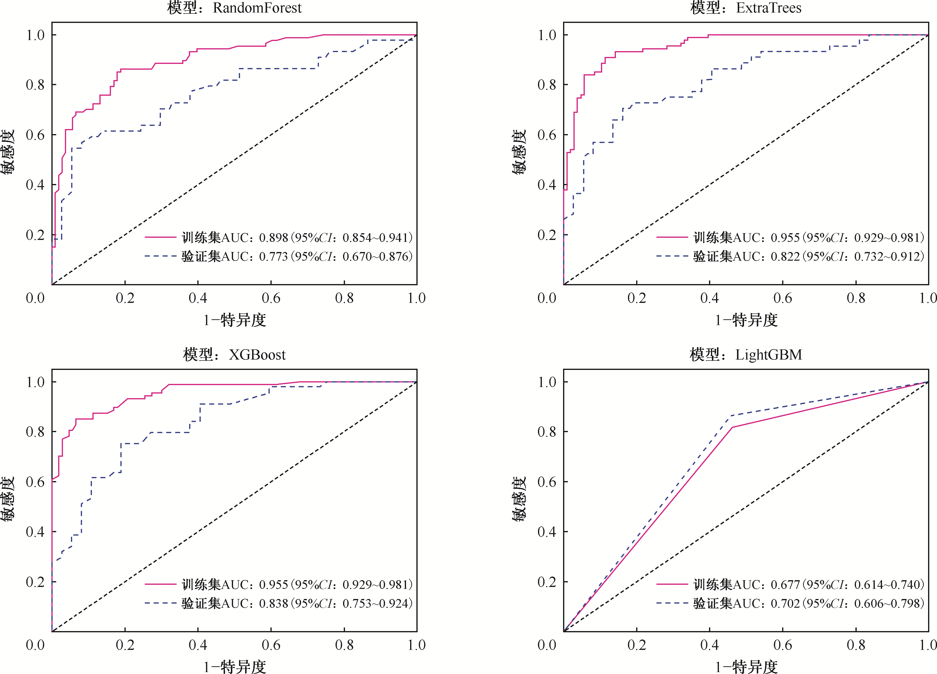
| 模型 | 准确率 | AUC值 | 95%CI值 | 敏感度 | 特异度 | 阳性预测值 |
|---|---|---|---|---|---|---|
| Random Forest | 0.829 | 0.898 | 0.854~0.941 | 0.851 | 0.811 | 0.787 |
| Extra Trees | 0.891 | 0.955 | 0.929~0.981 | 0.897 | 0.887 | 0.867 |
| XGBoost | 0.891 | 0.955 | 0.929~0.981 | 0.839 | 0.934 | 0.912 |
| LightGBM | 0.549 | 0.677 | 0.614~0.740 | 0.000 | 1.000 | 0.000 |
| 模型 | 准确率 | AUC值 | 95%CI值 | 敏感度 | 特异度 | 阳性预测值 |
|---|---|---|---|---|---|---|
| Random Forest | 0.716 | 0.773 | 0.670~0.876 | 0.523 | 0.946 | 0.920 |
| Extra Trees | 0.753 | 0.822 | 0.733~0.913 | 0.682 | 0.838 | 0.833 |
| XGBoost | 0.765 | 0.838 | 0.753~0.924 | 0.727 | 0.811 | 0.821 |
| LightGBM | 0.457 | 0.702 | 0.606~0.798 | 0.000 | 1.000 | 0.000 |
| [1] | 屈燕, 李涛, 马文斌, 等. 世界卫生组织《2025年全球结核病报告》解读[J]. 结核与肺部疾病杂志, 2025, 6(6): 613-623. doi:10.19983/j.issn.2096-8493.20250178. |
| [2] |
Garg R K, Somvanshi D S. Spinal tuberculosis: a review[J]. J Spinal Cord Med, 2011, 34(5): 440-454. doi:10.1179/2045772311y.0000000023.
pmid: 22118251 |
| [3] | Jin M, Fan Z, Gao R, et al. Research progress on complications of Brucellosis[J]. Front Cell Infect Microbiol, 2023, 13: 1136674. doi:10.3389/fcimb.2023.1136674. |
| [4] | Cordero M, Sánchez I. Brucellar and tuberculous spondylitis. A comparative study of their clinical features[J]. J Bone Joint Surg Br, 1991, 73(1): 100-103. doi:10.1302/0301-620x.73b1.1991738. |
| [5] |
Duarte R M, Vaccaro A R. Spinal infection: state of the art and management algorithm[J]. Eur Spine J, 2013, 22(12): 2787-2799. doi:10.1007/s00586-013-2850-1.
pmid: 23756630 |
| [6] | 奇丽霞, 张凤翔, 杜海, 等. 基于CT的影像组学列线图对布鲁氏菌性脊柱炎和脊柱结核的鉴别价值[J]. 中国实验诊断学, 2025, 29(4): 440-448. doi:10.3969/j.issn.1007-4287.2025.04.013. |
| [7] | 蔡一, 李睿涵, 伍倩, 等. 机器学习结合影像组学在结核性脊柱炎诊断与鉴别诊断的系统评价[J]. 实用放射学杂志, 2025, 41(8): 1348-1351,1360. doi:10.3969/j.issn.1002-1671.2025.08.020. |
| [8] | Wang W, Fan Z, Zhen J. MRI radiomics-based evaluation of tuberculous and brucella spondylitis[J]. J Int Med Res, 2023, 51(8): 3000605231195156. doi:10.1177/03000605231195156. |
| [9] |
Guo H, Lan S, He Y, et al. Differentiating brucella spondylitis from tuberculous spondylitis by the conventional MRI and MR T2 mapping: a prospective study[J]. Eur J Med Res, 2021, 26(1): 125. doi:10.1186/s40001-021-00598-4.
pmid: 34711265 |
| [10] | Mayerhoefer M E, Materka A, Langs G, et al. Introduction to Radiomics[J]. J Nucl Med, 2020, 61(4): 488-495. doi:10.2967/jnumed.118.222893. |
| [11] |
Lambin P, Rios-Velazquez E, Leijenaar R, et al. Radiomics: extracting more information from medical images using advanced feature analysis[J]. Eur J Cancer, 2012, 48(4): 441-446. doi:10.1016/j.ejca.2011.11.036.
pmid: 22257792 |
| [12] |
Kumar V, Gu Y, Basu S, et al. Radiomics: the process and the challenges[J]. Magn Reson Imaging, 2012, 30(9): 1234-1248. doi:10.1016/j.mri.2012.06.010.
pmid: 22898692 |
| [13] | 帕哈提·吐逊江, 杨来红, 何雄, 等. 影像组学在脊柱疾病中的应用[J]. 磁共振成像, 2022, 13(5): 162-166. doi:10.12015/issn.1674-8034.2022.05.035. |
| [14] | 帕哈提·吐逊江, 杨来红, 何雄, 等. 基于FS-T2WI序列联合机器学习对布鲁氏菌性脊柱炎与结核性脊柱炎的鉴别诊断[J]. 中华地方病学杂志, 2023, 42(5): 356-362. doi:10.3760/cma.j.cn231583-20220628-00237. |
| [15] | 何雄, 陈艳丽, 帕哈提·吐逊江, 等. MR T1WI影像组学对结核性脊柱炎与布鲁菌性脊柱炎的诊断价值[J]. 分子影像学杂志, 2023, 46(3):442-447. doi:10.12122/j.issn.1674-4500.2023.03.09. |
| [16] |
Wu S, Wei Y, Li H, et al. A Predictive Clinical-Radiomics Nomogram for Differentiating Tuberculous Spondylitis from Pyogenic Spondylitis Using CT and Clinical Risk Factors[J]. Infect Drug Resist, 2022, 15: 7327-7338. doi:10.2147/idr.s388868.
pmid: 36536861 |
| [17] |
Currie S, Galea-Soler S, Barron D, et al. MRI characteristics of tuberculous spondylitis[J]. Clin Radiol, 2011, 66(8): 778-787. doi:10.1016/j.crad.2011.02.016.
pmid: 21570065 |
| [18] | 刘涛, 孙建民, 崔新刚, 等. MRI及病理学鉴别早期化脓性脊柱炎及布氏杆菌脊柱炎中的应用及价值[J]. 中国组织工程研究, 2014(4): 499-504. doi:10.3969/j.issn.2095-4344.2014.04.002. |
| [19] | 陈鹰, 郭辉, 郑欢露, 等. 布鲁菌性脊柱炎临床诊断和治疗后分析[J]. 实用医学影像杂志, 2018, 19(6): 469-472. doi:10.16106/j.cnki.cn14-1281/r.2018.06.002. |
| [20] |
Yokota Y, Fushimi Y, Okada T, et al. Evaluation of image quality of pituitary dynamic contrast-enhanced MRI using time-resolved angiography with interleaved stochastic trajectories (TWIST) and iterative reconstruction TWIST (IT-TWIST)[J]. J Magn Reson Imaging, 2020, 51(5): 1497-1506. doi:10.1002/jmri.26962.
pmid: 31625655 |
| [21] | Lee S, Choi Y H, Cho Y J, et al. Quantitative evaluation of Crohn’s disease using dynamic contrast-enhanced MRI in children and young adults[J]. Eur Radiol, 2020, 30(6): 3168-3177. doi:10.1007/s00330-020-06684-1. |
| [22] |
Mui A W L, Lee A W M, Lee V H F, et al. Prognostic and therapeutic evaluation of nasopharyngeal carcinoma by dynamic contrast-enhanced (DCE), diffusion-weighted (DW) magnetic resonance imaging (MRI) and magnetic resonance spectroscopy (MRS)[J]. Magn Reson Imaging, 2021, 83: 50-56. doi:10.1016/j.mri.2021.07.003.
pmid: 34246785 |
| [23] | Yin P, Xu J, Sun X, et al. Intravoxel incoherent motion and dynamic contrast-enhanced magnetic resonance imaging for neoadjuvant chemotherapy response evaluation in patients with osteosarcoma[J]. Eur J Radiol, 2023, 162: 110790. doi:10.1016/j.ejrad.2023.110790. |
| [24] | Gleißner C, Kaczmarz S, Kufer J, et al. Hemodynamic MRI parameters to predict asymptomatic unilateral carotid artery stenosis with random forest machine learning[J]. Front Neuroimaging, 2022, 1: 1056503. doi:10.3389/fnimg.2022.1056503. |
| [25] | 张德唯, 王梓延, 马楚塬, 等. 增强CT影像组学特征构建随机森林机器学习模型鉴别诊断小细胞肺癌与非小细胞肺癌的应用价值[J]. 影像研究与医学应用, 2025, 9(7): 50-52. doi:10.20267/j.issn.2096-3807.2025.07.017. |
| [26] | 李立娟. 基于多器官融合和LightGBM的影像组学方法研究及其在食管静脉曲张中的应用[D]. 济南: 山东师范大学, 2021. |
| [27] | Greener J G, Kandathil S M, Moffat L, et al. A guide to machine learning for biologists[J]. Nat Rev Mol Cell Biol, 2022, 23(1): 40-55. doi:10.1038/s41580-021-00407-0. |
| [28] | 郑紫涵. 基于机器学习的CT影像组学模型术前预测肝细胞癌GPC3表达状态[D]. 大理: 大理大学, 2024. |
| [29] | Kucukakcali Z, Akbulut S, Colak C. Machine Learning-based Prediction of HBV-related Hepatocellular Carcinoma and Detection of Key Candidate Biomarkers[J]. Medeni Med J, 2022, 37(3): 255-263. doi:10.4274/MMJ.galenos.2022.39049. |
| [1] | Sun Zheng, Liu Jiongya, Chen Chi, Yu Quanji, Li Yanan, Chen Cheng, Zhu Limei. Study on the application of AI-assisted CT reading system in early detection of pulmonary tuberculosis in general hospitals [J]. Chinese Journal of Antituberculosis, 2026, 48(5): 586-593. |
| [2] | Fu Xuwen, He Weiyaozhen, Li Xiang. Imaging manifestations of male reproductive system tuberculosis [J]. Chinese Journal of Antituberculosis, 2026, 48(2): 188-196. |
| [3] | Imaging Professional Branch of Chinese Antituberculosis Association , Society of Tuberculosis, Chinese Medical Association , Standardization Professional Branch of Chinese Antituberculosis Association , Tuberculosis Control Professional Branch of Chinese Antituberculosis Association . Expert consensus on the application of artificial intelligence assisted image reading technology in the detection of pulmonary tuberculosis patients in chest imaging examination [J]. Chinese Journal of Antituberculosis, 2025, 47(9): 1093-1104. |
| [4] | Jiao Jiahuan, Sun Changfeng, Wu Gang, Huang Fuli, Sheng Yunjian. The value of machine learning algorithm-based diagnostic models in tuberculous pleural effusion [J]. Chinese Journal of Antituberculosis, 2025, 47(8): 1053-1061. |
| [5] | Zhu Qingdong, Zhao Chunyan, Xie Zhouhua, Song Shulin, Song Chang. Research progress on the application of artificial intelligence-based CT radiomics in the diagnosis and treatment response monitoring of tuberculosis [J]. Chinese Journal of Antituberculosis, 2025, 47(8): 1068-1076. |
| [6] | Abuduresuli Tu’ersun, Abudukeyoumujiang Abulizi, Patiman Maimaiti, Huang Chencui, Shen Lingyan, Mayidili Nijiati. Predicting pulmonary tuberculosis treatment outcomes using longitudinal chest CT radiomics and deep learning [J]. Chinese Journal of Antituberculosis, 2025, 47(8): 1044-1052. |
| [7] | Ning Fenggang, Fang Kun, Wang Jue, Lyu Yan, He Wei, Hou Dailun. Application of magnetic resonance 3D BRAVO enhanced scanning in the imaging diagnosis of intracranial tuberculosis [J]. Chinese Journal of Antituberculosis, 2025, 47(11): 1489-1494. |
| [8] | Li Tingting, Liu Huanqing, Lei Qian, You Zhuhong, Zhao Guolian. Application value of machine-learning-based diagnostic model on tuberculous pleurisy [J]. Chinese Journal of Antituberculosis, 2025, 47(11): 1508-1514. |
| [9] | Li Xiang, Pu Ying, Fu Xuwen, Qi Min, Wei Jialu, Cun Xinhua. Clinical and imaging characterization of fungal spondylitis misdiagnosed as spinal tuberculosis [J]. Chinese Journal of Antituberculosis, 2024, 46(9): 1109-1114. |
| [10] | Li Wenhan, Yang Jing, Li Chunhua. Research progress of artificial intelligence in pulmonary tuberculosis imaging diagnosis and drug resistance prediction [J]. Chinese Journal of Antituberculosis, 2024, 46(9): 1098-1103. |
| [11] | Liu Xueyan, Wang Fang, Li Chunhua, Tang Guangxiao, Zheng Jiaofeng, Wang Huiqiu, Li Yurui, Wang Jia’nan, Shu Weiqiang, Lyu Shengxiu. Construction and evaluation of a CT-based deep learning model for the auxiliary diagnosis of secondary tuberculosis [J]. Chinese Journal of Antituberculosis, 2024, 46(3): 279-287. |
| [12] | Qin Liyi, Lyu Pingxin, Guo Lin, Qian Lingjun, Xiao Qian, Yang Yang, Shang Yuanyuan, Jia Junnan, Chu Naihui, Liu Yuanming, Li Weimin. Deep learning to determine the healing status of pulmonary tuberculosis lesions on CT images [J]. Chinese Journal of Antituberculosis, 2024, 46(3): 272-278. |
| [13] | Gao Shan, Nie Wenjuan, Hou Dailun, Chu Naihui. Research progress on imaging manifestations of nontuberculosis mycobacterial pulmonary and application of new artificial intelligence technology [J]. Chinese Journal of Antituberculosis, 2024, 46(3): 362-366. |
| [14] | Liu Xin, Yu Qianhui. Diffusion weighted imaging with different b-values for the classification diagnosis of pulmonary tuberculosis and prediction of multidrug-resistance risk [J]. Chinese Journal of Antituberculosis, 2024, 46(11): 1356-1364. |
| [15] | Li Wenting, Wang Li, Fang Yong, Gu Jin, Sha Wei. The value of CT-based deep learning models in differentiating nontuberculous mycobacterial lung disease from pulmonary tuberculosis [J]. Chinese Journal of Antituberculosis, 2024, 46(10): 1236-1242. |
| Viewed | ||||||
|
Full text |
|
|||||
|
Abstract |
|
|||||
 京公网安备11010202007215号
Total visitors: Visitors of today: Now online:
京公网安备11010202007215号
Total visitors: Visitors of today: Now online:
 This work is licensed under Creative Commons Attribution 3.0 License.
