
Chinese Journal of Antituberculosis ›› 2021, Vol. 43 ›› Issue (4): 341-345.doi: 10.3969/j.issn.1000-6621.2021.04.008
• Original Articles • Previous Articles Next Articles
LIANG Rui-yun, FANG Wei-jun( ), REN Hui-li, LI Hui-ru, ZHANG Hui, LI Cheng-cheng
Received:2020-11-20
Online:2021-04-10
Published:2021-04-09
Contact:
FANG Wei-jun
E-mail:fangweijun9@sohu.com
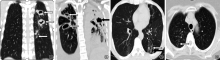
LIANG Rui-yun, FANG Wei-jun, REN Hui-li, LI Hui-ru, ZHANG Hui, LI Cheng-cheng. Analysis of CT features of drug-resistant pulmonary tuberculosis with cavity[J]. Chinese Journal of Antituberculosis, 2021, 43(4): 341-345. doi: 10.3969/j.issn.1000-6621.2021.04.008
Add to citation manager EndNote|Ris|BibTeX
URL: https://www.zgflzz.cn/EN/10.3969/j.issn.1000-6621.2021.04.008

| 组别 | 例数 | 空洞数量 | 空洞分布范围 | ||||
|---|---|---|---|---|---|---|---|
| 1个 | 2个 | ≥3个 | 1叶 | 2叶 | ≥3叶 | ||
| A组 | 56 | 8(14.29) | 17(30.36) | 31(55.36) | 15(26.79) | 15(26.79) | 26(46.43) |
| B组 | 56 | 15(26.79) | 23(41.07) | 18(32.14) | 22(39.29) | 19(33.93) | 15(26.79) |
| χ2值 | 2.681 | 1.400 | 6.132 | 1.978 | 0.676 | 4.655 | |
| P值 | 0.102 | 0.237 | 0.013 | 0.160 | 0.411 | 0.031 | |
| 空洞CT表现 | A组(153个) | B组(126个) | χ2值 | P值 | ||
|---|---|---|---|---|---|---|
| 个数 | 检出率(%) | 个数 | 检出率(%) | |||
| 类型 | ||||||
| 厚壁(>3mm) | 94 | 61.44 | 62 | 49.21 | 4.194 | 0.041 |
| 薄壁(≤3mm) | 22 | 14.38 | 38 | 30.16 | 10.192 | 0.001 |
| 无壁 | 37 | 24.18 | 26 | 20.63 | 0.498 | 0.481 |
| 形态 | ||||||
| 不规则形 | 74 | 48.37 | 61 | 48.41 | 0.000 | 0.994 |
| 圆形、类圆形 | 79 | 51.63 | 65 | 51.59 | 0.000 | 0.994 |
| 内壁 | ||||||
| 光滑 | 87 | 56.86 | 74 | 58.73 | 0.099 | 0.753 |
| 不光滑 | 42 | 27.45 | 33 | 26.19 | 0.056 | 0.813 |
| 壁结节 | 24 | 15.69 | 19 | 15.08 | 0.020 | 0.889 |
| 外壁 | ||||||
| 光滑 | 82 | 53.59 | 65 | 51.59 | 0.112 | 0.738 |
| 毛糙 | 27 | 17.65 | 21 | 16.67 | 0.047 | 0.829 |
| 棘状突起 | 5 | 3.27 | 8 | 6.35 | 1.477 | 0.224 |
| 内容物 | ||||||
| 无 | 74 | 48.37 | 65 | 51.59 | 0.287 | 0.592 |
| 结节 | 53 | 34.64 | 34 | 26.98 | 1.888 | 0.169 |
| 液气平 | 26 | 16.99 | 27 | 21.43 | 0.883 | 0.347 |
| 邻近胸膜受牵拉 | 59 | 38.56 | 47 | 37.30 | 0.047 | 0.829 |
| 引流支气管 | 43 | 28.10 | 51 | 40.48 | 4.734 | 0.030 |
| CT扫描征象 | A组(56例) | B组(56例) | χ2值 | P值 | ||
|---|---|---|---|---|---|---|
| 例数 | 检出率(%) | 例数 | 检出率(%) | |||
| 实变影 | 19 | 33.93 | 15 | 26.79 | 0.676 | 0.411 |
| 毁损肺 | 18 | 32.14 | 8 | 14.29 | 5.009 | 0.025 |
| 结节/团块影 | 17 | 30.36 | 15 | 26.79 | 0.175 | 0.676 |
| “树芽征” | 18 | 32.14 | 29 | 51.79 | 4.436 | 0.035 |
| 支气管扩张 | 22 | 39.29 | 19 | 33.93 | 0.346 | 0.556 |
| 胸腔积液 | 28 | 50.00 | 21 | 37.50 | 1.778 | 0.182 |
| 纵隔淋巴结肿大 | 31 | 55.36 | 27 | 48.21 | 0.572 | 0.449 |
| 胸廓塌陷 | 16 | 28.57 | 12 | 21.43 | 0.762 | 0.383 |
| [1] | World Health Organization Regional Office for Europe. Global Tuberculosis Control 2019:Epidemiology,Strategy and Financing. Geneva: World Health Organization, 2019. |
| [2] |
Engel NC. The making of a public health problem: multi-drug resistant tuberculosis in India. Health Policy Plan, 2013,28(4):375-385.
doi: 10.1093/heapol/czs069 URL pmid: 22865835 |
| [3] | 李春华, 赵攀, 吕圣秀, 等. 127例耐多药肺结核 CT 影像学改变与临床. 重庆医学, 2014,43(23):3078-3080. doi: 10.3969/j.issn.1671-8348.2014.23.039. |
| [4] | 余卫业, 谭卫国, 陆普选. 耐药肺结核的分类、分型及影像学表现. 新发传染病电子杂志, 2019,4(1):42-47. doi: 10.19871/j.cnki.xfcrbzz.2019.01.011. |
| [5] |
Chuchottaworn C, Thanachartwet V, Sangsayunh P, et al. Risk Factors for Muhidrug-Resistant Tuberculosis among Patients with Pulmonary Tuberculosis at the Central Chest Institute of Thailand. PLoS One, 2015,10(10):e0139986. doi: 10.1371/journal.pone.0139986.
doi: 10.1371/journal.pone.0139986 URL pmid: 26444421 |
| [6] | Li D, He W, Chen B, et al. Primary muhidrug-resistant tube-culosis versus drug-sensitive tuber culosis in non HIV-infected patients:Comparisons of CT findings. PLoS One, 2017,12(6):e0l76354. doi: 10.1371/journal.pone.0176354. |
| [7] | 中华人民共和国国家卫生和计划生育委员会. WS 288—2017肺结核诊断. 2017-11-09. |
| [8] | 周林, 夏强, 谢文君. 半巢式全自动实时荧光定量核酸扩增技术在淋巴结结核快速诊断中的应用. 中华临床感染病杂志, 2016,9(6):502-506. doi: 10.3760/cma.j.issn.1674-2397.2016.06.005. |
| [9] |
Shinu P, Nair A, Singh V, et al. Evaluation of rapid techniques for the detection of mycobacteria in sputum with scanty bacilli or clinically evident, smear negative cases of pulmonary and extra-pulmonary tuberculosis. Mem Inst Oswaldo Cruz, 2011,106(5):620-624.doi: 10.1590/s0074-02762011000500016.
doi: 10.1590/s0074-02762011000500016 URL pmid: 21894385 |
| [10] | 李成海, 周新华, 吕岩, 等. 不同耐药类型及药物敏感肺结核患者的CT征象分析. 中国防痨杂志, 2018,40(7):707-712. doi: 10.3969/j.issn.1000-6621.2018.07.008. |
| [11] | 董莘, 陈红兵, 洪剑, 等. 耐多药继发性肺结核的薄层CT特征. 中华临床医师杂志(电子版), 2013,7(21):9494-9497. doi: 10.3877/cma.j.issn.1674-0785.2013.21.023. |
| [12] | 代平, 欧光乾, 刘勇, 等. 薄壁囊腔类肺癌与薄壁空洞性肺结核MSCT诊断对比研究. 放射学实践, 2018,33(4):417-421. doi: 0.13609/j.cnki.1000-0313.2018.04.013. |
| [13] | 李利佳, 王迪, 刘倩颖, 等. 320层CT双入口灌注技术观察复治涂阳肺结核患者病灶灌注特点. 中国防痨杂志, 2016,38(5):622-626. doi: 10.3969/j.issn.1000-6621.2016.05.005. |
| [14] |
Wáng YXJ, Chung MJ, Skrahin A, et al. Radiological signs associated with pulmonary multi—drug resistant tuberculosis:an analysis of published evidences. Quant Imaging Med Surg, 2018,8(2):161-173. doi: 10.21037/qims.2018.03.06.
doi: 10.21037/qims.2018.03.06 URL pmid: 29675357 |
| [15] | 杨佳, 吕圣秀, 唐光孝, 等. 初治与复治耐多药肺结核患者的CT表现分析. 中国防痨杂志, 2020,42(1):38-43. doi: 10.3969/j.issn.1000-6621.2020.01.010. |
| [16] |
Hunter RI. Tuberculosis as a three-act play:A new paradigm for the pathogenesis of pulmonary tuberculosis. Tuberculosis (Edinb), 2016,97:8-17. doi: 10.1016/j.tube.2015.11.010.
doi: 10.1016/j.tube.2015.11.010 URL |
| [17] | 刘毅, 张亚莉, 张旭霞, 等. 结核分枝杆菌感染和免疫逃逸机制研究进展. 中华微生物学和免疫学杂志, 2015,35(5):398-400. doi: 10.3760/cma.j.issn.0254-5101.2015.05.015. |
| [18] | 吕岩, 李成海, 谢汝明, 等. 初治活动性继发性肺结核的HRCT影像研究. 中华实验和临床感染病杂志(电子版), 2015,9(5):71-75. doi: 10.3877/cma.j.issn.1674-1358.2015.05.011. |
| [19] | Jeon K, Choi WI, An JS, et al. Paradoxical response in HIV-negative patients with pleuraI tuberculosis:a retrospective muhicentre study. Int J Tubere Iung Dis, 2012,16(6):846-851. doi: 10.5588/ijtld.11.0642. |
| [20] |
Pereira M, Gazzoni FF, Marchiori E, et al. High-resolution CT timings of pulmonary Mycobacterium tuberculosis infection in renal transplant recipients. Br J Radiol, 2016,89(1058):20150686. doi: 10.1259/bjr.20150686.
doi: 10.1259/bjr.20150686 URL pmid: 26607644 |
| [1] | Li Yuhong, Mei Jinzhou, Su Wei, Ruan Yunzhou, Liu Yushu, Zhao Yanlin, Liu Xiaoqiu. Analysis of the treatment outcomes and influencing factors of rifampicin-resistant pulmonary tuberculosis patients aged 65 and above in China from 2015 to 2021 [J]. Chinese Journal of Antituberculosis, 2025, 47(4): 408-415. |
| [2] | Yang Ziyi, Chen Suting. Research progress on bedaquiline resistance and drug resistance diagnosis [J]. Chinese Journal of Antituberculosis, 2025, 47(3): 374-379. |
| [3] | Li Xuelian, Zhang Hongyan, Wang Jun, Wang Qingfeng, Ma Liping, Chu Naihui, Nie Wenjuan. Safety of extended delamanid use in drug-resistant tuberculosis patients [J]. Chinese Journal of Antituberculosis, 2025, 47(2): 164-168. |
| [4] | Xu Zian, Pu Feifei, Feng Jing, Xia Ping. Research progress of high-throughput sequencing technology in the diagnosis and treatment of osteoarticular tuberculosis [J]. Chinese Journal of Antituberculosis, 2025, 47(2): 224-230. |
| [5] | Fan Jun, Wang Heng, Lan Tinglong, Dong Weijie, Tang Kai, Li Yuan, Yan Guangxuan, Xu Shangsheng, Kang Zhigang, Qin Shibing. Clinical characteristics and surgical outcomes of 12 cases of non-tuberculous mycobacterial spondylitis [J]. Chinese Journal of Antituberculosis, 2025, 47(1): 87-95. |
| [6] | Geng Zimei, Wang Chaohong, Long Sibo, Zheng Maike, Shi Yiheng, Sun Yong, Zhao Yan, Wang Guirong. Analysis of bacteriological positivity and rifampicin resistance in patients with severe pulmonary tuberculosis [J]. Chinese Journal of Antituberculosis, 2024, 46(9): 1050-1055. |
| [7] | Wang Fei, Hua Duo, Guo Jianjian, Liu Chang, Han Lu, Ren Yi. Characteristic analysis of non-tuberculous mycobacterial pulmonary disease patients in Wuhan area from 2021 to 2023 [J]. Chinese Journal of Antituberculosis, 2024, 46(9): 1069-1076. |
| [8] | Palidanguli Abudureheman, Wang Senlu, Gulina Badeerhan, Wang Le, Zulikatiayi Abudula, Wang Xinqi, Maiwulajiang Yimamu, Wang Xijiang. Distribution of Mycobacterium tuberculosis genotypes in Kashgar region and their association with clinical characteristics of pulmonary tuberculosis patients [J]. Chinese Journal of Antituberculosis, 2024, 46(9): 1077-1082. |
| [9] | Yang Liangzi, Zhang Peize, Lu Shuihua. Interpretation of World Health Organization’s Co-administration of Treatment for Drug-resistant Tuberculosis and Hepatitis C: 2024 Update [J]. Chinese Journal of Antituberculosis, 2024, 46(8): 874-876. |
| [10] | Xue Yi, Liang Qian, Qi Haoran, Liang Ruixia, Huang Hairong. Reliability analysis of rifampicin-resistance detected by different diagnostics as a predictor for multidrug-resistant tuberculosis [J]. Chinese Journal of Antituberculosis, 2024, 46(8): 892-896. |
| [11] | Cai Qinghe, Fu Hui, Chen Ruiming, Fan Youming, Yang Qingwei. Analysis of the clinical characteristics and influencing factors of pulmonary tuberculosis patients with diabetes mellitus in Shantou City from 2016 to 2022 [J]. Chinese Journal of Antituberculosis, 2024, 46(8): 926-934. |
| [12] | Yu Lan, Chen Shuangshuang, Wang Nenhan, Tian Lili, Zhao Yanfeng, Fan Ruifang, Liu Haican, Li Chuanyou, Dai Xiaowei. Consistency between phenotypic resistance to fluoroquinolones and genetic mutations in rifampicin resistant Mycobacterium tuberculosis strains [J]. Chinese Journal of Antituberculosis, 2024, 46(8): 942-950. |
| [13] | Gao Lei, Liang Yaxue, Liu Shengsheng, Wang Hua. Analysis of treatment outcomes and influencing factors in 144 elderly patients with rifampicin drug-resistant pulmonary tuberculosis [J]. Chinese Journal of Antituberculosis, 2024, 46(7): 799-807. |
| [14] | Zhang Hongtai, Ren Yixuan, Hu peilei, Wang Nenhan, Li Jie, Tian Lili, Zhao Yanfeng, Chen Shuangshuang, Li Chuanyou. Comparison of microbiota diversity in the sputum of pulmonary tuberculosis patients with rifampicin resistance or sensitivity [J]. Chinese Journal of Antituberculosis, 2024, 46(6): 625-633. |
| [15] | Du Yuhua, Feng Yajuan, Lei Yu, Lai Keng, He Weiyun. Analysis of the detection and treatment of rifampicin-resistant pulmonary tuberculosis patients in Guangzhou during the “12th Five-Year Plan” and “13th Five-Year Plan” periods [J]. Chinese Journal of Antituberculosis, 2024, 46(6): 678-686. |
| Viewed | ||||||
|
Full text |
|
|||||
|
Abstract |
|
|||||
 京公网安备11010202007215号
Total visitors: Visitors of today: Now online:
京公网安备11010202007215号
Total visitors: Visitors of today: Now online:
 This work is licensed under Creative Commons Attribution 3.0 License.