
Chinese Journal of Antituberculosis ›› 2021, Vol. 43 ›› Issue (3): 268-273.doi: 10.3969/j.issn.1000-6621.2021.03.013
• Original Articles • Previous Articles Next Articles
LIN Jing, ZHANG Chen, DONG Yu-jie, LIU Zi-chen, LI Kun, CHE Nan-ying( )
)
Received:2020-08-31
Online:2021-03-10
Published:2021-03-03
Contact:
CHE Nan-ying
E-mail:cheny0448@163.com
LIN Jing, ZHANG Chen, DONG Yu-jie, LIU Zi-chen, LI Kun, CHE Nan-ying. Clinicopathological features of superficial lymphadenopathy caused by infectious diseases in HIV infection/AIDS patients[J]. Chinese Journal of Antituberculosis, 2021, 43(3): 268-273. doi: 10.3969/j.issn.1000-6621.2021.03.013
Add to citation manager EndNote|Ris|BibTeX
URL: http://www.zgflzz.cn/EN/10.3969/j.issn.1000-6621.2021.03.013
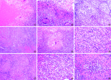

| 组织病理学特征 | MTB感染 (50例) | TM感染 (32例) | NTM感染 (3例)a | MTB并发TM 感染(2例)a | 隐球菌感染 (1例)a | χ2值b | P值b |
|---|---|---|---|---|---|---|---|
| 典型肉芽肿伴坏死 | 15(30.0) | 6(18.8) | 0 | 1 | 1 | 1.296 | 0.255 |
| 不典型肉芽肿伴坏死 | 16(32.0) | 2(6.2) | 0 | 1 | 0 | - | 0.006 |
| 组织细胞浸润伴坏死 | 0(0.0) | 6(18.8) | 1 | 0 | 0 | - | 0.002 |
| 仅见坏死 | 8(16.0) | 1(3.1) | 0 | 0 | 0 | - | 0.083 |
| 仅见组织细胞浸润 | 0(0.0) | 10(31.2) | 0 | 0 | 0 | - | <0.01 |
| 仅见不典型肉芽肿 | 3(6.0) | 4(12.5) | 2 | 0 | 0 | - | 0.423 |
| 仅见典型肉芽肿 | 3(6.0) | 2(6.2) | 0 | 0 | 0 | - | 1.000 |
| 淋巴结反应性增生 | 5(10.0) | 1(3.1) | 0 | 0 | 0 | - | 0.099 |
| 伴化脓性感染 | 14(28.0) | 2(6.2) | 1 | 0 | 0 | - | 0.013 |
| 可见多核巨细胞 | 8(16.0) | 2(6.2) | 0 | 0 | 1 | - | 0.302 |

| [1] |
Dimopoulos Y, Moysi E, Petrovas C. The Lymph Node in HIV Pathogenesis. Curr HIV/AIDS Rep, 2017,14(4):133-140. doi: 10.1007/s11904-017-0359-7.
doi: 10.1007/s11904-017-0359-7 URL pmid: 28685417 |
| [2] |
Wannakrairot P, Leong TY, Leong AS. The morphological spectrum of lymphadenopathy in HIV infected patients. Pathology, 2007,39(2):223-227. doi: 10.1080/0031302070 1230674.
doi: 10.1080/00313020701230674 URL pmid: 17454752 |
| [3] |
Özkan EA, Göret CC, Özdemir ZT, et al. Evaluation of peripheral lymphadenopathy with excisional biopsy: six-year experience. Int J Clin Exp Pathol, 2015,8(11):15234-15239.
URL pmid: 26823872 |
| [4] |
Sharma SK, Mohan A. Extrapulmonary Tuberculosis. Indian J Med Res, 2004,120(4):316-353.
URL pmid: 15520485 |
| [5] |
Gambhir S, Ravina M, Rangan K, et al. Imaging in extrapulmonary tuberculosis. Int J Infect Dis, 2017,56:237-247. doi: 10.1016/j.ijid.2016.11.003.
doi: 10.1016/j.ijid.2016.11.003 URL pmid: 27838445 |
| [6] |
Gosavi AV, Sulhyan KR, Shetty DS, et al. FNAC of lymph nodes in HIV positive patients-a diagnostic boon. J Am Soc Cytopathol, 2017,6(2):59-65. doi: 10.1016/j.jasc.2016.12.004.
doi: 10.1016/j.jasc.2016.12.004 URL pmid: 31042635 |
| [7] |
Mustafa T, Wiker HG, Merkve O, et a1. Differential expression of mycobacterial antigen MPT64, apoptosis and inflammatory markers in muhinucleated giant cells and epithelioid cells jn granulomas caused by Mycobacterium tuberculosis. Virchows Arch, 2008,452(4):449-456. doi: 10.1007/s00428-008-0575-z.
doi: 10.1007/s00428-008-0575-z URL pmid: 18266005 |
| [8] |
Mandal R, Mondal K, Datta S, et al. A clinicopathological study of peripheral lymph nodes in HIV-infected patients with special reference to CD4+ T-cell counts: experience from a tertiary care institution in Darjeeling (India) . Diagn Cytopathol, 2015,43(12):971-977. doi: 10.1002/dc.23379.
doi: 10.1002/dc.23379 URL pmid: 26457991 |
| [9] |
O’Connell ML, Birkenkamp KE, Kleiner DE, et al. Lung manifestations in an autopsy-based series of pulmonary or disseminated nontuberculous mycobacterial disease. Chest, 2012,141(5):1203-1209. doi: 10.1378/chest.11-0425.
URL pmid: 22194586 |
| [10] | 王宇轩, 王冲, 董宇杰, 等. 肺结核患者肺组织微生物组特征研究. 中国防痨杂志, 2018,40(11):1152-1158. doi: 10.3969/j.issn.1000-6621.2018.11.002. |
| [11] |
Shenoy R, Kapadi SN, Pai KP, et al. Fine needle aspiration diagnosis in HIV-related lymphadenopathy in Mangalore, India. Acta Cytol, 2002,46(1):35-29. doi: 10.1159/000326713.
doi: 10.1159/000326713 URL pmid: 11843556 |
| [12] | 董宇杰, 张莉, 王宇轩, 等. 免疫组织化学及PCR技术在淋巴结结核病理诊断中的应用价值. 中国防痨杂志, 2018,40(4):345-349. doi: 10.3969/j.issn.1000-6621.2018.04.002. |
| [13] |
Pinder SE, Colville A. Mycobacterial cervical lymphadenitis in children: can histological assessment help differentiate infections caused by non-tuberculous mycobacteria from Mycobacterium tuberculosis? Histopathology, 1993,22(1):59-64. doi: 10.1111/j.1365-2559.1993.tb00070.x.
doi: 10.1111/j.1365-2559.1993.tb00070.x URL pmid: 8436342 |
| [14] |
Evans MJ, Smith NM, Thornton CM, et al. Atypical mycobacterial lymphadenitis in children—a clinicopathological study of 17 cases. J Clin Pathol, 1998,51(12):925-927. doi: 10.1136/jcp.51.12.925.
doi: 10.1136/jcp.51.12.925 URL pmid: 10070335 |
| [15] | 孙磊, 张亮, 王玉亮, 等. 细针吸取细胞学在HIV感染者体表肿大淋巴结和肿块诊断中的应用. 临床与实验病理学杂志, 2011,27(10):1070-1074. |
| [16] |
Sfeir MM, Schuetz A, Van Besien K, et al. Mycobacterial spindle cell pseudotumour: epidemiology and clinical outcomes. J Clin Pathol, 2018,71(7):626-630. doi: 10.1136/jclinpath-2017-204777.
doi: 10.1136/jclinpath-2017-204777 URL pmid: 29367301 |
| [17] |
Dhibar DP, Sahu KK, Singh S, et al. Tubercular Mycobacterial Spindle Cell Pseudotumour: A Case Report. Iran J Med Sci, 2018,43(1):94-96.
URL pmid: 29398759 |
| [18] | Hu Y, Zhang J, Li X, et al. Penicillium marneffei infection: an emerging disease in mainland China. Mycopathologia, 2013,175(1/2):57-67. doi: 10.1007/s11046-012-9577-0. |
| [19] |
Le T, Wolbers M, Chi NH, et al. Epidemiology, seasonality, and predictors of outcome of AIDS-associated Penicillium marneffei infection in Ho Chi Minh City, Viet Nam. Clin Infect Dis, 2011,52(7):945-952. doi: 10.1093/cid/cir028.
doi: 10.1093/cid/cir028 URL pmid: 21427403 |
| [20] |
Boyce KJ, Andrianopoulos A. Morphogenetic circuitry regulating growth and development in dimorphic pathogen penicillium marneffei. Eukaryot Cell, 2013,12(2):154-160. doi: 10.1128/EC.00234-12.
doi: 10.1128/EC.00234-12 URL pmid: 23204189 |
| [21] | Chayakulkeeree M, Perfect JR. Cryptococcosis. Infect Dis Clin North Am, 2006,20(3):150-544,v-vi. doi: 10.1016/j.idc.2006.07.001. |
| [22] |
Srinivasan R, Gupta N, Shifa R, et al. Cryptococcal lympha-denitis diagnosed by fine needle aspiration cytology: a review of 15 cases. Acta Cytol, 2010,54(1):1-4. doi: 10.1159/000324958.
URL pmid: 20306981 |
| [23] | Ghosh A, Tilak R, Bhushan R, et al. Lymphnodal Co-infection of Cryptococcal and Histoplasma in HIV-infected Patients and Review of Published Reports. Mycopathologia, 2015,180(1/2):105-110. doi: 10.1007/s11046-015-9882-5. |
| [24] |
Mohanty SK, Vaiphei K, Dutta U, et al. Granulomatous cryptococcal lymphadenitis in immunocompetent individuals: report of two cases. Histopathology, 2003,42(1):96-97. doi: 10.1046/j.1365-2559.2003.01513_4.x.
doi: 10.1046/j.1365-2559.2003.01513_4.x URL pmid: 12493035 |
| [1] | YANG Ni, SU Qian, XIAO Yue, LU Jia, SONG Yang, RAO Zheng-yuan, XIA Lan, HE Jin-ge, CHEN Chuang, ZHANG Ling-lin. Analysis on treatment outcomes and influencing factors of 2158 patients with MTB/HIV co-infection [J]. Chinese Journal of Antituberculosis, 2021, 43(3): 274-279. |
| [2] | ZHANG Kai-yi, LYU Zheng-xuan, LIU Yong-li, ZHANG Le, ZHU Jiang-chun, YU Ming-li. A case of multisystemic disseminated infection caused by Mycobacterium abscess diagnostic and treatment process analysis [J]. Chinese Journal of Antituberculosis, 2021, 43(1): 66-71. |
| [3] | LI Ting, HE Jin-ge, SU Qian, LI Jing, LI Yun-kui, GAO Wen-feng, GAO Yuan, YANG Wen. Value of tuberculin test in screening tuberculosis infection in HIV infected/AIDS patients in Butuo County, Sichuan Province [J]. Chinese Journal of Antituberculosis, 2020, 42(9): 931-936. |
| [4] | LI Yun-kui, HE Jin-ge, SU Qian, LI Ting, LI Jing, GAO Wen-feng, YANG Wen, MAO Guang-yu. Value of tuberculin test in screening tuberculosis infection in HIV infected/AIDS patients in Butuo County, Sichuan Province [J]. Chinese Journal of Antituberculosis, 2020, 42(9): 937-941. |
| [5] | BAO Rui, LIU Xiao-yang, REN Peng, ZHANG Feng, LIANG Hai-yan, WANG Ru, FU Ling, GAN Di-shou. Analysis of surgical treatment characteristics in patients with spinal tuberculosis and HIV co-infection [J]. Chinese Journal of Antituberculosis, 2020, 42(6): 645-648. |
| [6] | The Joint Tuberculosis Professional Branch of Chinese Antituberculosis Association, the Western China Bone Tuberculosis Alliance, the North China Bone Tuberculosis Alliance, Editorial Board of Chinese Journal of Antituberculosis. Experts’ consensus on the diagnosis and treatment of patients with spinal tuberculosis complicated with HIV/AIDS [J]. Chinese Journal of Antituberculosis, 2020, 42(5): 418-424. |
| [7] | PU Yu, HUAN Ming-cang. Overview situation and progress of diagnosis and treatment in patients with spinal tuberculosis and HIV/AIDS co-infection [J]. Chinese Journal of Antituberculosis, 2020, 42(5): 428-435. |
| [8] | LI Bang-yin, PU Yu, HE Min, HE Lei, HUAN Ming-cang, CAI Yu-guo, LIU Lin, JIANG Xi. Clinical characteristics of HIV-positive spinal tuberculosis patients and effect analysis of strengthening perioperative management [J]. Chinese Journal of Antituberculosis, 2020, 42(5): 449-453. |
| [9] | ZHANG Xing,WANG Feng-ming,LYU Xu-feng,HUA Tian-qi,ZHANG Xue-jun,JIANG Jing-yi,DING Chen-li,ZHU Wei,XIA Guo-dong,JI Jun-min,ZHAO Fei. Analysis of influencing factors of Mycobacterium tuberculosis infection in HIV/AIDS patients [J]. Chinese Journal of Antituberculosis, 2020, 42(4): 360-365. |
| [10] | CHEN Qi-yi,LI Jing-jing,XU Yun-liang,LYU Zhi-bin,WEI Lian-gui,XU Dong-hai,XIE Ru-ming,CHEN Bu-dong. Magnetic resonance neuroimaging differential diagnosis of tuberculous meningitis and toxoplasmosis encephalopathy in HIV infected individuals [J]. Chinese Journal of Antituberculosis, 2020, 42(3): 222-226. |
| [11] | ZHANG Yun, JIANG Guang-lu, WANG Chong, DUAN Hong-fei, LIANG Qing-tao, LI Hua, YANG Yang, GUO Chao, SHAO Ling-ling, CHEN Xiao-you, YANG Xin-ting*. The diagnostic value of pleural biopsy by medical thoracoscopy in tuberculous pleural effusions [J]. Chinese Journal of Antituberculosis, 2020, 42(11): 1158-1164. |
| [12] | Yi ZHANG,Ke BI,Hui-ming ZHU,Yang CONG,Meng-jun SHEN,Hong-wei CHEN,Yin WANG. Diagnostic value of contrast-enhanced ultrasound-guided percutaneous lung puncture biopsy in culture negative presumptive tuberculosis patients [J]. Chinese Journal of Antituberculosis, 2019, 41(8): 828-832. |
| [13] | Yi HUANG,Han YANG,Xuan FENG,Yuan LIU,Yao-hui ZHANG,Qi ZHANG. Diagnostic value of ultrasound-guided pleural puncture in suspected tuberculosis pleurisy [J]. Chinese Journal of Antituberculosis, 2019, 41(8): 833-837. |
| [14] | Yi-min SUN,Zhen-qing WANG,Jia JIA,Hua-ping GU. Application value of thinprep cytologic test in the diagnosis of pulmonary tuberculosis [J]. Chinese Journal of Antituberculosis, 2019, 41(8): 882-886. |
| [15] | Wen-zhi ZHANG,Dong-ming SU,Jun MENG,Ning HE,Cai-fen WANG. Application and value of contrast-enhanced ultrasound in puncture biopsy of patients with chest wall tuberculosis [J]. Chinese Journal of Antituberculosis, 2019, 41(7): 715-718. |
| Viewed | ||||||
|
Full text |
|
|||||
|
Abstract |
|
|||||
 京公网安备11010202007215号
Total visitors: Visitors of today: Now online:
京公网安备11010202007215号
Total visitors: Visitors of today: Now online:
 This work is licensed under Creative Commons Attribution 3.0 License.