
Chinese Journal of Antituberculosis ›› 2024, Vol. 46 ›› Issue (12): 1548-1559.doi: 10.19982/j.issn.1000-6621.20240245
• Review Articles • Previous Articles Next Articles
Li Junliang1, Liu Xin2, Lin Zhiyuan1, Long Xianrong3, Jiang Zhihang1, Huo Yingyu4( )
)
Received:2024-06-13
Online:2024-12-10
Published:2024-12-03
Contact:
Huo Yingyu, Email: fosuhyy@163.com
Supported by:CLC Number:
Li Junliang, Liu Xin, Lin Zhiyuan, Long Xianrong, Jiang Zhihang, Huo Yingyu. The current application status of deep learning in Chest X-ray screening for lung diseases[J]. Chinese Journal of Antituberculosis, 2024, 46(12): 1548-1559. doi: 10.19982/j.issn.1000-6621.20240245
Add to citation manager EndNote|Ris|BibTeX
URL: https://www.zgflzz.cn/EN/10.19982/j.issn.1000-6621.20240245
| 文献编号 | 数据集 | 图像种类(病种) | 图片像素 | 数量 |
|---|---|---|---|---|
| [ | Shenzhen | 包含336张肺结核和326张无肺部病变的胸部X线摄片图像 | 3000×3000 | 662 |
| [ | Montgomery | 包含80张无肺部病变和58张肺结核的胸部X线摄片图像 | 4892×4020 | 138 |
| [ | ChestXray14 | 包含14种肺部病变图像(肺不张、实变、浸润、气胸、水肿、肺气肿、纤维化、胸腔积液、肺炎、胸膜增厚、心脏肥大、结节、肿块和疝气) | 1024×1024 | 112120 |
| [ | CheXpert | 包含14种类别图像(无肺部病变、纵隔扩大、心脏肥大、密度增高病灶、肺部病变、水肿、变实、肺炎、肺不张、气胸、胸腔积液、胸膜其他病变、骨折、辅助设备) | - | 224316 |
| [ | CoronaHack | 包括4种类别(无肺部病变、新型冠状病毒感染、病毒性肺炎、细菌性肺炎) | 384×127、 1214×937、 4248×3480 | 5933 |
| [ | COVID-ChestXray-15k | 包括3种图像(无肺部病变、肺炎、新型冠状病毒感染) | 1024×1024 | 15000 |
| [ | COVQU | 包含新型冠状病毒感染、密度增高病灶、病毒性肺炎、无肺部病变 | 1024×1024 | 21165 |
| [ | TBX11K | 包括无肺部病变、异常但不属于肺结核图像 | 512×512 | 11200 |
| [ | JSRT | 提供了100张恶性结节、54张良性结节和93张无结节的胸部X线摄片图像 | 2048×2048 | 247 |
| 方法 | 文献 | 预处理思想 | 数据 | 评估 |
|---|---|---|---|---|
| 数字图像处理 | ||||
| [ | 自适应直方图均衡化处理 | 病毒性肺炎:1495;细菌性肺炎:2779;无肺部病变:1583 | 准确率为98.32% | |
| [ | 基于K-Lerch超越函数模型的图像增强处理 | 无肺部病变:417+277;肺炎:250+196;新型冠状病毒感染:237+337a | X线摄片和CT图像准确率分别为98.60%和98.80% | |
| 预特征提取方法 | ||||
| [ | 主动形状模型进行肺野分割+主成分分析对肋骨建模 | 肺结节:548;无肺部病变:177 | 平均曲线下面积为0.815 | |
| [ | 以U型网络模型作为骨干模型的ResNet 18和EfficientNet B0的集成学习 | 新型冠状病毒感染:6032;无肺部病变:3136 | 准确率为98.20%,曲线下面积为0.998 | |
| [ | 使用四路卷积编码的改进U型网络模型对胸部X线摄片图像进行肺部区域分割 | ChestXray14 | 准确率为84.2% | |
| [ | 使用U型网络提取多尺度特征图,基于特征金字塔融合多尺度特征图,实现肺部轮廓分割 | 无肺部病变:8062;肺炎:5501;新型冠状病毒感染:562 | 准确率为91.3% |

| 文献 | 思想 | 数据 | 评估 | 优缺点 |
|---|---|---|---|---|
| [ | 含有5个卷积层、池化层及2个密集层的模型 | 新型冠状病毒感染:3616;肺炎:4273;无肺部病变:10192;肺结核:3500 | 98.63%的准确率和98.35%的召回率 | 模型结构简单且效果优秀,但鲁棒性差,层数较浅 |
| [ | 使用6种不同的卷积核并列进行特征提取,以极限学习机作为分类器 | ChestXray14数据集;病毒性肺炎:1493;肺结核:1036 | 90.92%的准确率和96.93%的曲线下面积 | 模型捕获多尺度特征强,轻量化程度高 |
| [ | VGG19+4个密集层 | 新型冠状病毒感染:3615;肺炎:11726;密度增高病灶:6012;肺癌:20000;无肺部病变:37247;肺结核:1400 | 96.48%的准确率、93.75%的召回率、99.82%的曲线下面积 | 使用较小的卷积块作特征提取,通过增加网络深度或加入注意力的方式丰富多层次的特征信息 |
| [ | VGG16+3个3×3的卷积块和1个全局平均池化层 | 无肺部病变:13672;肿块:5603;新型冠状病毒感染:15660;结节:6201;胸腔积液:13501;肺炎:9878;纤维化:3357;肺结核:3184;密度增高病灶:7179;气胸:6870 | 98.89%的准确率和99.87%的特异度 | 使用较小的卷积块作特征提取,通过增加网络深度或加入注意力的方式丰富多层次的特征信息 |
| [ | VGG16中加入了水平和垂直方向的坐标注意机制 | 深圳数据集 | 92.73%的准确率和97.71%的曲线下面积 | 使用较小的卷积块作特征提取,通过增加网络深度或加入注意力的方式丰富多层次的特征信息 |
| [ | 以ResNet50的4个具有残差结构的卷积块提取特征 | 肺炎胸部X线图像数据集;新型冠状病毒感染影像数据库 | 98.35%的准确率 | 以快捷连接的方式使模型更深、更复杂 |
| [ | DenseNet+由卷积构成的特征选择器+具有空间和通道编码能力的特征集成器 | ChestX-ray14数据集;CheXpert数据集 | 84.1%和92.1%的准确率 | 从多个通道分别获取丰富的特征信息,更具有鲁棒性 |
| [ | VGG16、Inception v3和ResNet 50网络的集成 | COVID-ChestXray数据集 | 特异度为98.54%,精确度为93.60% | 充分利用每个网络的优势,但模型的复杂度和训练难度更大 |
| [ | MobileNetV2、InceptionV3和VGG19的集成 | 密度增高病灶:6914;无肺部病变:11192;新型冠状病毒感染:11582;肺炎:10036;肺结核:2984 | 四分类和五分类的准确度分别为87.12%和91.71% | 充分利用每个网络的优势,但模型的复杂度和训练难度更大 |
| 文献 | 思想 | 数据 | 评估 | 优缺点 |
|---|---|---|---|---|
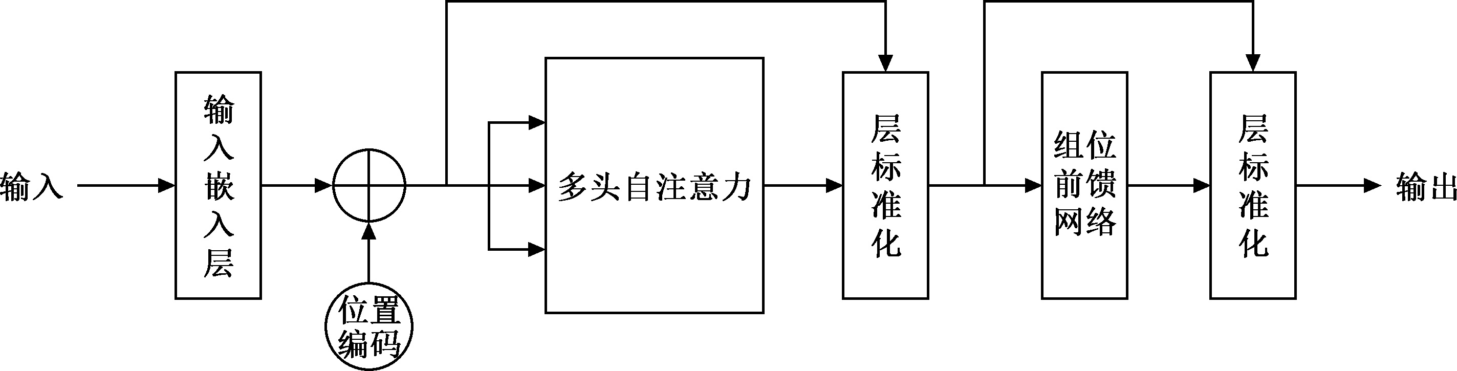
| [ | Vision Transformer | 无肺部病变:10314;新型冠状病毒感染:10819;肺炎:10702 | 98%的准确率和99%的曲线下面积 | 模型简单,易于实现,但是没有考虑局部信息的重要性 |
| [ | Vision Transformer+输入图像的3组2D卷积特征 | 无肺部病变:29477;肺炎:5852;肺结核:700;新型冠状病毒感染:20305 | 98.29%的平均准确率和98.95%的平均精确度 | 充分利用卷积的局部感受野和平移不变性,以及Transformer的全局注意力机制,对图像进行全局的语义建模和特征交互;Transformer的长程注意力机制容易导致图像块内部结构信息被破坏 |
| [ | Vision Transformer+2组不同卷积核大小的深度卷积 | 无肺部病变:10083+6893;新型冠状病毒感染:3466+7593;病毒性肺炎:1342+2618a | X线和CT图像的准确率分别达到97.25%和98.36% | 同上 |
| [ | Swin Transformer+2层密集层和2层Drop out层 | 新型冠状病毒感染:3616;密度增高病灶:6012;病毒性肺炎:1345;无肺部病变:10192 | 98%的召回率和96%的准确率 | 同上 |
| [ | 结合图像的多头注意力与标签注意力的双路径解码器 | ChestXray14数据集 | 83.1%的曲线下面积 | 特征图和标签向量进行信息交互,但是模型复杂、训练难度较高 |
| [ | Transformer图像编码+细节校正路径从X和Y像素方向的梯度信息+MagNet根据训练集分布调整测试集分布 | Synthesis-covid-cxr数据集 | 95.23%的准确率 | 能够根据数据特点自适应地调节测试数据分布,聚焦更重要的内容 |
| [ | pyramid vision Transformer变体模型 | ChestXray14数据集 | 83.0%的曲线下面积 | 位置编码的方式取代位置嵌入,减少注意力的计算量 |
| 文献 | 思想 | 数据 | 评估 | 优缺点 |
|---|---|---|---|---|
| [ | EfficientNet+Vision Transformer | 蒙哥马利数据集、深圳数据集、新型冠状病毒感染影像数据库 | 97.51%的准确率和98%的召回率 | 优点:能够捕获多尺度上下文信息 |
| [ | L和AB坐标+Inception V3+Transformer | 新型冠状病毒感染影像数据库、COVID-ChestX-ray-15k数据集 | 分别获得96.66%和97.87%的准确率 | 优点:能够捕获多尺度上下文信息 |
| [ | DenseNet与MaxViT组合成双通道模型 | COVID-QU-Ex数据集 | 95.61%的准确率和96%的召回率 | 优点:减少提取特征时信息的损失,增强有用的信息,抑制不重要的信息;缺点:模型参数量大 |
| [ | ResNet101+多级自注意力的Transformer | 无肺部病变:10192;新型冠状病毒感染:3615;密度增高病灶:6023;病毒性肺炎:1345 | 96.54%的准确率 | 优点:减少提取特征时信息的损失,增强有用的信息,抑制不重要的信息;缺点:模型参数量大 |
| [ | ResNet18作特征提取,提取的特征作为单独patch+Vision Transformer | 肺炎:11263;新型冠状病毒感染:11956;无肺部病变:10701 | 94.96%的准确率 | 优点:特征图不是裁切成多个patch,而是对整个特征图计算注意力,直接理解全图 |
| [ | VGG、Googlenet、Densenet集成学习+Vision Transformer | 新型冠状病毒感染影像数据库、COVID-ChestX-ray-15k数据集 | 分别获得98.00%和97.4%的平均精确度 | 优点:可有效获得不同层次之间由低阶到高阶的互补信息;缺点:特征图在多个级别上密集级联,易导致高计算资源需求 |
| [ | DenseNet121、VGG16、Effi-cientNet V2集成学习+Vision Transformer | 肺结核:57087;无肺部病变:18946 | 90.57%的准确度 | 优点:可有效获得不同层次之间由低阶到高阶的互补信息;缺点:特征图在多个级别上密集级联,易导致高计算资源需求 |
| [1] | 冯筠, 牛怡, 杨晨希, 等. 融合改进变分自编码器与影像组学的X光片肺部疾病筛查算法. 西北大学学报(自然科学版), 2023, 53(3): 313-324. doi:10.16152/j.cnki.xdxbzr.2023-03-001. |
| [2] | Ai T, Yang Z, Hou H, et al. Correlation of Chest CT and RT-PCR Testing for Coronavirus Disease 2019 (COVID-19) in China: A Report of 1014 Cases. Radiology, 2020, 296(2): E32-E40. doi:10.1148/radiol.2020200642. |
| [3] | 鲁慧民, 薛涵, 王奕龙, 等. 机器学习在影像组学分析中的应用综述. 计算机工程与应用, 2023, 59(17): 22-34. doi:10.3778/j.issn.1002-8331.2210-0435. |
| [4] | 王瑜, 吴江, 伍东升. 卷积神经网络在职业性尘肺病影像学诊断中的应用研究进展. 生物医学工程学杂志, 2024, 41(2): 413-420. doi:10.7507/1001-5515.202309079. |
| [5] | Santosh KC, Allu S, Rajaraman S, et al. Advances in Deep Learning for Tuberculosis Screening using Chest X-rays: The Last 5 Years Review. J Med Syst, 2022, 46(11): 82. doi:10.1007/s10916-022-01870-8. |
| [6] | Liang S, Ma J, Wang G, et al. The Application of Artificial Intelligence in the Diagnosis and Drug Resistance Prediction of Pulmonary Tuberculosis. Front Med (Lausanne), 2022, 9: 935080. doi:10.3389/fmed.2022.935080. |
| [7] | 李新, 陈帆, 郝海江, 等. 深度学习在肺炎检测中的研究综述. 桂林理工大学学报, 2020, 40(4): 859-866. doi:10.3969/j.issn.1674-9057.2020.04.027. |
| [8] | 国家呼吸内科医疗质量控制中心, 中华医学会结核病学分会, 中国防痨协会结核病控制专业分会, 等. 综合医疗机构肺结核早期发现临床实践指南. 结核与肺部疾病杂志, 2024, 5(1): 1-14. doi:10.19982/j.issn.1000-6621.20230428. |
| [9] | 杨红杰, 齐菲, 张红梅, 等. 肺癌合并肺结核的诊断和临床治疗现状. 结核与肺部疾病杂志, 2024, 5(3): 273-278. doi:10.19983/j.issn.2096-8493.2024054. |
| [10] | Ghiasi MM, Zendehboudi S. Application of decision tree-based ensemble learning in the classification of breast cancer. Comput Biol Med, 2021, 128: 104089. doi:10.1016/j.compbiomed.2020.104089. |
| [11] |
Chung SW, Han SS, Lee JW, et al. Automated detection and classification of the proximal humerus fracture by using deep learning algorithm. Acta Orthop, 2018, 89(4): 468-473. doi:10.1080/17453674.2018.1453714.
pmid: 29577791 |
| [12] | Mayidili N, Jie M, Chuling H, et al. Artificial Intelligence Assisting the Early Detection of Active Pulmonary Tuberculosis from Chest X-Rays: A Population-Based Study. Front Mol Biosci, 2022: 9874475. doi:10.3389/fmolb.2022.874475. |
| [13] | Faiz KA, Tripti P, Belay T, et al. Computer-aided reading of tuberculosis chest radiography: moving the research agenda for-ward to inform policy. Eur Respir J, 2017, 50(1): 1700953. doi:10.1183/13993003.00953-2017. |
| [14] | Khan MA, Quasim MT, Alghamdi NS, et al. A secure framework for authentication and encryption using improved ECC for IoT-based medical sensor data. IEEE Access, 2020, 8: 52018-52027. doi:10.1109/access.2020.2980739. |
| [15] | Gao XW, James-Reynolds C, Currie E. Analysis of tuberculosis severity levels from CT pulmonary images based on enhanced residual deep learning architecture. Neurocomputing, 2020, 392: 233-244. doi:10.1016/j.neucom.2018.12.086. |
| [16] | Luján-García EJ, Yáñez-Márquez C, Villuendas-Rey Y, et al. A Transfer Learning Method for Pneumonia Classification and Visualization. Applied Sciences, 2020, 10(8): 2908. doi:10.3390/app10082908. |
| [17] | Morís DI, de Moura J, Novo J, et al. Unsupervised contrastive unpaired image generation approach for improving tuberculosis screening using chest X-ray images. Pattern Recognition Letters, 2022, 164: 60-66. doi:10.1016/j.patrec.2022.10.026. |
| [18] | Jaeger S, Candemir S, Antani S, et al. Two public chest X-ray datasets for computer-aided screening of pulmonary diseases. Quant Imaging Med Surg, 2014, 4(6): 475-477. doi:10.3978/j.issn.2223-4292.2014.11.20. |
| [19] | Shamrat FJM, Azam S, Karim A, et al. High-precision multiclass classification of lung disease through customized MobileNetV2 from chest X-ray images. Comput Biol Med, 2023, 155: 106646. doi:10.1016/j.compbiomed.2023.106646. |
| [20] | Cho K, Kim KD, Nam Y, et al. CheSS: Chest X-Ray Pre-trained Model via Self-supervised Contrastive Learning. J Digit Imaging, 2023, 36(3): 902-910. doi:10.1007/s10278-023-00782-4. |
| [21] | Chen H, Zhang T, Chen R, et al. A Novel COVID-19 Image Classification Method Based on the Improved Residual Network. Electronics, 2023, 12: 80. doi:10.3390/electronics12010080. |
| [22] | Badawi A, Elgazzar K. Detecting coronavirus from chest X-rays using transfer learning. Covid, 2021, 1(1): 403-415. doi:10.3390/covid1010034. |
| [23] | Rahman T, Khandakar A, Qiblawey Y, et al. Exploring the effect of image enhancement techniques on COVID-19 detection using chest X-ray images. Comput Biol Med, 2021, 132: 104319. doi:10.1016/j.compbiomed.2021.104319. |
| [24] |
Shiraishi J. Development of a digital image database for chest radiographs with and without a lung nodule: ROC analysis on radiologists’ performance in detection of pulmonary nodules. Am J Roentgenol, 2000, 174(1): 71-74. doi:10.2214/ajr.174.1.1740071.
pmid: 10628457 |
| [25] | Nahiduzzaman M, Goni MOF, Anower MS, et al. A novel method for multivariant pneumonia classification based on hybrid CNN-PCA based feature extraction using extreme learning machine with CXR images. IEEE Access, 2021, 9: 147512-147526. doi:10.1109/access.2021.3123782. |
| [26] | Al-Sheikh MH, Al Dandan O, Al-Shamayleh AS, et al. Multi-class deep learning architecture for classifying lung diseases from chest X-Ray and CT images. Scientific Reports, 2023, 13(1): 19373. doi:10.1038/s41598-023-46147-3. |
| [27] | Li X, Shen L, Xie X, et al. Multi-resolution convolutional networks for chest X-ray radiograph based lung nodule detection. Artif Intell Med, 2020, 103: 101744. doi:10.1016/j.artmed.2019.101744. |
| [28] | Rajaraman S, Cohen G, Spear L, et al. DeBoNet: A deep bone suppression model ensemble to improve disease detection in chest radiographs. PLoS One, 2022, 17(3): e0265691. doi:10.1371/journal.pone.0265691. |
| [29] | Chowdary GJ, Kanhangad V. A Dual-Branch Network for Diagnosis of Thorax Diseases From Chest X-Rays. IEEE J Biomed Health Inform, 2022, 26(12): 6081-6092. doi:10.1109/JBHI.2022.3215694. |
| [30] | 武卓越, 田雪琴, 侯潇芮, 等. 基于卷积神经网络的肺野分割和肺炎筛查. 西北大学学报(自然科学版), 2022, 52(4): 571-580. doi:10.16152/j.cnki.xdxbzr.2022-04-006. |
| [31] | Ahmed MS, Rahman A, AlGhamdi F, et al. Joint Diagnosis of Pneumonia, COVID-19, and Tuberculosis from Chest X-ray Images: A Deep Learning Approach. Diagnostics (Basel), 2023, 13(15): 2562. doi:10.3390/diagnostics13152562. |
| [32] | Nahiduzzaman M, Goni MOF, Hassan R, et al. Parallel CNN-ELM: A multiclass classification of chest X-ray images to identify seventeen lung diseases including COVID-19. Expert Syst Appl, 2023, 229: 120528. doi:10.1016/j.eswa.2023.120528. |
| [33] | Alshmrani GMM, Ni Q, Jiang R, et al. A deep learning architecture for multi-class lung diseases classification using chest X-ray (CXR) images. Alexandria Engineering Journal, 2023, 64: 923-935. doi:10.1016/j.aej.2022.10.053. |
| [34] | Shamrat FMJM, Azam S, Karim A, et al. LungNet22: A Fine-Tuned Model for Multiclass Classification and Prediction of Lung Disease Using X-ray Images. J Pers Med, 2022, 12(5): 680. doi:10.3390/jpm12050680. |
| [35] | Xu W, Fu YL, Zhu D. ResNet and its application to medical image processing: Research progress and challenges. Comput Methods Programs Biomed, 2023, 240: 107660. doi:10.1016/j.cmpb.2023.107660. |
| [36] | Farhan AMQ, Yang S. Automatic lung disease classification from the chest X-ray images using hybrid deep learning algorithm. Multimed Tools Appl, 2023, 82(25): 38561-38587. doi:10.1007/s11042-023-15047-z. |
| [37] | Huang H, Li Y, Wu R, et al. Benign-malignant classification of pulmonary nodule with deep feature optimization framework. Biomed Signal Process Control, 2022, 76: 103701. doi:10.1016/j.bspc.2022.103701. |
| [38] | Reddy ASK, Rao KNB, Soora NR, et al. Multi-modal fusion of deep transfer learning based COVID-19 diagnosis and classification using chest x-ray images. Multimed Tools Appl, 2023, 82(8): 12653-12677. doi:10.1007/s11042-022-13739-6. |
| [39] | Türk F, Kökver Y. Detection of Lung Opacity and Treatment Planning with Three-Channel Fusion CNN Model. Arab J Sci Eng, 2023, 14: 1-13. doi:10.1007/s13369-023-07843-4. |
| [40] | Qi Xuanhao, Zhi Min. Review of Attention Mechanisms in Image Processing. Front Comput Sci, 2024, 18(2): 345-362. doi:10.3778/j.issn.1673-9418.2305057. |
| [41] | Xu T, Yuan Z. Convolution neural network with coordinate attention for the automatic detection of pulmonary tuberculosis images on chest x-rays. IEEE Access, 2022, 10: 86710-86717. doi:10.1109/access.2022.3199419. |
| [42] | Guan Q, Huang Y, Luo Y, et al. Discriminative Feature Learning for Thorax Disease Classification in Chest X-ray Images. IEEE Trans Image Process, 2021, 30: 2476-2487. doi:10.1109/TIP.2021.3052711. |
| [43] | 卢玲, 漆为民. 基于Transformer的脊椎CT图像分割. 中国图象图形学报, 2023, 28(11): 3618-3628. doi:10.11834/jig.221084. |
| [44] | Wang B, Zhang D, Tian Z. CoroTrans-CL: A novel transformer-based continual deep learning model for image recognition of coronavirus infections. Electronics, 2023, 12(4): 866. doi:10.3390/electronics12040866. |
| [45] | Han K, Wang Y, Chen H, et al. A survey on vision transformer. IEEE Trans Pattern Anal Mach Intell, 2022, 45(1): 87-110. doi:10.1109/TPAMI.2022.3152247. |
| [46] | Shome D, Kar T, Mohanty SN, et al. COVID-Transformer: Interpretable COVID-19 Detection Using Vision Transformer for Healthcare. Int J Environ Res Public Health, 2021, 18(21): 11086. doi:10.3390/ijerph182111086. |
| [47] | Okolo GI, Katsigiannis S, Ramzan N. IEViT: An enhanced vision transformer architecture for chest X-ray image classification. Comput Methods Programs Biomed, 2022, 226: 107141. doi:10.1016/j.cmpb.2022.107141. |
| [48] | Hao Y, Zhang C, Li X. DBM-ViT: A multiscale features fusion algorithm for health status detection in CXR/CT lungs images. Biomed Signal Process Control, 2024, 87: 105365. doi:10.1016/j.bspc.2023.105365. |
| [49] | Jiang X, Zhu Y, Cai G, et al. Mxt: A new variant of pyramid vision transformer for multi-label chest x-ray image classification. Cognitive Computation, 2022, 14(4): 1362-1377. doi:10.1007/s12559-022-10032-4. |
| [50] | Jiang X, Zhu Y, Liu Y, et al. TransDD: A transformer-based dual-path decoder for improving the performance of thoracic dis-eases classification using chest X-ray. Biomed Signal Process Control, 2024, 91: 105937. doi:10.1016/j.bspc.2023.105937. |
| [51] | An K, Zhang Y. A Self-Supervised Detail-Sensitive ViT-Based Model for COVID-19 X-ray Image Diagnosis: SDViT. Ap-plied Sciences, 2022, 13(1): 454. doi:10.3390/app13010454. |
| [52] | Chen Y, Feng J, Liu J, et al. Detection and classification of Lung Cancer cells using swin transformer. J Cancer Ther, 2022, 13(7): 464-475. doi:10.4236/jct.2022.13704. |
| [53] | Ma Y, Lv W. Identification of Pneumonia in Chest X-Ray Image Based on Transformer. Int J Antennas Propag, 2022, 2022(1): 5072666. doi:10.1155/2022/5072666. |
| [54] | Almalki EY, Zaffar M, Irfan M, et al. A Novel-based Swin Transfer Based Diagnosis of COVID-19 Patients. Intelligent Automation Soft Computing, 2023, 35(1): 163-180. doi:10.32604/iasc.2023.025580. |
| [55] | Duong LT, Le NH, Tran TB, et al. Detection of tuberculosis from chest X-ray images: Boosting the performance with vision transformer and transfer learning. Expert Syst Appl, 2021, 184: 115519. doi:10.1016/j.eswa.2021.115519. |
| [56] | Ukwuoma CC, Qin Z, Agbesi VK, et al. Dual_Pachi: Attention-based dual path framework with intermediate second order-pooling for Covid-19 detection from chest X-ray images. Comput Biol Med, 2022, 151(Pt A): 106324. doi:10.1016/j.compbiomed.2022.106324. |
| [57] | Bangalore Vijayakumar S, Chitty-Venkata KT, Arya K, et al. ConVision Benchmark: A Contemporary Framework to Benchmark CNN and ViT Models. AI, 2024, 5(3): 1132-1171. doi:10.3390/ai5030056. |
| [58] | Chen S, Ren S, Wang G, et al. Interpretable CNN-Multilevel Attention Transformer for Rapid Recognition of Pneumonia From Chest X-Ray Images. IEEE J Biomed Health Inform, 2024, 28(2): 753-764. doi:10.1109/JBHI.2023.3247949. |
| [59] | Wang T, Nie Z, Wang R, et al. PneuNet: deep learning for COVID-19 pneumonia diagnosis on chest X-ray image analysis using Vision Transformer. Med Biol Eng Comput, 2023, 61(6): 1395-1408. doi:10.1007/s11517-022-02746-2. |
| [60] | Ukwuoma CC, Qin Z, Heyat MBB, et al. Automated Lung-Related Pneumonia and COVID-19 Detection Based on Novel Feature Extraction Framework and Vision Transformer Approaches Using Chest X-ray Images. Bioengineering (Basel), 2022, 9(11): 709. doi:10.3390/bioengineering9110709. |
| [61] | Rajaraman S, Zamzmi G, Folio LR, et al. Detecting Tuberculosis-Consistent Findings in Lateral Chest X-Rays Using an Ensemble of CNNs and Vision Transformers. Front Genet, 2022, 13: 864724. doi:10.3389/fgene.2022.864724. |
| [1] | Song Feier, Mao Yanjun, Xia Qiuyue, Zhou Yang, Lin Huan. The prevalence and influencing factors of post-tuberculosis lung disease: A Meta-analysis [J]. Chinese Journal of Antituberculosis, 2025, 47(3): 322-330. |
| [2] | Tan Shouyong. Research progress on comprehensive treatment beyond antibiotic therapy for nontuberculous mycobacterium pulmonary disease [J]. Chinese Journal of Antituberculosis, 2024, 46(8): 967-970. |
| [3] | Chai Dongyu, Qin Shuyi, Zhang Ronghua, Zou Nannan, Wang Xin. Analysis of risk factors for viral pneumonia combined with invasive pulmonary mycosis [J]. Chinese Journal of Antituberculosis, 2024, 46(7): 750-755. |
| [4] | Han Wenya, Zhou Yangyu, Wang Meifang, Xue Xinying. Progress in clinical diagnosis and treatment of pulmonary cryptococcosis [J]. Chinese Journal of Antituberculosis, 2024, 46(7): 830-838. |
| [5] | WU Chao-ling, DENG Guo-fang, FU Liang, YUAN Xiao-liang. Research progress of exhaled volatile organic compounds on the diagnosis of pulmonary infectious diseases [J]. Chinese Journal of Antituberculosis, 2022, 44(5): 505-511. |
| [6] | LIANG Rui-yun, FANG Wei-jun, REN Hui-li, LI Hui-ru, ZHANG Hui. Study on CT manifestations of non-tuberculous mycobacterium pulmonary disease patients with and without diabetes mellitus [J]. Chinese Journal of Antituberculosis, 2020, 42(9): 962-967. |
| [7] | ZHANG Ming-hui,ZHANG Qiu-di,ZHANG Su-juan,SUN Yi-fang. Study on chest CT findings of 55 patients with HIV-negative pulmonary cryptococcosis [J]. Chinese Journal of Antituberculosis, 2020, 42(3): 233-239. |
| [8] | Rong-zhen ZHOU,Xiu-li WU,Jian WANG,Hai YANG,Wen-bin JI. CT features analysis of Mycobacterium avium-intracellulare complex lung disease with cavities [J]. Chinese Journal of Antituberculosis, 2019, 41(9): 1009-1014. |
| [9] | Ya-ni XUE,Mei ZHANG,Cun-long LI. Prediction and analysis of national tuberculosis epidemic based on grey model [J]. Chinese Journal of Antituberculosis, 2019, 41(7): 782-789. |
| [10] | Duo LI,Kun FANG,Jue WANG,Zhen ZHOU,Ping-xin LYU. Analysis of CT image classification and clinical characteristics of nontuberculous mycobacterial pulmonary disease [J]. Chinese Journal of Antituberculosis, 2019, 41(2): 202-209. |
| [11] | Hui-li REN,Pin-ru CHEN,Hua CHEN,Jin-xing HU,Wen LIU,Wei-jun FANG. The comparison of CT imaging features between Mycobacterium avium-intracellulare complex and Mycobacterium chelonei and abscessus pulmonary diseases combined with bronchiectasis [J]. Chinese Journal of Antituberculosis, 2019, 41(2): 195-201. |
| [12] | Fang HUANG,Bo WANG,Guo-lian ZHAO,Hai-dong WANG,Li-yun DANG. Diagnosis value of T lymphocyte detection for non-tuberculous mycobacteria diseases [J]. Chinese Journal of Antituberculosis, 2019, 41(12): 1263-1268. |
| [13] | LIN Xue,JIA Hui-jun,ZHANG Hui,REN Hui-li,LIU Wen. The value of high resolution CT in the diagnosis and treatment of nontuberculous mycobacterial pulmonary diseases [J]. Chinese Journal of Antituberculosis, 2018, 40(12): 1361-1365. |
| [14] | ZHU Gui-yun,LI Xiao-xia,KANG Li-fei,CHEN Ning,YANG Yong-hui. A case of pulmonary cryptococcosis with mediastinal tuberculous lymphadenitis——report of case and review of literature [J]. Chinese Journal of Antituberculosis, 2018, 40(11): 1231-1234. |
| [15] | ZHOU Zhen,Lv Yan,XIE Ru-ming,ZHOU Xin-hua,HE Wei,XU Jin-ping. Analysis on characteristics of CT imaging for local pulmonary consolidation lesions [J]. Chinese Journal of Antituberculosis, 2014, 36(3): 149-154. |
| Viewed | ||||||
|
Full text |
|
|||||
|
Abstract |
|
|||||
 京公网安备11010202007215号
Total visitors: Visitors of today: Now online:
京公网安备11010202007215号
Total visitors: Visitors of today: Now online:
 This work is licensed under Creative Commons Attribution 3.0 License.