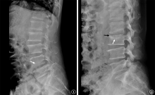
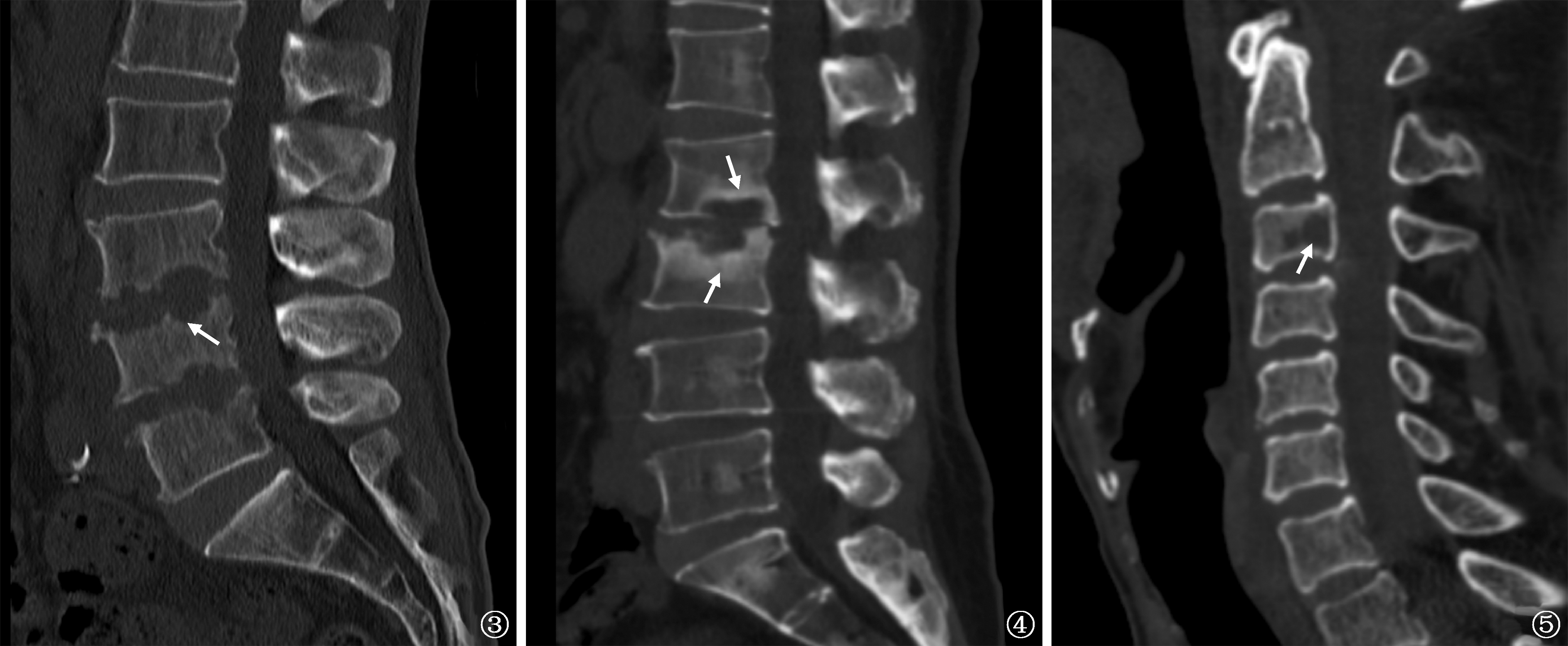
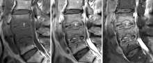
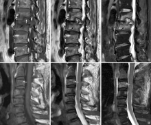
| [1] |
Saeed K, Esposito S, Ascione T, et al. Hot topics on vertebral osteomyelitis from the International Society of Antimicrobial Chemotherapy. Int J Antimicrob Agents, 2019, 54(2):125-133. doi:10.1016/j.ijantimicag.2019.06.013.
|
| [2] |
Ganesh D, Gottlieb J, Chan S, et al. Fungal Infections of the Spine. Spine (Phila Pa 1976), 2015, 40(12):E719-E728. doi:10.1097/BRS.0000000000000903.
|
| [3] |
刘斌峰, 杨光, 高延征. 真菌性脊柱炎的研究进展. 中国脊柱脊髓杂志, 2021, 31(10):951-955. doi:10.3969/j.issn.1004-406X.2021.10.12.
|
| [4] |
Caldera G, Lemus MAC, Cobar A, et al. Fungal Spondylodiscitis: Review. J Spine, 2016,2016:1-6.
|
| [5] |
Arbelaez A, Restrepo F, Castillo M. Spinal infections: clinical and imaging features. Top Magn Reson Imaging, 2014 23(5):303-314. doi:10.1097/RMR.0000000000000032.
|
| [6] |
Duarte RM, Vaccaro AR. Spinal infection: state of the art and management algorithm. Eur Spine J, 2013, 22(12):2787-2799. doi:10.1007/s00586-013-2850-1.
|
| [7] |
Wang C, Zhang L, Zhang H, et al. Sequential endoscopic and robot-assisted surgical solutions for a rare fungal spondylodiscitis, secondary lumbar spinal stenosis, and subsequent discal pseudocyst causing acute cauda equina syndrome: a case report. BMC Surgery, 2022, 22(1):34. doi:10.1186/s12893-022-01493-3.
|
| [8] |
Gamaletsou MN, Rammaert B, Bueno MA, et al. Aspergillus osteomyelitis: epidemiology, clinical manifestations, management, and outcome. J Infect, 2014, 68(5):478-493. doi:10.1016/j.jinf.2013.12.008.
|
| [9] |
Martinez-Del-Campo E, Kalb S, Rangel-Castilla L, et al. Spinal Coccidioidomycosis: A Current Review of Diagnosis and Management. World Neurosurg, 2017, 108:69-75. doi:10.1016/j.wneu.2017.08.103.
|
| [10] |
Ganesh D, Gottlieb J, Chan S, et al. Fungal Infections of the Spine. Spine (Phila Pa 1976), 2015, 40(12):E719-728. doi:10.1097/BRS.0000000000000903.
|
| [11] |
Adelhoefer SJ, Gonzalez MR, Bedi A, et al. Candida spondy-lodiscitis: a systematic review and meta-analysis of seventy two studies. Int Orthop, 2024, 48(1):5-20. doi:10.1007/s00264-023-05989-2.
|
| [12] |
Dai G, Wang T, Yin C, et al. Aspergillus spondylitis: case series and literature review. BMC Musculoskelet Disord, 2020, 21(1):572. doi:10.1186/s12891-020-03582-x.
|
| [13] |
Stolberg-Stolberg J, Horn D, Ro?lenbroich S, et al. Management of destructive Candida albicans spondylodiscitis of the cervical spine: a systematic analysis of literature illustrated by an unusual case. Eur Spine J, 2017, 26(4):1009-1018. doi:10.1007/s00586-016-4827-3.
|
| [14] |
Li WJ, Guo YL, Liu TJ, et al. Diagnosis of pneumocystis pneumonia using serum (1-3)-beta-D-Glucan: a bivariate meta-analysis and systematic review. J Thorac Dis, 2015, 7(12):2214-2225. doi:10.3978/j.issn.2072-1439.2015.12.27.
|
| [15] |
刘春, 朱超, 臧雨峰, 等. 57例脊柱感染手术患者NGS检测分析. 实用骨科杂志, 2022, 28(6):509-511,536.
|
| [16] |
张舒, 周梦诗, 李睿, 等. 基于不同通用培养基的真菌培养组学方法研究. 中国热带医学, 2023, 23(8):783-789. doi:10.13604/j.cnki.46-1064/r.2023.08.01.
|
| [17] |
Garg RK, Somvanshi DS. Spinal tuberculosis: a review. J Spinal Cord Med, 2011, 34(5):440-454. doi:10.1179/2045772311Y.0000000023.
|
| [18] |
周津如, 陈秀杰, 朱宇智, 等. AIDS相关性中枢神经系统新型隐球菌病的MRI表现. 中国医学计算机成像杂志, 2022, 28(2):168-172. doi:10.3969/j.issn.1006-5741.2022.02.012.
|
| [19] |
楼敏超, 虞晓菁, 胡红杰. 真菌性脊柱炎的磁共振成像表现分析. 中华医学杂志, 2021, 101(15):1102-1105. doi:10.3760/cma.j.cn112137-20200817-02407.
|
| [20] |
姚黎明, 董昭良, 王连波, 等. 原发性非特异性脊柱感染与脊柱结核的临床特征分析. 中国防痨杂志, 2021, 43(6):612-618. doi:10.3969/j.issn.1000-6621.2021.06.016.
|
| [21] |
宁锋钢, 赵泽钢, 周新华, 等. 193例脊椎结核的MRI表现分析. 中国防痨杂志, 2014, 36(3):161-165. doi:10.3969/j.issn.1000-6621.2014.03.004.
|

 )
)
 京公网安备11010202007215号
Total visitors: Visitors of today: Now online:
京公网安备11010202007215号
Total visitors: Visitors of today: Now online:
 This work is licensed under Creative Commons Attribution 3.0 License.