
Chinese Journal of Antituberculosis ›› 2018, Vol. 40 ›› Issue (5): 499-505.doi: 10.3969/j.issn.1000-6621.2018.05.012
• Original Articles • Previous Articles Next Articles
Fang LI,Wei HE( ),Xin-hua ZHOU,Chun-sheng ZHAO,Yan LYU,Cheng-hai LI,Dong-po. WANG
Received:2018-03-21
Online:2018-05-10
Published:2018-06-12
Contact:
Wei HE
E-mail:hewei7734@sina.com
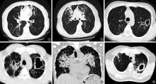
Fang LI,Wei HE,Xin-hua ZHOU,Chun-sheng ZHAO,Yan LYU,Cheng-hai LI,Dong-po. WANG. The similarities and differences of HRCT imaging features between nontuberculous mycobacterial lung diseases and active pulmonary tuberculosis[J]. Chinese Journal of Antituberculosis, 2018, 40(5): 499-505. doi: 10.3969/j.issn.1000-6621.2018.05.012
Add to citation manager EndNote|Ris|BibTeX
URL: http://www.zgflzz.cn/EN/10.3969/j.issn.1000-6621.2018.05.012
| 病变分布规律 | NTM肺病组(74例) | 肺结核组(100例) | χ2值 | P值 | ||
|---|---|---|---|---|---|---|
| 例数 | 构成比(%) | 例数 | 构成比(%) | |||
| 单或双侧分布 | 0.095 | 0.758 | ||||
| 单侧 | 12 | 16.2 | 18 | 18.0 | ||
| 双侧 | 62 | 83.8 | 82 | 82.0 | ||
| 优势部位 | ||||||
| 上叶 | 44 | 59.5 | 82 | 82.0 | 10.817 | 0.001 |
| 中叶和(或)舌叶 | 12 | 16.2 | 5 | 5.0 | 6.069 | 0.014 |
| 下叶 | 15 | 20.2 | 11 | 11.0 | 2.876 | 0.090 |
| 双侧肺门旁 | 3 | 4.1 | 2 | 2.0 | 0.652 | 0.350 |

| 组别 | CT分型 | 合计 | ||||
|---|---|---|---|---|---|---|
| 空洞为主型 | 结节-支气管扩张型 | 结节-肿块型 | 混合型 | 其他a | ||
| NTM肺病组 | 22(29.7) | 38(51.4) | 6(8.1) | 2(2.7) | 6(8.1) | 74(100.0) |
| 肺结核组 | 30(30.0) | 14(14.0) | 21(21.0) | 10(10.0) | 25(25.0) | 100(100.0) |
| 合计 | 52(29.9) | 52(29.9) | 27(15.5) | 12(6.9) | 31(17.8) | 174(100.0) |
| χ2值 | 0.001 | 28.316 | 5.392 | 3.527 | 8.288 | 34.505 |
| P值 | 0.969 | 0.000 | 0.020 | 0.060 | 0.004 | 0.000 |
| HRCT表现 | NTM肺病组(74例) | 肺结核组(100例) | χ2值 | P值 | ||
|---|---|---|---|---|---|---|
| 例数 | 发生率(%) | 例数 | 发生率(%) | |||
| 肺实变 | 50 | 67.6 | 86 | 86.0 | 8.465 | 0.004 |
| 单发或多发结节(直径<3cm) | 61 | 82.4 | 91 | 90.0 | 2.826 | 0.093 |
| 球性病变及团块影(直径>3cm) | 4 | 5.4 | 12 | 12.0 | 2.215 | 0.137 |
| 空洞 | 41 | 55.4 | 68 | 68.0 | 3.252 | 0.071 |
| 上叶空洞 | 36 | 48.6 | 40 | 40.0 | 1.293 | 0.255 |
| 支气管扩张 | 69 | 93.2 | 61 | 61.0 | 23.403 | 0.000 |
| 树芽征 | 47 | 63.5 | 58 | 58.0 | 0.540 | 0.462 |
| 小叶中心结节影 | 59 | 79.7 | 81 | 81.0 | 0.044 | 0.835 |
| 肺索条影 | 57 | 77.0 | 66 | 66.0 | 2.496 | 0.114 |
| 并发叶性肺不张 | 4 | 5.4 | 22 | 22.0 | 9.215 | 0.002 |
| 淋巴结肿大 | 17 | 23.0 | 34 | 34.0 | 2.496 | 0.114 |
| 淋巴结钙化 | 7 | 9.5 | 33 | 33.0 | 13.312 | 0.001 |
| 胸腔积液 | 15 | 20.3 | 34 | 34.0 | 3.963 | 0.047 |
| 胸膜增厚 | 66 | 89.2 | 85 | 85.0 | 0.651 | 0.420 |
| CT表现特点 | NTM肺病组(69例) | 肺结核组(61例) | χ2值 | P值 | ||
|---|---|---|---|---|---|---|
| 例数 | 构成比(%) | 例数 | 构成比(%) | |||
| 分布 | ||||||
| 一叶分布 | 11 | 15.9 | 29 | 47.5 | 15.176 | 0.000 |
| 两叶分布 | 10 | 14.5 | 17 | 27.9 | 3.520 | 0.061 |
| 多叶多段分布 | 48 | 69.6 | 15 | 24.6 | 26.221 | 0.000 |
| 优势部位 | ||||||
| 上叶 | 27 | 39.1 | 43 | 70.5 | 12.813 | 0.000 |
| 舌叶和(或)中叶 | 27 | 39.1 | 8 | 13.1 | 11.138 | 0.001 |
| 下叶 | 15 | 21.7 | 10 | 16.4 | 0.596 | 0.440 |
| 类型 | ||||||
| 柱状扩张 | 30 | 43.5 | 19 | 31.1 | 2.096 | 0.148 |
| 囊状扩张 | 10 | 14.5 | 20 | 32.8 | 6.104 | 0.013 |
| 静脉曲张样扩张 | 5 | 7.2 | 10 | 16.4 | 2.654 | 0.103 |
| 混合型 | 24 | 34.8 | 12 | 19.7 | 3.692 | 0.055 |
| 空洞特点 | NTM肺病组(41例) | 肺结核组(68例) | χ2值 | P值 | ||
|---|---|---|---|---|---|---|
| 例数 | 发生率(%) | 例数 | 发生率(%) | |||
| 薄壁空洞 | 12 | 29.2 | 19 | 27.9 | 0.022 | 0.882 |
| 厚壁空洞 | 27 | 65.8 | 32 | 47.1 | 3.639 | 0.056 |
| 无壁空洞 | 1 | 2.4 | 8 | 11.8 | 2.937 | 0.087 |
| 混合空洞 | 1 | 2.4 | 9 | 13.2 | 2.400 | 0.121 |
| 空洞位于肺野周边邻近胸膜 | 39 | 95.1 | 42 | 61.8 | 14.909 | 0.000 |
| 空洞并见引流支气管 | 37 | 90.2 | 46 | 67.6 | 7.191 | 0.070 |
| 空洞周围可见卫星灶 | 35 | 85.4 | 51 | 75.0 | 1.165 | 1.165 |
| [1] |
Winthrop KL, Mcnelley E, Kendall B , et al. Pulmonary nontuberculous mycobacterial disease prevalence and clinical features: an emerging public health disease. Am J Respir Crit Care Med, 2010,182(7):977-982.
doi: 10.1164/rccm.201003-0503OC URL |
| [2] |
Prevots DR, Shaw PA, Strickland D , et al. Nontuberculous mycobacterial lung disease prevalence at four integrated health care delivery systems. Am J Respir Crit Care Med, 2010,182(7):970-976.
doi: 10.1164/rccm.201002-0310OC URL |
| [3] |
尤正千, 朱晓华 . 肺非结核分支杆菌病的CT影像表现. 中国临床医学影像杂志, 2005,16(3) 141-143.
doi: 10.3969/j.issn.1008-1062.2005.03.007 URL |
| [4] |
马玙 . 关注非结核分枝杆菌肺病的诊断与治疗. 中华结核和呼吸杂志, 2011,34(8):566-568.
doi: 10.3760/cma.j.issn.1001-0939.2011.08.005 URL |
| [5] | 中华医学会结核病学分会. 非结核分支杆菌病诊断与处理指南. 中华结核和呼吸杂志, 2000,23(11):650-653. |
| [6] |
戴洁, 史景云, 梁丽 , 等. 非结核分枝杆菌肺病的CT表现与继发性肺结核CT表现比较. 中国防痨杂志, 2014,36(8):706-709.
doi: 10.3969/j.issn.1000-6621.2014.08.021 URL |
| [7] |
Polverosi R, Guarise A, Balestro E , et al. High-resolution CT of nontuberculous mycobacteria pulmonary infection in immunocompetent, non-HIV-positive patients. Radiol Med, 2010,115(2):191-204.
doi: 10.1007/s11547-009-0479-2 URL |
| [8] |
Kim TS, Koh WJ, Han J , et al. Hypothesis on the evolution of cavitary lesions in nontuberculous mycobacterial pulmonary infection: thin-section CT and histopathologic correlation. AJR Am J Roentgenol, 2005,184(4):1247-1252.
doi: 10.2214/ajr.184.4.01841247 URL |
| [9] |
Aksamit TR, Philley JV, Griffith DE . Nontuberculous mycobacterial (NTM) lung disease: the top ten essentials. Respir Med, 2014,108(3):417-425.
doi: 10.1016/j.rmed.2013.09.014 URL pmid: 24484653 |
| [10] | 孙勤, 沙巍 . 非结核分枝杆菌肺病与肺结核患者的临床特征对比分析. 中国防痨杂志, 2011,33(2):120-122. |
| [11] |
Mitchell JD, Bishop A, Cafaro A , et al. Anatomic lung resection for nontuberculous mycobacterial disease. Ann Thorac Surg, 2008,85(6):1887-1892.
doi: 10.1016/j.athoracsur.2008.02.041 URL pmid: 18498789 |
| [12] |
张贤兰, 梁敏青, 肖芃 . 49 例非结核分枝杆菌肺病临床分析. 中国防痨杂志, 2008,30(3):245-246.
doi: 10.3969/j.issn.1000-6621.2008.03.026 URL |
| [13] | 姚景江, 贺亚琼, 张亚林 . 非结核分枝杆菌肺病的临床与MSCT表现. 中国医学影像技术, 2017,33(3):414-418. |
| [14] | 宁峻岩, 刘新忠, 徐伟 . 非结核分枝杆菌肺病与培阳肺结核的影像学表现对照分析. 临床放射学杂志, 2015,34(9):1406-1409. |
| [15] |
Watanabe H, Uruma T, Seita I , et al. Solitary pulmonary casea-ting granulomas: A 5 year retrospective singlecenter analysis. Mol Clin Oncol, 2017,6(6):839-845.
doi: 10.3892/mco.2017.1244 URL pmid: 5451854 |
| [16] | 余庭山, 沈晓兰, 龙显荣 , 等. 非结核分枝杆菌肺病与耐多药肺结核的CT影像对比分析. 天津医药, 2017,45(6):628-631. |
| [17] | 薛卉, 邢志珩, 秦超 , 等. 非结核分枝杆菌肺病患者的胸部CT影像学特点分析. 中国全科医学, 2016,19(21):2572-2576. |
| [18] | 吴龙章, 蔡杏珊, 关玉华 , 等. 66例非结核分支杆菌肺病的临床分析. 中国防痨杂志, 2003,25(4):257-259. |
| [1] | LIU Xiao-li, LEI Li-mei, GUO Zhou-li, HUANG Yin, XU Jing, ZHAO Xia, WANG Yan, FU Li. Study on the relationship of stigma and social support of tuberculosis patients [J]. Chinese Journal of Antituberculosis, 2020, 42(9): 1002-1008. |
| [2] | Academic Working Committee of Chinese Antituberculosis Association, Editorial Board of Chinese Journal of Antituberculosis . Expert consensus of clinical application of fixed-dose combination formulations [J]. Chinese Journal of Antituberculosis, 2020, 42(9): 885-893. |
| [3] | JIN Hong-jian. The construction of tuberculosis prevention and control service system at county level in China needs to be strengthened urgently —— Comments and suggestions of an old tuberculosis control and prevention worker [J]. Chinese Journal of Antituberculosis, 2020, 42(9): 896-902. |
| [4] | ZHANG Can-you, XIA Hui, CHENG Jun. Testing and reporting requirements for Class Ⅱ biosafety cabinet in tuberculosis laboratory [J]. Chinese Journal of Antituberculosis, 2020, 42(9): 903-909. |
| [5] | ZHOU Lin, LIU Er-yong, MENG Qing-lin, CHEN Ming-ting, ZHOU Xin-hua, GAO Wei-wei, LIN Ming-gui, XIE Ru-ming. Evaluation of the quality of pulmonary tuberculosis diagnosis after the implementation of the newly revised WS 288-2017 Diagnosis for pulmonary tuberculosis standards [J]. Chinese Journal of Antituberculosis, 2020, 42(9): 910-915. |
| [6] | LIU Er-yong, WANG Qian, ZHOU Lin, ZHANG Guo-qin, ZHANG Xiu-lei, MA Yong-cheng, YANG Shu-min, WANG Cui, MENG Qing-lin, CHEN Ming-ting, LIN Ming-gui, TU De-hua.. Analysis of diagnostic quality of pulmonary tuberculosis with negative etiology in some areas of China [J]. Chinese Journal of Antituberculosis, 2020, 42(9): 916-920. |
| [7] | MENG Qing-lin, LI Jin-lan, LIN Ding-wen, MA Yong-cheng, HOU Shuang-yi, LIU Nian-qiang, ZHOU Lin. Analysis of the awareness about knowledge on the updated TB diagnosis standard among the practitioners in TB control institutions [J]. Chinese Journal of Antituberculosis, 2020, 42(9): 921-925. |
| [8] | WANG Qian, ZHOU Lin, LIU Er-yong, ZHAO Yan-lin, LI Tao, CHEN Ming-ting, YANG Li-jia, WANG Jia.. A survey on the diagnostic ability of tuberculosis in the county-level medical institutions in China [J]. Chinese Journal of Antituberculosis, 2020, 42(9): 926-930. |
| [9] | LI Ting, HE Jin-ge, SU Qian, LI Jing, LI Yun-kui, GAO Wen-feng, GAO Yuan, YANG Wen. Value of tuberculin test in screening tuberculosis infection in HIV infected/AIDS patients in Butuo County, Sichuan Province [J]. Chinese Journal of Antituberculosis, 2020, 42(9): 931-936. |
| [10] | LI Yun-kui, HE Jin-ge, SU Qian, LI Ting, LI Jing, GAO Wen-feng, YANG Wen, MAO Guang-yu. Value of tuberculin test in screening tuberculosis infection in HIV infected/AIDS patients in Butuo County, Sichuan Province [J]. Chinese Journal of Antituberculosis, 2020, 42(9): 937-941. |
| [11] | SU Qian, XIA Yong, LU Jia, WANG Dan-xia, HE Jin-ge. Analysis on the epidemiological characteristics of pulmonary tuberculosis among children aged 0-14 in Sichuan Province from 2009 to 2018 [J]. Chinese Journal of Antituberculosis, 2020, 42(9): 942-947. |
| [12] | DENG Ya-li, ZHANG Tian-hua, LIU Wei-ping, ZHANG Hong-wei, MA Yu, LI Peng.. Temporal and spatial clustering analysis of pulmonary tuberculosis incidence in Shaanxi Province from 2014 to 2018 [J]. Chinese Journal of Antituberculosis, 2020, 42(9): 948-955. |
| [13] | DONG Xiao, ZHAO Zhen, LIU Nian-qiang, WANG Sen-lu, CUI Yan. Analysis of the finding characteristics of pulmonary tuberculosis in the elderly population in Xinjiang Uygur Autonomous Region during 2009—2017 [J]. Chinese Journal of Antituberculosis, 2020, 42(9): 956-961. |
| [14] | LIANG Rui-yun, FANG Wei-jun, REN Hui-li, LI Hui-ru, ZHANG Hui. Study on CT manifestations of non-tuberculous mycobacterium pulmonary disease patients with and without diabetes mellitus [J]. Chinese Journal of Antituberculosis, 2020, 42(9): 962-967. |
| [15] | MA Ting-long, HAN Yi, CHENG Xu, LIU Zhi-dong. Clinical observation on treatment effectiveness of transdermal ultrasound-mediated drug delivery combined with oral anti-tuberculosis drug in patients with chest wall tuberculosis [J]. Chinese Journal of Antituberculosis, 2020, 42(9): 968-972. |
| Viewed | ||||||
|
Full text |
|
|||||
|
Abstract |
|
|||||
 京公网安备11010202007215号
Total visitors: Visitors of today: Now online:
京公网安备11010202007215号
Total visitors: Visitors of today: Now online:
 This work is licensed under Creative Commons Attribution 3.0 License.