
Chinese Journal of Antituberculosis ›› 2020, Vol. 42 ›› Issue (8): 832-837.doi: 10.3969/j.issn.1000-6621.2020.08.010
• Original Articles • Previous Articles Next Articles
CHEN Feng-fang*, MA Jun, HUANG Jin, YIN Hong-yun, SHA Wei, YANG Guang-hong, FENG Yong-hong( )
)
Received:2020-03-28
Online:2020-08-10
Published:2020-08-10
Contact:
FENG Yong-hong
E-mail:feng_yonghong@tongji.edu.cn
CHEN Feng-fang, MA Jun, HUANG Jin, YIN Hong-yun, SHA Wei, YANG Guang-hong, FENG Yong-hong. Preliminary study on establishing the diagnostic model for smear negative tuberculosis and stage Ⅰ/Ⅱ sarcoidosis with hilar/mediastinal lymphadenopathy and positive T-SPOT.TB results[J]. Chinese Journal of Antituberculosis, 2020, 42(8): 832-837. doi: 10.3969/j.issn.1000-6621.2020.08.010
Add to citation manager EndNote|Ris|BibTeX
URL: http://www.zgflzz.cn/EN/10.3969/j.issn.1000-6621.2020.08.010
| 观察项目 | 纵隔淋巴结结核组(100例) | 结节病组(50例) | 统计检验值 | P值 |
|---|---|---|---|---|
| 性别 | χ2=0.349 | 0.555 | ||
| 男 | 41(41.00) | 18(36.00) | ||
| 女 | 59(59.00) | 32(64.00) | ||
| 年龄(岁,$\bar{x}$±s) | 54.36±11.15 | 54.56±9.51 | t=0.109 | 0.914 |
| BMI($\bar{x}$±s) | 21.45±2.60 | 24.88±2.82 | t=6.636 | 0.000 |
| 症状与体征 | ||||
| 咳嗽 | 27(27.00) | 32(64.00) | χ2=19.124 | 0.000 |
| 咳痰 | 14(14.00) | 12(24.00) | χ2=2.326 | 0.127 |
| 胸痛 | 5(5.00) | 5(10.00) | χ2=1.339 | 0.247 |
| 咯血 | 2(2.00) | 0(0.00) | χ2=1.014 | 0.314 |
| 发热 | 22(22.00) | 5(10.00) | χ2=3.252 | 0.071 |
| 观察指标 | 纵隔淋巴结结核组(100例) | 结节病组(50例) | 检验值 | P值 |
|---|---|---|---|---|
| WBC[×109/L,M(Q1,Q3)] | 5.77(5.04,7.29) | 5.03(4.20,6.08) | Z=-3.154 | 0.002 |
| Neu[×109/L,M(Q1,Q3)] | 3.56(2.98,4.78) | 3.15(2.48,3.90) | Z=-2.372 | 0.018 |
| Lym[×109/L,M(Q1,Q3)] | 1.56(1.16,1.92) | 1.39(1.03,1.56) | Z=-2.293 | 0.022 |
| Mon[×109/L,M(Q1,Q3)] | 0.47(0.38,0.59) | 0.41(0.31,0.48) | Z=-2.861 | 0.004 |
| NLR[M(Q1,Q3)] | 2.41(1.74,3.45) | 2.42(1.68,3.36) | Z=-0.155 | 0.876 |
| MPV[fl,M(Q1,Q3)] | 10.20(9.60,11.10) | 10.70(9.90,11.50) | Z=-2.235 | 0.025 |
| CRP[mg/L,M(Q1,Q3)] | 5.20(3.15,18.40) | 3.10(2.38,4.10) | Z=-4.040 | 0.000 |
| ESR[mm/1h,M(Q1,Q3)] | 41.00(21.00,69.00) | 15.00(8.00,29.00) | Z=-5.386 | 0.000 |
| SACE[IU/L,M(Q1,Q3)] | 36.50(28.00,46.00) | 54.50(38.00,69.25) | Z=-4.287 | 0.000 |
| 血钙(mmol/L,$\bar{x}$±s) | 2.26±0.12 | 2.29±0.12 | t=1.421 | 0.157 |
| sIL-2R[U/ml,M(Q1,Q3)] | 650.00(494.25,1067.50) | 791.50(546.75,1262.25) | Z=-1.425 | 0.154 |
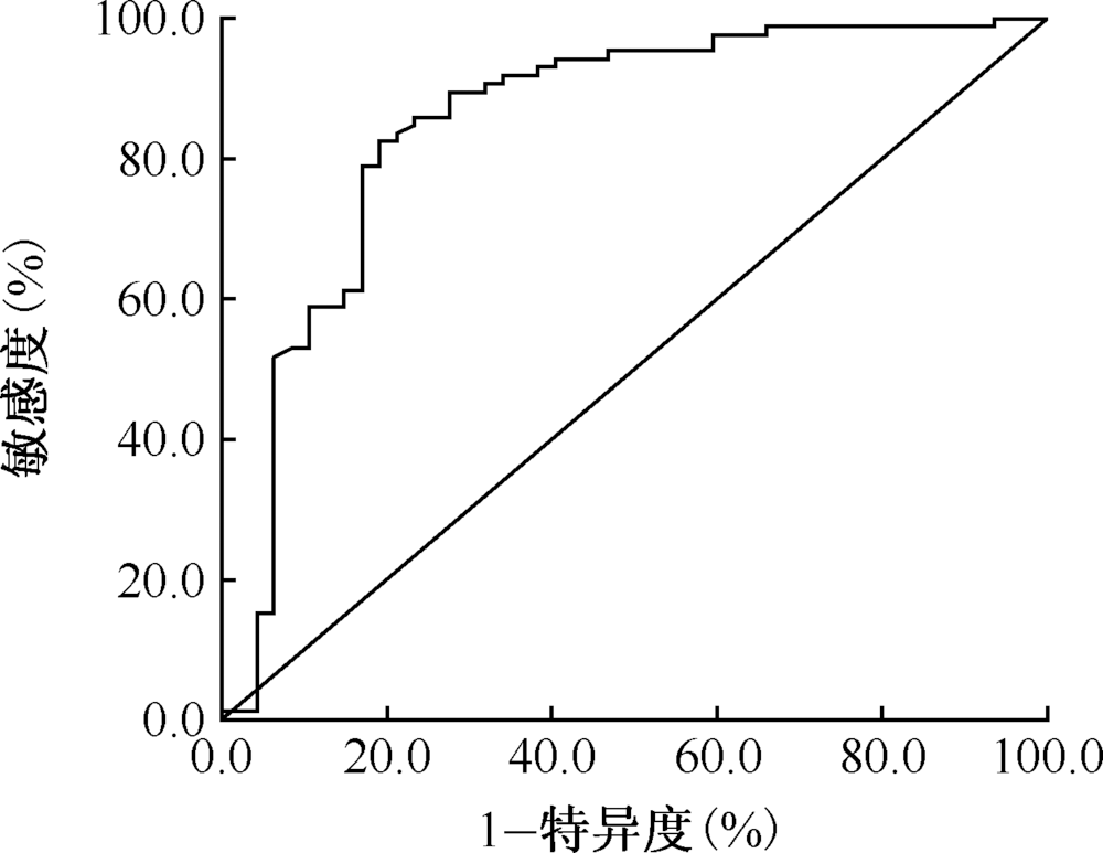
| 实验室指标 | 临界值 | AUC | P值 | 95%CI | 敏感度 (%) | 特异度 (%) | |
|---|---|---|---|---|---|---|---|
| 上限 | 下限 | ||||||
| MPV (fl) | 10.25 | 0.600 | 0.052 | 0.503 | 0.698 | 70.0 | 51.8 |
| Lym (×109/L) | 1.56 | 0.627 | 0.020 | 0.525 | 0.728 | 51.1 | 78.0 |
| Neu (×109/L) | 2.79 | 0.642 | 0.009 | 0.539 | 0.746 | 80.4 | 48.8 |
| Mon (×109/L) | 0.34 | 0.655 | 0.004 | 0.554 | 0.756 | 85.9 | 39.0 |
| WBC (×109/L) | 4.72 | 0.679 | 0.001 | 0.576 | 0.782 | 84.8 | 53.7 |
| CRP (mg/L) | 3.25 | 0.712 | 0.000 | 0.623 | 0.802 | 63.0 | 73.2 |
| SACE (IU/L) | 53.00 | 0.724 | 0.000 | 0.630 | 0.819 | 52.0 | 89.4 |
| ESR (mm/1h) | 31.00 | 0.781 | 0.000 | 0.698 | 0.865 | 67.4 | 82.9 |
| 变量 | β值 | $S_{\bar{x}}$值 | Wald χ2值 | P值 | OR(95%CI)值 |
|---|---|---|---|---|---|
| SACE (IU/L) | -0.033 | 0.014 | 5.826 | 0.016 | 0.968(0.942~0.994) |
| ESR (mm/1h) | 0.036 | 0.015 | 5.583 | 0.018 | 1.036(1.006~1.068) |
| Lym (×109/L) | 1.501 | 0.744 | 4.074 | 0.044 | 4.487(1.044~19.279) |
| 常量 | -0.418 | 3.162 | 0.018 | 0.895 | 0.658 |

| [1] |
Scadding JG. Prognosis of intrathoracic sarcoidosis in England. A review of 136 cases after five years’ observation. Br Med J, 1961,2(5261):1165-1172.doi: 10.1136/bmj.2.5261.1165.
doi: 10.1136/bmj.2.5261.1165 URL pmid: 14497750 |
| [2] | 王昌国, 曾大雄, 蒋军红, 等. 气道内超声特征在纵隔淋巴结结核与结节病鉴别诊断中的作用. 中国内镜杂志, 2017,23(8):1-6.doi: 10.3969/j.issn.1007-1989.2017.08.001. |
| [3] | 尹洪云, Tan W, 马俊, 等. 结核感染T细胞斑点试验在不同患者群中的检测结果分析. 中国防痨杂志, 2018,40(4):358-364.doi: 10.3969/j.issn.1000-6621.2018.04.004. |
| [4] | 阴晴, 刘大东, 邢虎, 等. 应用Logistic回归模型和ROC曲线评价外周血感染性指标在脓毒症中的诊断价值. 中国老年学杂志, 2019,39(12):2955-2957.doi: 10.3969/j.issn.1005-9202.2019.12.042. |
| [5] |
Ying C, Jingchun Y, Hua H, et al. The Value of Contrast-Enhanced Ultrasonography Combined with Real-Time Strain Elastography in the Early Diagnosis of Prostate Cancer. Aging Dis, 2018,9(3):480-488.doi: 10.14336/AD.2017.0704.
doi: 10.14336/AD.2017.0704 URL pmid: 29896435 |
| [6] | 邵建国, 罗蕾蕾, 陈琳, 等. 多项腹水指标 Logistic 回归模型对腹水性质的鉴别诊断价值. 胃肠病学, 2012,17(3):156-160.doi: CNKI:SUN:WIEC.0.2012-03-007. |
| [7] | 王志刚, 陈丁莉, 李守霞, 等. 采用Logistic回归模型联合的肿瘤标志物在胃癌诊断中的价值. 标记免疫分型与临床, 2019,26(11):1898-1902.doi: 10.11748/bjmy.issn.1006-1703.2019.11.024. |
| [8] | 中华医学会呼吸病学分会间质性肺疾病学组, 中国医师协会呼吸医师分会间质性肺疾病工作委员会. 中国肺结节病诊断和治疗专家共识. 中华结核和呼吸杂志, 2019,42(9):685-693.doi: 10.3760/cma.j.issn.1001-0939.2019.09.007. |
| [9] | 刘淼淼. 中性粒细胞与淋巴细胞比值在肺结节病与肺结核鉴别诊断中的应用. 郑州:郑州大学, 2018.doi: CNKI:CDMD:2.1018.105818. |
| [10] | 杨景茹. NLR和TB-IGRA在肺结节病与肺结核病鉴别诊断中的应用价值. 济南:山东大学, 2015.doi: 10.7666/d.Y2793447. |
| [11] |
Dastoori M, Fedele S, Leao JC, et al. Sarcoidosis-a clinically orientated review. J Oral Pathol Med, 2013,42(4):281-289.doi: 10.1111/j.1600-0714.2012.01198.x.
doi: 10.1111/j.1600-0714.2012.01198.x URL pmid: 22845844 |
| [12] |
翁跃颂, 王华英, 吕定丰, 等. 结节病患者辅助性T细胞17和调节性T细胞与疾病活动的关系. 浙江大学学报(医学版), 2015,44(2):154-161.doi: 10.3785/j.issn.1008-9292.2015.03.006.
doi: 10.3785/j.issn.1008-9292.2015.03.006 URL |
| [13] |
Hyldgaard C, Kaae S, Riddervold M, et al. Value of s-ACE, BAL lymphocytosis, and CD4+/CD8+ and CD103+CD4+/CD4+ T-cell ratios in diagnosis of sarcoidosis. Eur Respir J, 2012,39(4):1037-1039.doi: 10.1183/09031936.00144311.
doi: 10.1183/09031936.00144311 URL pmid: 22467726 |
| [14] |
Ungprasert P, Carmona EM, Crowson CS, et al. Diagnostic Utility of Angiotensin-Convert ing Enzyme in Sarcoidosis: A Population-Based Study. Lung, 2016,194(1):91-95.doi: 0.1 007/s00408-015-9826-3.
URL pmid: 26563332 |
| [15] |
Kargaltseva NM, Kotcherovets VI, Mironov AY, et al. Inflammation markers and bloodstream infection (review of literature). Klin Lab Diagn, 2019,64(7):435-442.doi: 10.18821/0869-2084-2019-64-7-435-442.
doi: 10.18821/0869-2084-2019-64-7-435-442 URL pmid: 31408597 |
| [16] |
Enver Yalnız, Gülistan Karadeniz, Fatma Demirci Üçsular, et al. Predictive value of platelet-to-lymphocyte ratio in patients with sarcoidosis. Biomark Med, 2019,13(3):197-204.doi: 10.2217/bmm-2018-0252.
doi: 10.2217/bmm-2018-0252 URL pmid: 30604642 |
| [17] |
Groen-Hakan F, Eurelings L, Rothova A, et al. Lymphopaenia as a predictor of sarcoidosis in patients with a first episode of uveitis. Br J Ophthalmol, 2019,103(9):1296-1300.doi: 10.1136/bjophthalmol-2018-313212.
doi: 10.1136/bjophthalmol-2018-313212 URL pmid: 30442816 |
| [18] |
Jones NP, Tsierkezou L, Patton N. Lymphopenia as a predictor of sarcoidosis in patients with uveitis. Br J Ophthalmol, 2016,100(10):1393-1396.doi: 10.1136/bjophthalmol-2015-307455.
doi: 10.1136/bjophthalmol-2015-307455 URL pmid: 26729767 |
| [19] |
Sweiss NJ, Rafah S, Seema G, et al. Significant CD4, CD8, and CD19 lymphopenia in peripheral blood of sarcoidosis patients correlates with severe disease manifestations. PLoS One, 2010,5(2):e9088.doi: 10.1371/journal.pone.0009088.
doi: 10.1371/journal.pone.0009088 URL pmid: 20140091 |
| [20] |
Du SS, Zhao MM, Zhan Y, et al. Screening for Differentially Expressed Proteins Relevant to the Differential Diagnosis of Sarcoidosis and Tuberculosis. PLoS One, 2015,10(9):e0132466.doi: 10.1371/journal.pone.0132466.
doi: 10.1371/journal.pone.0132466 URL pmid: 26368286 |
| [1] | DUAN Li-ming, DING Chao, LIU Yu-gang, WEI Lin, GU Zhen-ning. Application value of video-assisted thoracoscopic surgery in the treatment of chronic tuberculous empyema [J]. Chinese Journal of Antituberculosis, 2020, 42(8): 850-853. |
| [2] | CHEN Zhen-hua,LIU Bin-bin,CHEN Zhong-nan,TAN Yun-hong. Establishment and preliminary evaluation of a diagnostic model for the new patients with pathogen-negative pulmonary tuberculosis [J]. Chinese Journal of Antituberculosis, 2020, 42(3): 266-271. |
| [3] | MA Gai-xia,TIAN Li-ming,WANG Yu,QIU Lei,XUE Ling-na,LU Zhen-hui,ZHANG Hui-yong,GUO Xiao-yan. Systematic evaluation and meta-analysis of integrated traditional Chinese and western medicine in the treatment of MDR-PTB [J]. Chinese Journal of Antituberculosis, 2020, 42(2): 95-100. |
| [4] | GU Ji-xiu, LI Qing, MA ling, LI Yin-hua, WANG Dong-dong, SI Hong-yan. Application of next-generation sequencing technology in the diagnosis of drug-resistant tuberculosis [J]. Chinese Journal of Antituberculosis, 2020, 42(11): 1203-1208. |
| [5] | WANG Ze-ming, SUN Lin, SHEN A-dong. Current status and thinking of clinical practice guidelines of tuberculosis in children [J]. Chinese Journal of Antituberculosis, 2020, 42(10): 1025-1028. |
| [6] | BAI Yun-song, ZHANG Xue-jun, CAO Jun, FENG Lei, QI Xin-yu, GUO Dong, YAO Zi-ming, LI Cheng-xin. Analysis of clinical efficacy of stageⅠanterior-posterior approach surgery for lumbar tuberculosis in children [J]. Chinese Journal of Antituberculosis, 2020, 42(10): 1048-1052. |
| [7] | MAO Yi, ZENG Yi-lan, WU Gui-hui, JIANG Zhi-hao, FAN Lin. Correlation analysis between clinical characteristics and flow cytometry cells subsets in peripheral blood of patients with pulmonary tuberculosis [J]. Chinese Journal of Antituberculosis, 2020, 42(10): 1080-1086. |
| [8] | NING Feng-gang,ZHOU Xin-hua,HOU Dai-lun,LYU Ping-xin,LYU Yan,HE Wei. MR imaging features of hematogeneous pulmonary tuberculosis accompanied with intracranial tuberculosis in adult patients [J]. Chinese Journal of Antituberculosis, 2020, 42(1): 19-25. |
| [9] | REN Hang-kong,DUAN Li-ming,ZHEN Qin-fang. The value of two techniques in detecting chest wall tuberculosis from different surgical specimens [J]. Chinese Journal of Antituberculosis, 2020, 42(1): 74-78. |
| [10] | Hong-wei ZHANG,Jing XU,Yu MA,Ya-li DENG. Evaluating the implementation effect of tuberculosis patients health management services for Basic Public Health Projects in Shaanxi Province [J]. Chinese Journal of Antituberculosis, 2019, 41(9): 951-956. |
| [11] | Xi CHEN,Zhong-quan LIU,Bin WANG,Hui ZHU,Lei FU,Yuan-yuan LI,Yu LU. Evaluation of antituberculosis activities of 14 antituberculosis drugs in macrophage [J]. Chinese Journal of Antituberculosis, 2019, 41(9): 993-998. |
| [12] | Kai-wen WU,Ke BI,Yi ZHANG,Meng-jun SHEN,Yang CONG,Yin WANG. The value of conventional ultrasound combined with contrast-enhanced ultrasonography in the differential diagnosis of pleural-based pulmonary tuberculosis and bacterial pneumonia [J]. Chinese Journal of Antituberculosis, 2019, 41(8): 822-827. |
| [13] | Yi ZHANG,Ke BI,Hui-ming ZHU,Yang CONG,Meng-jun SHEN,Hong-wei CHEN,Yin WANG. Diagnostic value of contrast-enhanced ultrasound-guided percutaneous lung puncture biopsy in culture negative presumptive tuberculosis patients [J]. Chinese Journal of Antituberculosis, 2019, 41(8): 828-832. |
| [14] | Xiao-wei DAI,Shuang-shuang CHEN,Xin-yu YANG,Yan-feng ZHAO,Li-li TIAN,Jun-li YI,Hao CHEN,Bei-chuan DING. Evaluation of clinical diagnostic efficacy of four detection methods for Mycobacterium tuberculosis [J]. Chinese Journal of Antituberculosis, 2019, 41(8): 876-881. |
| [15] | Hai-ming YE,Wen-si CHEN,Ju-fang ZHANG,Yu-fang LIAO,Jie XU,Jie CHEN,Sheng-nan ZHANG. Analysis on the effect of pilot work of grading diagnosis and treatment and comprehensive prevention service model of tuberculosis in Futian District, Shenzhen [J]. Chinese Journal of Antituberculosis, 2019, 41(8): 900-904. |
| Viewed | ||||||
|
Full text |
|
|||||
|
Abstract |
|
|||||
 京公网安备11010202007215号
Total visitors: Visitors of today: Now online:
京公网安备11010202007215号
Total visitors: Visitors of today: Now online:
 This work is licensed under Creative Commons Attribution 3.0 License.