结核性胸膜炎超声诊断、分型及介入治疗专家共识(2022年版)
Expert consensus on ultrasound diagnosis, classification and interventional therapy of tuberculous pleurisy (2022 Edition)
结核性胸膜炎超声诊断、分型及介入治疗专家共识(2022年版) |
| 中华医学会结核病学分会超声专业委员会, 中国医师协会介入医师分会超声介入专业委员会单位 |
|
Expert consensus on ultrasound diagnosis, classification and interventional therapy of tuberculous pleurisy (2022 Edition) |
| Ultrasound Professional Committee of Tuberculosis Branch of Chinese Medical Association, Interventional Ultrasound Professional Committee of Interventional Physician Branch of Chinese Medical Doctor Association Danwei |
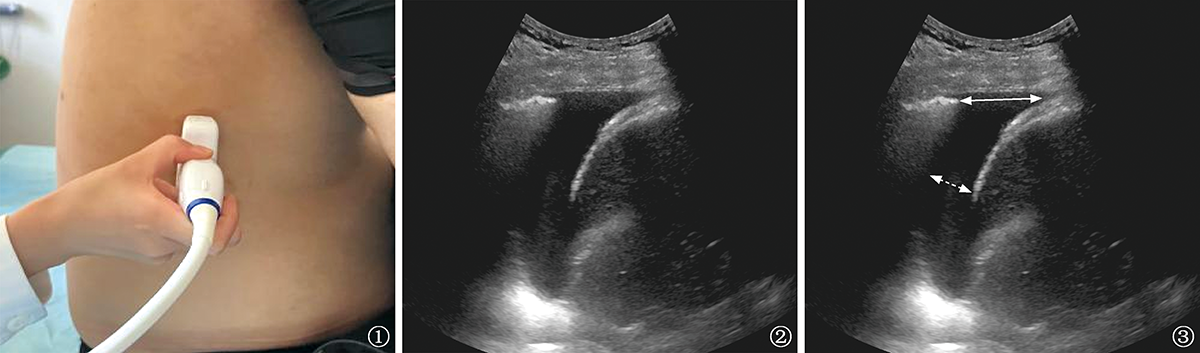
| 图1~3 患者,男性, 38岁,临床诊断胸膜炎,采用坐位进行胸腔积液超声评估。 |
|
|