Email Alert | RSS    帮助

中国防痨杂志 ›› 2022, Vol. 44 ›› Issue (3): 289-293.doi: 10.19982/j.issn.1000-6621.20210561

• 临床病例讨论 • 上一篇    下一篇

分子生物学假阳性致肺癌性淋巴管炎误诊为肺结核的临床诊治过程分析

杨澄清1, 梅春林1, 杜荣辉1(), 雷美2, 秦立新3   

  1. 1武汉市肺科医院呼吸与危重症医学科,武汉 430030
    2武汉市肺科医院病理科,武汉 430030
    3武汉市肺科医院影像科,武汉 430030
  • 收稿日期:2021-09-18 出版日期:2022-03-10 发布日期:2022-03-08
  • 通信作者: 杜荣辉 E-mail:bluesearh006@sina.com
  • 基金资助:
    湖北省卫生健康科研基金(WJ2019H320);武汉市医学科研项目(WX19A05);武汉医学中青年骨干人才培养工程(201987)

Analysis of diagnosis and treatment for a pulmonary lymphangitic carcinomatosis misdiagnosed as pulmonary tuberculosis caused by false positive molecular biological test

YANG Cheng-qing1, MEI Chun-lin1, DU Rong-hui1(), LEI Mei2, QIN Li-xin3   

  1. 1Department of Pulmonary and Critical Care Mdicine,Wuhan Pulmonary Hospital,Wuhan 430030,China
    2Department of Pathology,Wuhan Pulmonary Hospital,Wuhan 430030,China
    3Department of Radiology,Wuhan Pulmonary Hospital,Wuhan 430030,China
  • Received:2021-09-18 Online:2022-03-10 Published:2022-03-08
  • Contact: DU Rong-hui E-mail:bluesearh006@sina.com
  • Supported by:
    Health Commission of Hubei Province Scientific Research Project(WJ2019H320);Wuhan Medical Research Project(WX19A05);Wuhan Municipal Health Commission Medical Talent Training Project(201987)

摘要:

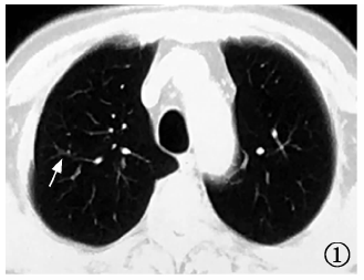
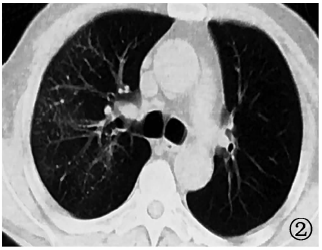
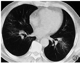
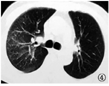
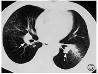
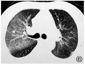
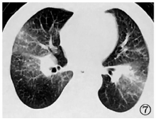
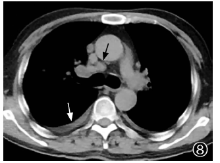
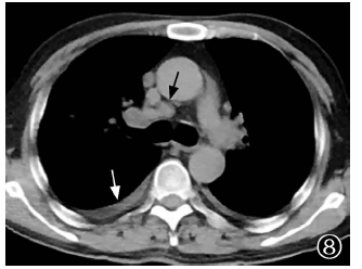
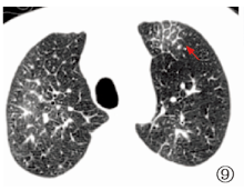
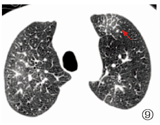
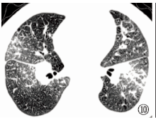
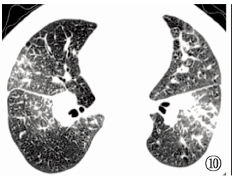
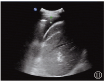
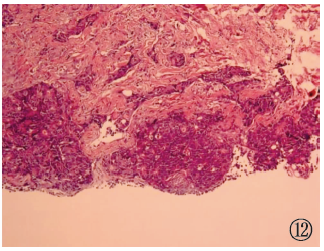
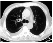
2019年4月17日,武汉市肺科医院呼吸科收治1例69岁男性因外院结核分子生物学阳性诊断为肺结核的肺癌性淋巴管炎患者。该患者因“发现肺部阴影3个月,间断咳嗽2个月”入院。既往有结肠癌病史。胸部CT扫描显示双肺弥漫性沿支气管血管束分布结节伴磨玻璃影,伴小叶间隔增厚,双肺门及纵隔淋巴结肿大,双侧胸腔积液,在外院行肺泡灌洗液(BALF)GeneXpert MTB/RIF(简称“ GeneXpert”)检出MTB(极低)及TB-PCR检出MTB,诊断为肺结核,转诊至武汉市肺科医院。入院后完善结核相关检查,PPD皮肤试验阴性,γ干扰素释放试验阴性,胸部CT影像学特征不符合肺结核改变,临床怀疑BALF结核病病原分子生物学检测假阳性,建议患者复查BALF、GeneXpert及经气管镜超声引导针吸活检术(EBUS-TBNA),患者拒绝。给予诊断性胸腔穿刺,抽出胸腔积液20ml,送检显示,癌胚抗原(118.4μg/L)明显升高,提示恶性胸腔积液;最后行内科胸腔镜胸膜活检,提示转移性低分化腺癌,结合胸部CT表现,诊断为肺癌性淋巴管炎。患者后因病情恶化死亡。笔者认为,影像学表现为沿支气管血管束分布结节伴小叶间隔增厚及纵隔淋巴结肿大时需鉴别肺癌性淋巴管炎。影像学表现与肺结核不相符时,分子生物学阳性诊断肺结核需谨慎,以避免误诊误治。

关键词: 结核, 癌性淋巴管炎, 分子生物学, 诊断

Abstract:

On April 17, 2019, a 69-year-old male patient with pulmonary lymphangitic carcinomatosis was admitted to the Respiratory Department of Wuhan Pulmonary Hospital,and diagnosed as pulmonary tuberculosis due to the positive tuberculosis molecular biological result in other hospital. The patient was hospitalized because of “pulmonary shadow for 3 months and intermittent cough for 2 months”. He had previously been diagnosed with colon cancer. Chest CT scan revealed diffusing nodules along bronchovascular bundles with ground glass shadows, interlobular septal thickening, bilateral hilar and mediastinal lymph node enlargement and bilateral pleural effusion. In bronchoalveolar lavage fluid (BALF), MTB (extremely low) was detected by GeneXpert MTB/RIF (referred to as “GeneXpert”) and MTB was detected by TB-PCR in previous hospital. Therefore, he was diagnosed as tuberculosis, then transferred to Wuhan Pulmonary Hospital. After admission, relevant examinations were completed. PPD skin test and interferon gamma release test was negative, and chest CT imaging features were not consistent with pulmonary tuberculosis. We suspected it was a false positive molecular biological result. Therefore, GeneXpert and tracheal endoscopic ultrasound-guided needle aspiration biopsy (EBUS-TBNA) were recommended. However, the patient refused. The doctor performed a pleural puncture for diagnosis, and 20 ml pleural effusion was extracted. The examination showed that the carcinoembryonic antigen (118.4 μg/L) was significantly elevated, indicating malignant pleural effusion. Finally, medical thoracoscopic pleural biopsy revealed metastatic poorly differentiated adenocarcinoma. Combined with chest CT findings, the patient was diagnosed as pulmonary lymphangitic carcinomatosis who later died due to deterioration of his condition. Therefore, the authors suggest that radiographic findings of nodules along the bronchovascular bundle with thickening of interlobular septa and mediastinal lymph node enlargement should be considered as pulmonary lymphangitic carcinomatosis for differential diagnosis. When imaging findings are not consistent with tuberculosis and molecular biological test is positive, we should be cautious about diagnosing tuberculosis to avoid misdiagnosis and mistreatment.

Key words: Tuberculosis, Pulmonary lymphangitic carcinomatosis, Molecular biology, Diagnosis

中图分类号: