Email Alert | RSS    帮助

中国防痨杂志 ›› 2020, Vol. 42 ›› Issue (11): 1153-1157.doi: 10.3969/j.issn.1000-6621.2020.11.003

• 论著 • 上一篇    下一篇

胸膜结核瘤的MRI表现特征分析

尹曲华, 蒋智善, 聂赣娟, 姚其能()   

  1. 410013 长沙,湖南省胸科医院放射科(尹曲华、蒋智善、姚其能),病理科(聂赣娟)
  • 收稿日期:2020-04-16 出版日期:2020-11-10 发布日期:2020-11-13
  • 通信作者: 姚其能 E-mail:1409088478@qq.com

Analysis of MRI characteristics of pleural tuberculoma

YIN Qu-hua, JIANG Zhi-shan, NIE Gan-juan, YAO Qi-neng()   

  1. Department of Radiology, Hunan Chest Hospital, Changsha 410013,China
  • Received:2020-04-16 Online:2020-11-10 Published:2020-11-13
  • Contact: YAO Qi-neng E-mail:1409088478@qq.com

摘要:

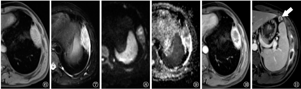
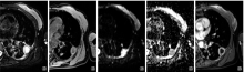
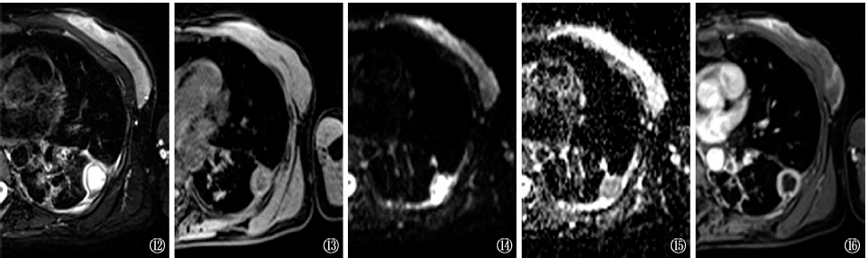
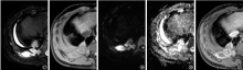
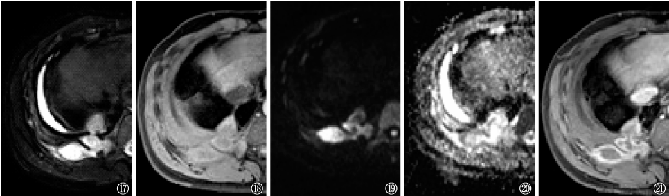
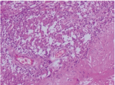
目的 探讨胸膜结核瘤的MRI表现特征,以提高其诊断水平。方法 收集湖南省胸科医院2018年1月至2019年12月经病理学和(或)病原学证实或临床诊断(依据临床症状体征、免疫学检查结果,以及诊断性抗结核药品治疗有效进行综合诊断)的87例胸膜结核瘤患者。将其中资料完整的60例患者作为研究对象,男41例,女19例;年龄13~78岁,中位年龄27岁。其中8例胸膜病变经手术后病理检查确诊,43例经胸膜活检病理检查确诊,9例为临床诊断患者。所有患者均进行了结核病相关实验室检查、胸部CT平扫、MRI平扫及增强扫描检查,分析评价患者的临床及胸部MRI表现特征。结果 60例患者MRI表现为单发病灶47例(78.3%),多发病灶13例(21.7%)。共74个病灶,其中右下肺37个(50.0%)病灶,34个病灶(45.9%)呈类圆形;51个病灶(68.9%)与胸膜呈宽基底相贴,边缘光滑,病灶基底部胸膜有移行性增厚。60例患者中,13例(21.7%)为未成熟结核瘤,T1WI呈等或稍低信号,T2WI、表现弥散系数(ADC)图像呈稍高信号,弥散加权成像(DWI)呈等信号,弥散不受限;增强检查呈斑点状强化或结节状均匀强化。29例(48.3%)为中心凝固性坏死结核瘤,T1WI呈等或稍低信号,T2WI、ADC 图像呈混杂高信号,DWI呈等信号,弥散不受限,增强检查呈不均匀结节状强化或环形强化;18例(30.0%)为中心液性坏死结核瘤,T1WI呈低信号,T2WI、ADC图像呈高信号, DWI呈混杂高信号,弥散受限,增强检查呈环形强化。2例(3.3%)可见多个结核瘤灶融合,形成脓肿,破溃至胸膜外脂肪间隙和(或)胸壁,ADC图像呈低信号,DWI呈高信号,弥散受限,增强检查呈环形和分隔样强化。结论 胸膜结核瘤具有一定的MRI表现特征,MRI在判断胸膜结核瘤累及范围及其所处病理阶段有优势。

关键词: 结核瘤,结核, 胸膜, 磁共振成像, 疾病特征

Abstract:

Objective To explore the characteristics of MRI imaging of pleural tuberculoma, so as to improve the imaging diagnosis of this disease. Methods Clinical and imaging data of 87 patients diagnosed of pleural tuberculoma by pathology and/or bacteriology or clinically confirmed (based on clinical manifestations, immunological tests, response to diagnostic anti-TB treatment) in Hunan Chest Hospital from January 2018 to December 2019 were retrospectively analyzed. Among them, 60 patients (41 males, 19 females, 13-78 years old, median age 27 years) with complete information were selected, which included 8 cases confirmed by surgical pathology and 43 cases by percutaneous pleural puncture, 9 cases according to clinical diagnosis standard. They were all tested with tuberculosis related laboratory examinations, as well as CT scan, MRI scan and enhanced MRI examination. Results Among 60 patients, there were 47 (78.3%) with single lesions and 13 (21.7%) with multiple lesions; 37 lesions (37/74, 50.0%) occupied the lower right lobes. Rounded shadows were the most common signs, which were present in 34 lesions (45.9%).Most of lesions (68.9%) were close to the pleura with a wide base and smooth edges. Thirteen cases (21.7%) were immature tuberculomas, showed iso-intensity or slight hypo-intensity signal on T1WI, DWI; slight hyper-intensity signal on T2WI,ADC; nodular enhancement after contrast injection. Twenty-nine cases (48.3%) were mature tuberculomas with caseous necrosis, showed iso-intensity or slight hypo-intensity signal on T1WI, DWI and variegated hyper-intensity signal on T2WI,ADC,and nodular or annular enhancement. Eighteen cases (30.0%) were mature tuberculomas with liquefactive necrosis, showed hypo-intensity signal on T1WI and high hyper-intensity signal on T2WI,DWI,ADC,and annular enhancement after contrast injection. Multiple tuberculomas fused to form an abscess that ruptured into the extra-pleural fat gap and/or the chest wall in 2 cases (3.3%), showed high hyper-intensity signal on DWI and hypo-intensity signal ADC, and variegated-annular enhancement after contrast injection. Conclusion Pleural tuberculosis has certain characteristics of MRI which have advantages in determining the scope of pleural tuberculosis and its pathological stage.

Key words: Tubereuloma, Tuberculosis, pleural, Magnetic resonance imaging, Disease attributes