Email Alert | RSS    帮助

中国防痨杂志 ›› 2020, Vol. 42 ›› Issue (3): 245-248.doi: 10.3969/j.issn.1000-6621.2020.03.012

• 论著 • 上一篇    下一篇

颈部淋巴结结核的二维超声声像图表现及病理对照分析

姜爽爽,郑海伦,曹丽娅,罗萍()   

  1. 100035 北京结核病控制研究所门诊部
  • 收稿日期:2019-10-29 出版日期:2020-03-10 发布日期:2020-03-18
  • 通信作者: 罗萍 E-mail:pluo12@126.com

Comparative analysis of two-dimensional ultrasonography and pathology in cervical lymph node tuberculosis

JIANG Shuang-shuang,ZHENG Hai-lun,CAO Li-ya,LUO Ping()   

  1. Department of Outpatient, Beijing Research Institute for Tuberculosis Control, Beijing 100035, China
  • Received:2019-10-29 Online:2020-03-10 Published:2020-03-18
  • Contact: Ping LUO E-mail:pluo12@126.com

摘要:

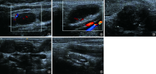
目的 分析颈部淋巴结结核的二维超声声像图表现,并与病理结果对照。方法 搜集2015年9月至2019年8月就诊于北京结核病控制研究所的淋巴结结核患者63例,其中,男16例(25.4%),女47例(74.6%);年龄18~70岁,中位年龄(四分位数)为29(25,43)岁;共累及101枚淋巴结。回顾性对照分析研究对象颈部淋巴结结核超声声像图表现与病理结果。结果 63例颈部淋巴结结核患者双侧受累者27例(42.9%),左侧受累者14例(22.2%),右侧受累者22例(34.9%)。根据国际通用7分区法,101枚病变淋巴结中位于颌下及颏下(Ⅰ区)3枚(2.9%),位于胸锁乳突肌周围(Ⅱ、Ⅲ、Ⅳ区)84枚(83.2%),位于锁骨上区及颈后三角区(Ⅴ区)14枚(13.9%),位于颈前区(Ⅵ区)0枚,位于上纵隔(Ⅶ区)0枚;病变淋巴结多呈椭圆形和类圆形,大部分为散在分布,少部分相互融合。病变淋巴结按超声声像图表现可分为5种类型:实质炎症型18枚(17.8%)、干酪坏死型39枚(38.6%)、周围炎症型19枚(18.8%)、脓肿窦道型6枚(6.0%)、愈合钙化型19枚(18.8%)。结论 颈部淋巴结结核的二维超声声像图表现与其病理变化密切相关,超声声像图可直观反映病变的各个阶段,可作为该病的首选影像学检查方法。

关键词: 结核, 淋巴结, 超声检查, 疾病特征, 病理状态, 体征和症状

Abstract:

Objective This study aimed to analyze the two-dimensional ultrasonographic manifestations of cervical lymph node tuberculosis and then compare with pathological results. Methods A total of 101 lymph nodes in 63 patients with cervical lymph node tuberculosis who were admitted to Beijing Research Institute for Tuberculosis Control from September 2015 to August 2019 were collected, including 16 males (25.4%) and 47 females (74.6%); with age from 18 to 70 years, and the median age (quartile) was 29 (25, 43) years. The ultrasonographic manifestations and pathological results of patients with cervical lymph node tuberculosis were retrospectively analyzed. Results In 63 patients, 27 cases (42.9%) involved bilateral neck, 14 cases (22.2%) involved left, and 22 cases (34.9%) involved right. According to the international general 7 partition method, among these 101 involved lymph nodes, 3 (2.9%) lymph nodes were located in the submandibular and submental region (level Ⅰ), 84 (83.2%) lymph nodes were located around the sternocleidomastoid muscles (level Ⅱ, Ⅲ,Ⅳ), 14 (13.9%) lymph nodes were located in the supraclavicular region and posterior triangle (level Ⅴ), and none was located in the anterior region of neck (level Ⅵ) and the high mediastinum region (level Ⅶ). The involved lymph nodes were mostly elliptic and quasi-circular, and most of them were scattered, while a few of them were fused with each other. According to the ultrasonographic manifestations, 101 involved lymph nodes could be divided into five types, including 18 (17.8%) of solid inflammatory type, 39 (38.6%) of caseous necrosis type, 19 (18.8%) of peripheral inflammatory type, 6 (6.0%) of abscess sinus type, and 19 (18.8%) of healing calcification type. Conclusion The two-dimensional ultrasonographic manifestations of cervical lymph node tuberculosis are closely related to its pathological changes The ultrasound sonogram can intuitively reflect the various stages of the lesion, and can be used as the preferred imaging method for the disease.

Key words: Tuberculosis, lymph node, Ultrasonography, Disease attributes, Pathological conditions, signs and symptoms