Email Alert | RSS    帮助

中国防痨杂志 ›› 2020, Vol. 42 ›› Issue (2): 101-107.doi: 10.3969/j.issn.1000-6621.2020.02.005

• 论著 • 上一篇    下一篇

杨梅素通过磷脂酰肌醇3-激酶/蛋白激酶B/哺乳动物雷帕霉素靶蛋白信号通路诱导MTB感染巨噬细胞发生自噬的研究

孙锦霞,张晴雯,李银虹,姜昕()   

  1. 201203上海中医药大学基础医学院免疫学与病原生物学教研室
  • 收稿日期:2019-11-13 出版日期:2020-02-10 发布日期:2020-02-19
  • 通信作者: 姜昕 E-mail:jiangxingao@163.com
  • 基金资助:
    国家自然科学基金(81873069);上海市自然科学基金(18ZR1439700);上海中医药大学预算内项目(2015YSN03);“十三五”国家科技重大专项(2018ZX10725-509)

Study of autophagy induced by myricetin in MTB infected macrophages through PI3K/Akt/mTOR signaling pathway

SUN Jin-xia,ZHANG Qing-wen,LI Yin-hong,JIANG Xin()   

  1. Department of Immunology and Pathology, School of Basic Medicine, Shanghai University of Traditional Chinese Medicine, Shanghai 201203, China
  • Received:2019-11-13 Online:2020-02-10 Published:2020-02-19
  • Contact: Xin JIANG E-mail:jiangxingao@163.com

摘要:

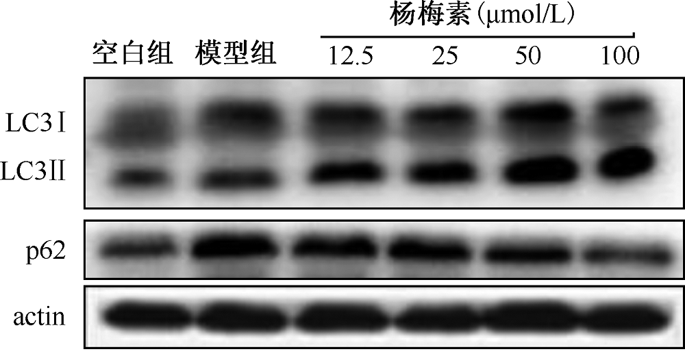
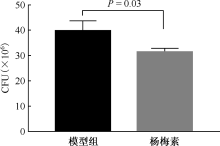
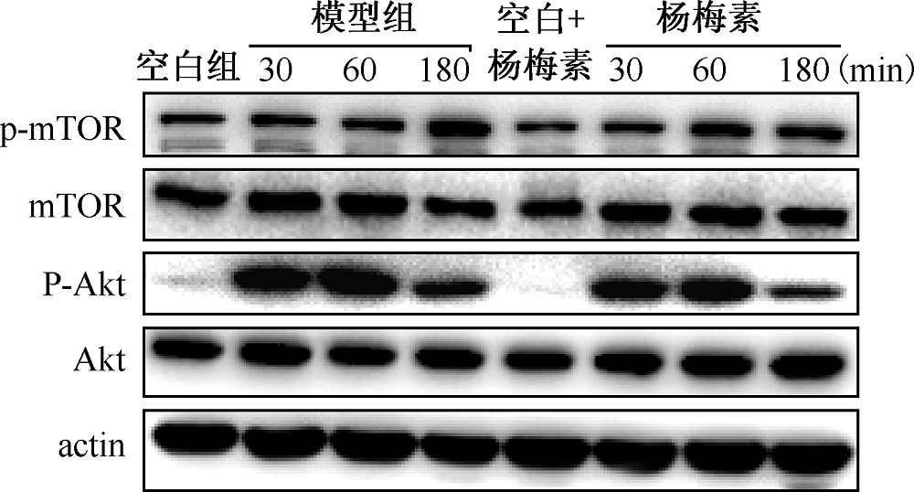
目的 对杨梅素通过磷脂酰肌醇3-激酶/蛋白激酶B/哺乳动物雷帕霉素靶蛋白(PI3K/Akt/mTOR)信号通路诱导MTB感染的巨噬细胞发生自噬进行研究,从而探讨杨梅素抗结核作用的机理。方法 用CCK8法检测杨梅素对细胞增殖的影响,确定安全的用药范围;以H37Ra菌株感染的小鼠巨噬细胞Raw 264.7为模型组,并设空白组和药物处理组。按感染复数(MOI,即细菌∶细胞=10∶1)加入模型组、药物处理组,共孵育4h后,磷酸盐缓冲液(PBS)洗3次以弃掉未进入胞内的MTB。药物处理组分别用不同浓度(12.5、25、50、100μmol/L)的杨梅素作用24h, Western blot法检测自噬相关蛋白即“微管相关蛋白1轻链3-Ⅱ(LC3-Ⅱ)和p62”表达水平的变化,并以此筛选出杨梅素促进自噬的最佳作用浓度;100μmol/L杨梅素作用于感染细胞72h后,0.1%聚乙二醇辛基苯基醚(Triton X-100)冰上裂解细胞10min,菌落形成单位(CFU)法检测巨噬细胞胞内荷菌量;杨梅素作用感染细胞不同时间(30、60、180min)后Western blot测定PI3K/Akt/mTOR信号通路中Akt、mTOR的磷酸化水平。以Image J软件做蛋白定量分析,用GraphPad Prism 7.0制图,采用单因素方差分析(ANOVA)进行统计分析,以P<0.05为差异有统计学意义。结果 杨梅素在100μmol/L浓度以下细胞生存率在90%左右,对细胞毒性较小;Western blot结果显示:与模型组(0.52±0.01)相比,杨梅素不同浓度(12.5、25、50、100μmol/L)处理均能促进LC3Ⅱ的表达(0.59±0.02、 0.65±0.01、 0.71±0.01、 0.83±0.01),差异有统计学意义(t=2.97、P=0.04,t=7.91、P=0.00,t=9.77、P=0.00,t=16.37、P=0.00);而较模型组p62蛋白(0.86±0.02),药物处理亦能抑制p62的表达(0.72±0.01、0.86±0.00、0.60±0.02、0.58±0.01),25μmol/L杨梅素处理组差异无统计学意义(t=0.81、P=0.46),12.5、50、100μmol/L处理组差异均有统计学意义(t=6.50、P=0.00,t=9.53、P=0.00,t=12.01、P=0.00);杨梅素促进自噬的最佳药物浓度为100μmol/L;100μmol/L杨梅素作用于感染细胞72h后,对胞内MTB的抑制率为21.02%;模型组细胞在MTB感染后30、60、180min时,PI3K/Akt/mTOR通路中Akt蛋白的磷酸化(p-Akt)水平(1.23±0.01、1.52±0.01、0.74±0.02)明显增加,而杨梅素作用相同的时间后,可明显抑制Akt蛋白的磷酸化(0.99±0.01、0.96±0.01、0.43±0.01),差异有统计学意义(t=27.60、P=0.00,t=30.06、P=0.00,t=18.60、P=0.00);而模型组磷酸化mTOR(p-mTOR)蛋白水平仅在MTB感染后180min(0.57±0.00)明显增加(t=94.61、P=0.00),杨梅素作用180min亦能抑制mTOR蛋白的磷酸化(0.46±0.01),差异有统计学意义(t=21.60、P=0.00)。结论 杨梅素通过抑制Akt和mTOR蛋白的磷酸化来抑制PI3K/Akt/mTOR通路,从而诱导MTB感染的巨噬细胞发生自噬来杀灭胞内的MTB。

关键词: 分枝杆菌,结核, 巨噬细胞, 自噬, 磷脂酰肌醇3-激酶/蛋白激酶B/哺乳动物雷帕霉素靶蛋白, 杨梅素

Abstract:

Objective To explore the anti-tuberculosis mechanism of myricetin by studying the autophagy of MTB-infected macrophages induced by myricetin through PI3K/Akt/mTOR signaling pathway. Methods CCK8 method was used to detect the effect of myricetin on cell proliferation and determine the safe dosage range. Murine Raw 264.7 macrophages infected with H37Ra strain was used as the model group, meanwhile we set a blank group and a myricetin group. The cells were infected with MTB H37Ra (Multiplicity of infection=10). After 4 h of co-incubation, cells were washed three times with PBS to discard the MTB that did not enter the cells. In order to determine the optimal concentration of myricetin which promoted autophagy, the protein levels of LC3Ⅱ and p62 were detected by western blot in MTB-infected macrophages treated by different concentrations (12.5, 25, 50, 100 μmol/L) of myricetin for 24 h. The intracellular bacterial load of macrophages after treated with myricetin for 72 h were checked using colony-forming unit (CFU) method. Phosphorylations of Akt and mTOR in PI3K/Akt/mTOR signaling pathway were tested with western blot method for those infected macrophages treated with myricetin for 30, 60 and 180 min. Protein quantitative analysis was performed with Image J software. The results were analyzed using GraphPad Prism 7.0, one-way analysis of variance (ANOVA) was used for pairwise comparison of data, and P<0.05 was considered statistically significant. Results The survival rate of infected macrophages treated with myricetin below 100 μmol/L was about 90%, indicating minor toxic to cells. Western blot showed that compared with model group (0.52±0.01), LC3 Ⅱ expression (0.59±0.02, 0.65±0.01, 0.71±0.01, 0.83±0.01) were changed significantly with different concentrations of myricetin (12.5, 25, 50, 100 μmol/L). Differences were statistically significant (t=2.97,P=0.04,t=7.91,P=0.00,t=9.77,P=0.00,t=16.37,P=0.00); Comparing with the model group (0.86±0.02), different concentrations of myricetin treatment also inhibited the expression of p62 (0.72±0.01, 0.85±0.00, 0.60±0.02, 0.58±0.01). Differences were statistically significant (t=6.50, P=0.00,t=9.53, P=0.00,t=12.01, P=0.00) for concentrations of 12.5, 50, 100 μmol/L respectively, while the group treated with 25 μmol/L (t=0.81,P=0.46) was not statistically significant. The optimal drug concentration of myricetin to promote autophagy was 100 μmol/L. The inhibition rate of intracellular MTB was 21.02% when infected macrophages were treated with myricetin for 72 h. The phosphorylation of Akt in PI3K/Akt/mTOR pathway (1.23±0.01, 1.52±0.01, 0.74±0.02) was significantly increased in the model group at 30, 60, and 180 min after MTB infection, while the phosphorylation of Akt was significantly inhibited by myricetin at the same time (0.99±0.01, 0.96±0.01, 0.43±0.01), with statistically significant differences (t=27.60, P=0.00, t=30.06, P=0.00,t=18.60, P=0.00). However, the protein level of phosphorylated mTOR (p-mTOR) in the model group only increased significantly after MTB infection for 180 min (0.57±0.00), and myricetin also inhibited the phosphorylation of mTOR protein for 180 min (0.46±0.01), with a statistically significant difference (t=21.60, P=0.00). Conclusion Myricetin inhibited the PI3K/Akt/mTOR pathway by inhibiting the phosphorylation of Akt and mTOR proteins, thus inducing autophagy in MTB-infected macrophages to kill intracellular MTB.

Key words: Mycobacterium tuberculosis, Macrophages, Autophagy, PI3K/Akt/mTOR, Myricetin