Email Alert | RSS    帮助

中国防痨杂志 ›› 2020, Vol. 42 ›› Issue (1): 44-47.doi: 10.3969/j.issn.1000-6621.2020.01.011

• 论著 • 上一篇    下一篇

20例女性乳腺结核患者的CT特征分析

王惠秋,吕圣秀(),李春华,舒伟强,杨佳   

  1. 400036 重庆市公共卫生医疗救治中心医学影像科
  • 收稿日期:2019-12-04 出版日期:2020-01-10 发布日期:2020-01-08
  • 通信作者: 吕圣秀 E-mail:598341390@qq.com

Analysis of CT manifestations in 20 female patients with mammary tuberculosis

WANG Hui-qiu,LYU Sheng-xiu(),LI Chun-hua,SHU Wei-qiang,YANG Jia   

  1. Department of Medical Imaging,Chongqing Public Health Medical Center,Chongqing 400036, China
  • Received:2019-12-04 Online:2020-01-10 Published:2020-01-08
  • Contact: Sheng-xiu LYU E-mail:598341390@qq.com

摘要:

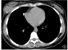
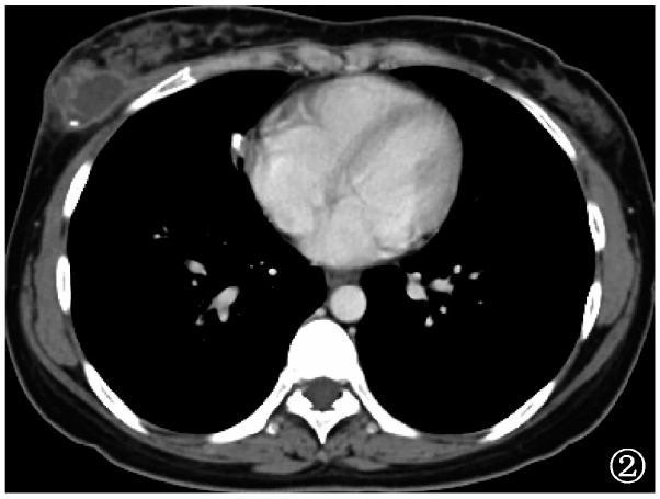
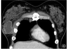
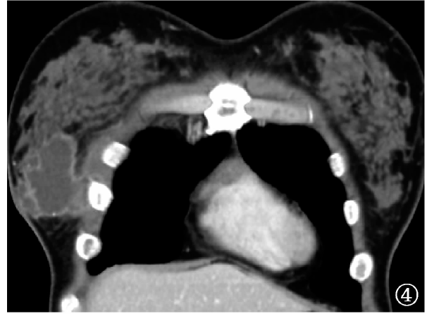
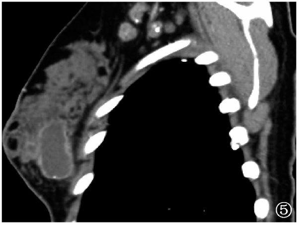
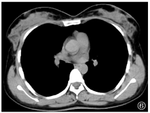
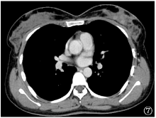
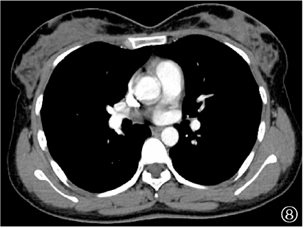
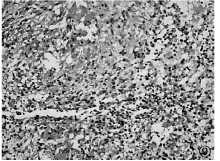
目的 分析女性乳腺结核的CT征象,探讨CT扫描对此类患者的诊断价值。方法 收集重庆市公共卫生医疗救治中心2015年8月至2019年5月行胸部CT检查时发现乳腺病变并经手术病理证实的20例女性乳腺结核患者,分析其CT表现及邻近组织及器官受累情况。结果 20例乳腺结核中12例发生于右乳,8例发生于左乳;13例为单发,7例为多发;16例呈结节状,1例呈团块状,3例呈片状;病灶大小1.0cm×1.5cm~5.1cm×3.2cm。CT平扫显示,11例病灶呈等密度,9例呈稍低密度,4例病灶内见结节状钙化,17例显示边界模糊;12例邻近皮肤呈均匀性增厚。CT增强扫描显示,15例病灶表现为环形强化,3例表现为不均匀强化,2例表现为均匀强化,2例伴窦道形成;其中13例并发同侧或双侧腋窝淋巴结结核,伴环形强化;5例并发同侧胸壁结核,伴边缘强化;10例并发肺结核。结论 乳腺结核的特征性CT表现为环形强化,同时常并发同侧腋窝淋巴结结核、胸壁结核及肺结核。

关键词: 结核, 乳腺疾病, 体层摄影术,螺旋计算机, 诊断显像

Abstract:

Objective The paper was aimed to analyze CT signs of mammary tuberculosis in females and investigate the diagnostic value of CT scan for these patients. Methods A total of 20 female patients with mammary tuberculosis, who were found with mammary lesions when undergoing chest CT and confirmed by surgical pathology in Chongqing Public Health Medical Center from August 2015 to May 2019, were enrolled, and then the CT manifestations and involvement of adjacent tissues and organs were analyzed. Results Of the 20 cases of mammary tuberculosis, 12 were presented with lesions in the right breasts, while 8 in the left breasts; there were 13 with single lesion and 7 with multiple lesions; the numbers of nodular, lump and flake morphologies were 16, 1 and 3, respectively; and the lesion size was measured with 1.0 cm×1.5 cm-5.1 cm×3.2 cm. Based on plain CT scan, there were 11 lesions with equal density,9 with slightly lower density, 4 with nodular calcification,17 with blurred borders, and 12 with uniform thickening of adjacent skin. According to CT enhancement scan, there were 15 lesions with ring enhancement, 3 with uneven enhancement, 2 with uniform enhancement, and 2 with accompanied sinus formation. Among them, 13 cases were accompanied by ipsilateral or bilateral axillary lymph node tuberculosis with annular enhancement, 5 cases had ipsilateral chest wall tuberculosis with marginal enhancement, and 10 cases had pulmonary tuberculosis. Conclusion The characteristic CT manifestation of mammary tuberculosis is ring enhancement, which is often accompanied by ipsilateral axillary lymph nodes, chest wall tuberculosis, and pulmonary tuberculosis.

Key words: Tuberculosis, Breast diseases, Tomography,spiral computed, Diagnostic imaging