Email Alert | RSS    帮助

中国防痨杂志 ›› 2020, Vol. 42 ›› Issue (1): 19-25.doi: 10.3969/j.issn.1000-6621.2020.01.007

• 论著 • 上一篇    下一篇

成年血行播散性肺结核并发颅内结核的临床及头颅MRI表现特征

宁锋钢,周新华,侯代伦(),吕平欣,吕岩,贺伟   

  1. 101149 首都医科大学附属北京胸科医院影像科
  • 收稿日期:2019-12-05 出版日期:2020-01-10 发布日期:2020-01-08
  • 通信作者: 侯代伦 E-mail:hodelen@126.com
  • 基金资助:
    北京市结核病与胸部肿瘤研究所人才项目(2019-3)

MR imaging features of hematogeneous pulmonary tuberculosis accompanied with intracranial tuberculosis in adult patients

NING Feng-gang,ZHOU Xin-hua,HOU Dai-lun(),LYU Ping-xin,LYU Yan,HE Wei   

  1. Department of Medical Imaging,Beijing Chest Hospital,Capital Medical University,Beijing 101149,China
  • Received:2019-12-05 Online:2020-01-10 Published:2020-01-08
  • Contact: Dai-lun HOU E-mail:hodelen@126.com

摘要:

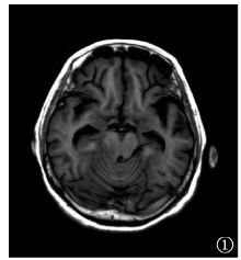
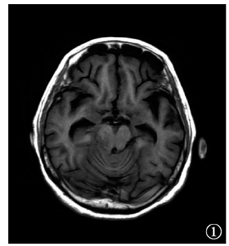
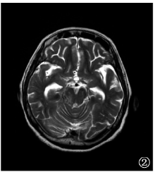
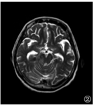
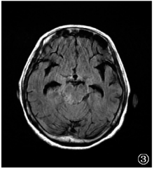
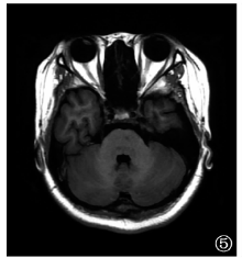
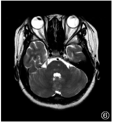
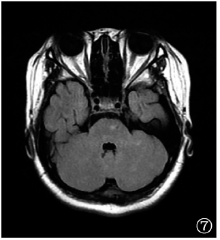
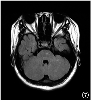
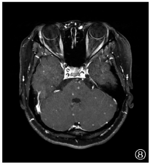
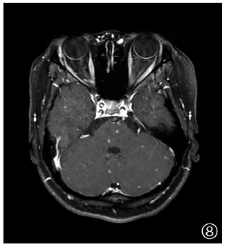
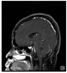
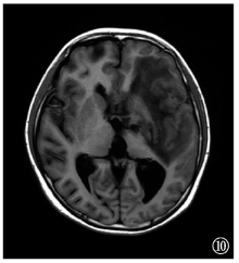
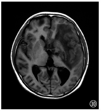
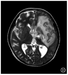
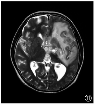
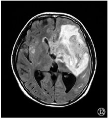
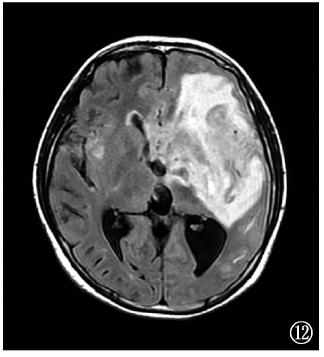
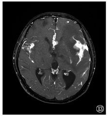
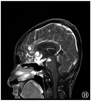
目的 探讨成年血行播散性肺结核并发颅内结核的临床及头颅MRI表现特征,以减少漏诊与误诊。方法 收集2015年2月至2019年7月首都医科大学附属北京胸科医院临床确诊的53例成年血行播散性肺结核并发颅内结核患者的临床资料,纳入资料完整的48例患者作为研究对象,所有患者均进行了结核病相关实验室检查(包括脑脊液检测)、头颅MR平扫及增强扫描检查,分析评价患者的临床及头颅MRI表现特征。结果 48例患者中,24例(50.0%)存在结核中毒症状和呼吸系统症状,36例(75.0%)出现发热、头痛,29例(60.4%)具有神经系统症状和体征;胸部CT平扫均可见两肺弥漫性粟粒状影;脑脊液常规和生化检查异常者45例(93.8%),其中蛋白升高43例(89.6%),葡萄糖含量降低38例(79.2%),氯化物降低37例(77.1%);所有患者均行腰椎穿刺术检查,颅内压增高[>180mm H2O (1mm H2O=0.0098kPa)]者31例(64.6%)。48例患者行头颅MR增强扫描,8例(16.7%)未发现明确结核病灶,40例有脑实质和(或)脑膜结核病变,分别为单纯脑膜结核9例(18.8%)、单纯脑实质结核19例(39.6%)、混合型颅内结核12例(25.0%);而48例行头颅MR平扫的患者仅35例显示颅内结核病灶,除8例增强MR未显示的患者外,仍有5例未发现结核病灶。25例患者抗结核药物治疗3个月后进行了头颅MR复查,其中11例较前好转,7例较前加重,5例出现部分病灶好转和部分病灶加重,2例未见变化。结论 血行播散性肺结核并发颅内结核患者临床多见发热、头痛、神经系统症状和体征、脑脊液常规和生化检查指标异常、颅内压增高。MR头颅扫描对该病的发现率较高,尤以MR增强扫描更为明显,是发现和诊断颅内结核的重要技术。

关键词: 结核,肺, 结核,粟粒性, 结核, 中枢神经系统, 疾病特征, 磁共振成像, 评价研究

Abstract:

Objective To investigate the MR imaging features of hematogeneous pulmonary tuberculosis accompanied with intracranial tuberculosis in adult patients, in order to reduce the misdiagnosis and missed diagnosis. Methods Clinical and imaging data of 53 patients diagnosed of hematogeneous pulmonary tuberculosis accompanied with intracranial tuberculosis in Beijing Chest Hospital affiliated to Capital Medical University from February 2015 to July 2019 were retrospectively analyzed. And of them, 48 patients with complete information were selected. They were all tested with laboratory examination related tuberculosis (including cerebrospinal fluid examination), as well as MR scan and enhanced MR examination; the results were investigated was and evaluated. Results Of the 48 cases, symptoms of tuberculosis and respiratory symptoms were found in 24 cases (50.0%), 36 (75.0%) were with fever and headache, symptoms and signs of nervous system were found in 29 (60.4%) cases; diffuse miliary shadow of lungs was found by chest CT; cerebrospinal fluid examination were abnormal in 45 cases (93.8%), and protein elevated in 43 (89.6%), glucose (38, 79.2%) and chloride (37, 77.1%) reduced; all the patients underwent lumbar puncture, and intracranial pressure increased (>180 mm H2O (1 mm H2O=0.0098 kPa)) in 31 (64.6%). All the 48 patients were examined by cranial enhanced MRI. In 8 cases (16.7%), no tuberculosis lesion was found; brain parenchyma and/or meningeal tuberculosis were fond in 40 patients, including simple meningeal tuberculosis (n=9, 18.8%), simple parenchymal tuberculosis (n=19, 39.6%) and mixed intracranial tuberculosis (n=12, 25.0%). However, by MR, cranial lesions were found just in 35 patients; of the other 13 patients, 8 were found by enhanced MRI, and still no lesion was found in 5 cases. Of the 25 patients re-examined by MRI after 3-month anti-tuberculosis treatment, 11 improved, 7 worsened, 5 partially improved, and 2 without any change. Conclusion Fever, headache, symptoms and signs of nervous system, abnormal cerebrospinal fluid and biochemical examination, and increased intracranial pressure are often found in patients of hematogeneous pulmonary tuberculosis accompanied with intracranial tuberculosis, and MR scan could find most of the abnormalities. Enhanced MR could supply even more information. MR scan is an important examination for the diagnosis of intracranial tuberculosis.

Key words: Tuberculosis,pulmonary, Tuberculosis,miliary, Tuberculosis,central nervous system, Disease attributes, Magnetic resonance imaging, Evaluation studies