Email Alert | RSS    帮助

中国防痨杂志 ›› 2021, Vol. 43 ›› Issue (8): 832-837.doi: 10.3969/j.issn.1000-6621.2021.08.015

• 论著 • 上一篇    下一篇

动脉栓塞介入治疗肺结核并发肺动脉假性动脉瘤伴大咯血三例并文献复习

陈珊珊, 唐晓军, 唐小莉, 戴广川, 曾谊(), 张侠()   

  1. 211131 南京中医药大学附属南京医院 南京市第二医院 南京市公共卫生医疗中心
  • 收稿日期:2021-04-15 出版日期:2021-08-10 发布日期:2021-07-30
  • 通信作者: 曾谊,张侠 E-mail:njyy002@njucm.edu.cn;njyy043@njucm.edu.cn

Endovascular embolization for pulmonary artery pseudoaneurysms associated with pulmonary tuberculosis with massive hemoptysis:three case reports and literature review

CHEN Shan-shan, TANG Xiao-jun, TANG Xiao-li, DAI Guang-chuan, ZENG Yi(), ZHANG Xia()   

  1. Nanjing Hospital Affiliated of Nanjing University of Chinese Medicine, the Second Hospital of Nanjing, Nanjing Municipal Public Health Medical Center, Nanjing 211131, China
  • Received:2021-04-15 Online:2021-08-10 Published:2021-07-30
  • Contact: ZENG Yi,ZHANG Xia E-mail:njyy002@njucm.edu.cn;njyy043@njucm.edu.cn

摘要:

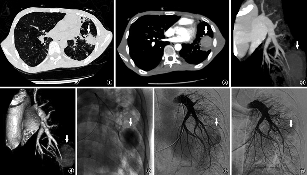
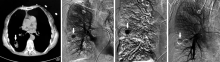
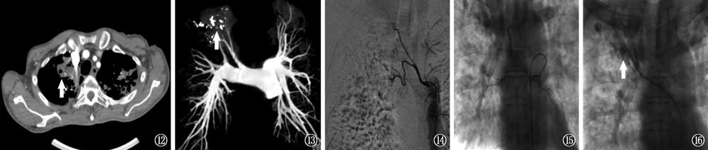
目的 总结和分析动脉栓塞介入治疗肺结核相关肺动脉假性动脉瘤(pulmonary artery pseudoaneurysms,PAP)伴大咯血的临床特征。方法 对南京市第二医院行动脉介入栓塞治疗的3例肺结核并发PAP伴大咯血患者的临床表现、治疗及预后进行报道。并以“pulmonary tuberculosis”和“pulmonary artery pseudoaneurysms”或“Rasmussen’s aneurysm”为检索词检索PubMed数据库,检索时间为2011年1月至2020年12月,剔除综述类文献、重复发表及资料不全等文献,纳入肺结核并发PAP患者临床资料完整的文章,收集患者的临床特征、影像学表现、PAP分型、诊治方法及预后,并进行文献复习。结果 病例1~3均确诊为肺结核并发大咯血,影像学提示肺结核空洞性病灶,且经胸部CT血管造影(CTA)及肺动脉造影均发现PAP继发于结核空洞壁上的肺动脉血管。其中,病例1,男,24岁,以“痰中带血7d,加重3d(咯血量>500ml/d,意识不清)”为主要表现,行支气管动脉栓塞术后再次大咯血,再行左胸廓内动脉和左胸外侧动脉、左侧肺动脉栓塞术后咯血停止;病例2,女,65岁,以“咯血5d(100ml/次,2次/d)伴胸闷”为主要表现,行支气管动脉栓塞术后再次大咯血,再行右侧肺动脉栓塞术后咯血停止;病例3,男,58岁,以“咳嗽、胸闷、盗汗4个月,咯血3d(300ml)”为主要表现,行双侧支气管动脉栓塞术后仍有咯血,再行左右支气管动脉、右上肺动脉栓塞术后咯血停止。通过筛选共获得21篇文献,42例肺结核并发PAP患者,与本文3例患者共计45例,其中男性36例,女9例,中位年龄为52.0(35.0,63.0)岁。45例患者均进行了CTA,PAP的检出率为100.0%;41例进行了肺动脉造影,PAP的检出率为75.6%(31例)。41例患者接受了血管介入治疗,其中26例行肺动脉栓塞治疗,7例行支气管动脉栓塞治疗,8例接受了支气管动脉栓塞+肺动脉栓塞治疗,术后有效止血40例,1例患者因大咯血窒息死亡。术后40例获得随访,2例复发,分别经外科切除术和再次行支气管动脉栓塞而治愈。结论 PAP是导致肺结核大咯血的主要原因之一,肺动脉介入栓塞治疗仍是PAP并发大咯血的首选干预措施。当支气管动脉栓塞效果不佳或咯血近期复发时,应及时进行胸部CTA检查,以排除PAP可能。

关键词: 咯血, 放射学,介入性, 栓塞,治疗性, 肺动脉, 动脉瘤,假性

Abstract:

Objective To summarize and analyze the clinical characteristics of pulmonary tuberculosis-associated pulmonary artery pseudoaneurysms (PAP) with massive hemoptysis in the treatment of arterial embolization intervention. Methods The clinical manifestations, treatment and prognosis of 3 patients with PAP complicated with pulmonary tuberculosis and massive hemoptysis who underwent interventional embolization in Nanjing Second Hospital were reported. PubMed database was searched with key words of “pulmonary tuberculosis” and “pulmonary artery pseudoaneurysms” or “Rasmussen’s aneurym” from January 2011 to December 2020. Reviews, repeated publications and articles with incomplete data were excluded, and researches with complete clinical data of patients with tuberculosis complicated with PAP were included. The clinical features, imaging manifestations, PAP typing, methods of diagnosis and treatment, and prognosis of the patients were collected to review the literature. Results Three cases were diagnosed with pulmonary tuberculosis complicated with massive hemoptysis. Radiology suggested cavity lesion of pulmonary tuberculosis, and chest CT angiography (CTA) and pulmonary angiography showed that PAP were secondary to pulmonary artery vessels on the pulmonary cavity wall of tuberculosis. Case 1, male, 24 years old, with blood in sputum for 7 days and aggravation for 3 days (hemoptysis volume >500 ml/d, unconscious) as the main manifestations. The patient had massive hemoptysis again after bronchial artery embolization and hemoptysis was stopped after embolization of left internal thoracic artery, left lateral thoracic artery and left pulmonary artery. Case 2, female, 65 years old, with hemoptysis for 5 days (100 ml/time, twice/day) and chest tipiness as the main presentation. Massive hemoptysis was relapsed after bronchial artery embolization and was stopped until right pulmonary artery embolization surgery. Case 3, male, 58 years old, presented with cough, chest tiresomy and night sweats for 4 months and hemoptysis for 3 days (300 ml). Hemoptysis still occurred after bilateral bronchial artery embolization, and was stopped after embolization of left and right bronchial arteries and right upper pulmonary artery. Through PubMed database searching, a total of 21 literatures were obtained, of which 42 patients with tuberculosis complicated with PAP. A total of 45 patients were included with the 3 patients in this paper, including 36 males and 9 females, with a median age of 52.0 (35.0,63.0) years. CTA was performed in all patients, and the detection rate of PAP was 100.0%. Pulmonary arteriography was performed in 41 cases and the detection rate of PAP was 75.6% (31/41). Forty-one patients received vascular interventional therapy, including 26 cases of pulmonary artery embolization, 7 cases of bronchial artery embolization, 8 cases of bronchial artery embolization and pulmonary artery embolization. Forty cases were of effective hemostasis after surgery and 1 patient died of asphyxia due to massive hemoptysis. Forty cases were followed up after surgery, of which 2 patients relapsed and were cured by surgical resection and bronchial artery embolization. Conclusion PAP is one of the main causes of massive hemoptysis in pulmonary tuberculosis, and pulmonary vascular interventional embolization is still the preferred intervention for PAP complicated with massive hemoptysis. When bronchial artery embolization is not effective or hemoptysis recedes recently, chest CTA examination should be performed in time to rule out the possibility of PAP.

Key words: Hemoptysis, Radiology,interventional, Embolization,therapeutic, Pulmonary artery, Aneurysm,false