Email Alert | RSS    帮助

中国防痨杂志 ›› 2020, Vol. 42 ›› Issue (6): 549-557.doi: 10.3969/j.issn.1000-6621.2020.06.004

• 论著 • 上一篇    下一篇

治疗失败耐多药肺结核患者CT征象演变的回顾性分析

许岩, 路希维(), 蔡春葵, 孙诗学, 顾晓峰, 于洋, 李刚, 王颖   

  1. 116033 大连市结核病医院
  • 收稿日期:2020-04-18 出版日期:2020-06-10 发布日期:2020-06-11
  • 通信作者: 路希维 E-mail:yiluxiwei@126.com
  • 基金资助:
    大连市科技创新基金项目(2018J13SN110)

Retrospective study of CT imaging evolution of treatment failure patients with multidrug-resistant tuberculosis

XU Yan, LU Xi-wei(), CAI Chun-kui, SUN Shi-xue, GU Xiao-feng, YU Yang, LI Gang, WANG Ying   

  1. Dalian Tuberculosis Hospital, Dalian 116033, China
  • Received:2020-04-18 Online:2020-06-10 Published:2020-06-11
  • Contact: LU Xi-wei E-mail:yiluxiwei@126.com

摘要:

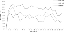
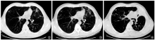
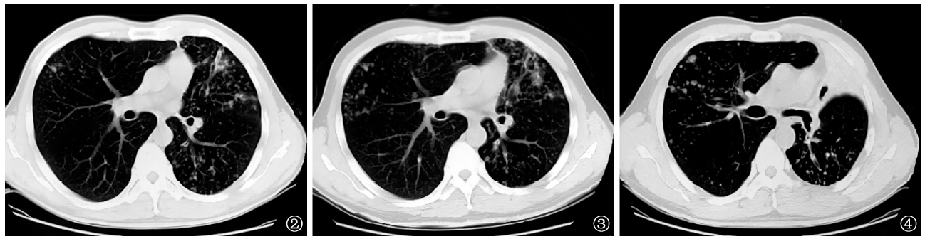
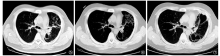
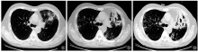
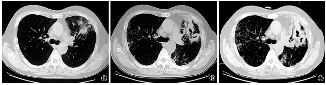
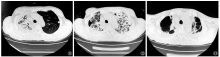
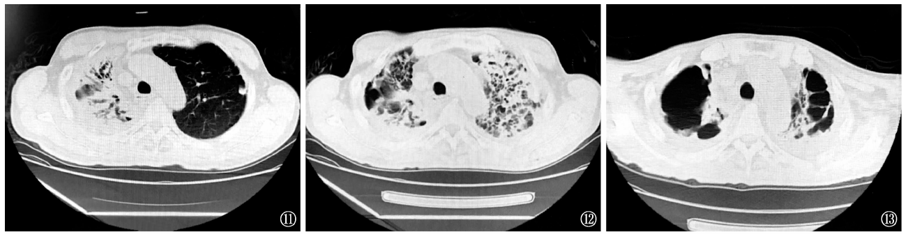
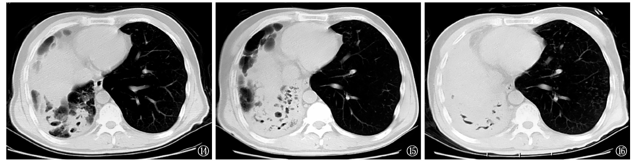
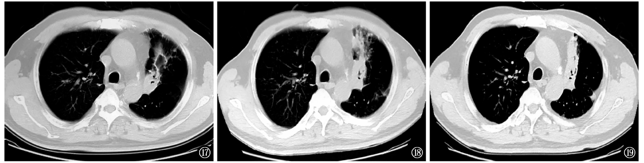
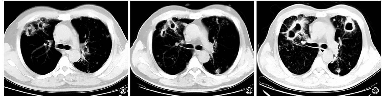
目的 研究治疗失败耐多药肺结核(MDR-PTB)患者的影像学演变规律,为MDR-PTB影像学疗效评价提供依据。方法 收集大连市结核病医院治疗失败的MDR-PTB 患者56例,疗程中每3个月进行一次CT检查,观察56例MDR-PTB患者的肺CT征象的演变与转归表现。根据CT征象将病程演变分为3种类型:吸收-进展型21例(37.5%,21/56);吸收好转型20例(35.7%,20/56);间歇进展型15例(26.8%,15/56)。计数资料采用χ 2检验,以P<0.05为差异有统计学意义。结果 (1)第3、6、24个月的痰菌阴性率在吸收-进展型患者中占比为81.0%(17/21)、61.9%(13/21)、28.6%(6/21);吸收-好转型患者中为90.0%(18/20)、85.0%(17/20)、60.0%(12/20);间歇进展型患者中为40.0%(6/15)、26.7%(4/15)、20.0%(3/15)。三型患者第3、6、24个月痰菌阴性率比较,χ 2值分别为11.953、12.248、6.994, P值分别为0.003、0.002、0.030,差异均有统计学意义。(2)56例患者的肺CT征象:树芽征、磨玻璃状影、结节状影的检出率在治疗前分别为67.9%(38/56)、21.4%(12/56)和80.4%(45/56);在治疗后分别为8.9%(5/56)、1.8%(1/56)和60.7%(34/56),χ 2值分别为41.108、10.530、5.198, P值分别为0.000、0.001、0.023,差异均有统计学意义。支气管聚拢迂曲、肺纤维化、毁损肺、支气管扩张治疗,治疗前分别为21.4%(12/56)、5.4%(3/56)、5.4%(3/56)和8.9%(5/56);治疗后分别为41.1%(23/56)、17.9%(10/56)、23.2%(13/56)和37.5%(21/56),χ 2值分别为5.029、4.264、7.292、12.823, P值分别为0.025、0.039、0.007、0.000,差异均有统计学意义。(3)在固有病变中,空洞(37.8%,108/286)、结节状病灶(25.9%,74/286)、肺实变(16.4%,47/286)和树芽征(10.8%,31/286)多见;而在新增病变中树芽征(46.9%,23/49)和结节状病灶(40.8%,20/49)多见。新增树芽征与无新增树芽征患者中“干酪性肺实变伴空洞”的检出率分别为47.4%(9/19)和18.9%(7/37),χ 2=4.979 P=0.026,差异有统计学意义。结论 通过CT征象连续观察,可对治疗失败进行预测,从而提示临床及时调整化疗方案,以减少不可逆肺损伤的发生。

关键词: 结核, 肺, 结核, 抗多种药物性, 治疗失败, 体层摄影术, X线计算机, 诊断显像, 回顾性研究

Abstract:

Objective To investigate imaging evolution of treatment failure patients with multidrug-resistant tuberculosis, and provide the evidence for the evaluation. Methods Fifty-six cases of MDR-PTB patients who failed to be treated in Dalian tuberculosis hospital were collected. CT examination was carried out every 3 months in the course of treatment. The evolution and outcome of lung CT signs of 56 MDR-PTB patients were observed. According to CT signs, the course of disease can be divided into three types: absorption progressive type in 21 cases (37.5%, 21/56); absorption transforming type in 20 cases (35.7%, 20/56); intermittent progressive type in 15 cases (26.8%, 15/56). χ 2 test was used to count the data, with a statistical significance of P<0.05. Results (1) At the 3rd, 6th and 24th month, the negative rate of sputum bacteria was 81.0% (17/21), 61.9% (13/21), 28.6% (6/21) in the absorption progressive type, 90.0% (18/20), 85.0% (17/20), 60.0% (12/20) in the absorption transformation type, and 40.0% (6/15), 26.7% (4/15), 20.0% (3/15) in the intermittent progressive type. There were significant differences in the sputum negative rate of the 3rd, 6th and 24th month among three types (χ 2=11.953,12.248,6.994, P=0.003,0.002,0.030). (2) In 56 patients, the detection rates of tree bud sign, ground glass shadow and nodular shadow were 67.9% (38/56), 21.4% (12/56) and 80.4% (45/56) before treatment, and 8.9% (5/56), 1.8% (1/56) and 60.7% (34/56) after treatment, there were statistically significant differences between before and after treatment (χ 2=41.108,10.530,5.198, P=0.000,0.001,0.023). After treatment, the number of bronchus clustered, fibrosis, damaged lung and bronchiectasis increased significantly, which were 21.4% (12/56), 5.4% (3/56), 5.4% (3/56) and 8.9% (5/56) before treatment, 41.1% (23/56), 17.9% (10/56), 23.2% (13/56) and 37.5% (21/56) after treatment, there were significant differences between before and after treatment.(χ 2=5.029,4.264,7.292,12.823, P=0.025,0.039,0.007,0.000).(3) In the inherent lesions, cavity 37.8% (108/286),nodule 25.9% (74/286),consolidation 16.4% (47/286) and tree in bud 10.8% (31/286) were common; in the new lesions, tree in bud 46.9% (23/49) and nodule 40.8% (20/49) were common. The detection rate of caseous consolidation with cavities in tree in bud cases was 47.4% (9/19) and that in non-tree in bud cases was 18.9% (7/37),which was statistically significant (χ 2=4.979, P=0.026). Conclusion Through continuous observation of CT imaging, treatment failure can be predicted, which suggests that clinical chemotherapy should be adjusted in time to reduce the incidence of irreversible lung injury.

Key words: Tuberculosis, pulmonary, Tuberculosis, multidrug-resistant, Treatment failure, Tomography, X-ray computed, Diagnostic imaging, Retrospective studies