Email Alert | RSS    帮助

中国防痨杂志 ›› 2020, Vol. 42 ›› Issue (1): 38-43.doi: 10.3969/j.issn.1000-6621.2020.01.010

• 论著 • 上一篇    下一篇

初治与复治耐多药肺结核患者的CT表现分析

杨佳,吕圣秀,唐光孝,舒伟强,王惠秋,杨长萍,刘雪艳,李春华()   

  1. 400036 重庆市公共卫生医疗救治中心医学影像科
  • 收稿日期:2019-10-16 出版日期:2020-01-10 发布日期:2020-01-08
  • 通信作者: 李春华 E-mail:250367603@qq.com
  • 基金资助:
    重庆市卫生和计划生育委员会医学科研项目(2017MSXM120)

Analysis of CT findings in patients with initial and retreated multidrug-resistant tuberculosis

YANG Jia,LYU Sheng-xiu,TANG Guang-xiao,SHU Wei-qiang,WANG Hui-qiu,YANG Chang-ping,LIU Xue-yan,LI Chun-hua()   

  1. Department of Medical Radiology, Chongqing Public Health Medical Center, Chongqing 400036, China
  • Received:2019-10-16 Online:2020-01-10 Published:2020-01-08
  • Contact: Chun-hua LI E-mail:250367603@qq.com

摘要:

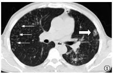
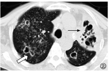
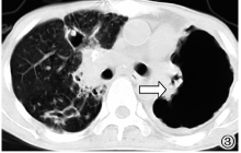
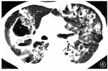
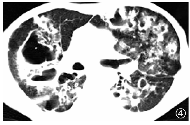
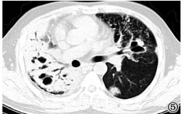
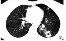
目的 探讨初治与复治耐多药肺结核患者的CT表现。 方法 搜集2016年1月至2019年1月重庆市公共卫生医疗救治中心经药物敏感性试验确诊的186例耐多药肺结核患者,其中初治患者80例,作为初治组;复治患者106例,作为复治组。对比两组患者病变范围、病灶形态(树芽征、腺泡结节、斑片状影、大片状影、条索状影、钙化影)、空洞数量及形态、空洞壁、支气管扩张等CT表现,对CT表现等计数资料进行统计学分析,采用χ 2检验,以P<0.05为差异有统计学意义。 结果 复治组肺毁损者占20.8%(22/106)、病灶范围≥3叶者占 90.6%(96/106)、病灶位于中叶及舌段者占85.8%(91/106),以及出现索条状影者占54.7%(58/106)、钙化灶者占27.4%(29/106)、支气管扩张者占77.4%(82/106),与初治组比较 [分别为6.2%(5/80)、71.2%(57/80)、72.5%(58/80)、23.8%(19/80)、10.0%(8/80)、31.2%(25/80)],差异均有统计学意义(χ 2值分别为7.730、11.656、5.098、18.021、8.621、39.670,P值均<0.05)。复治组发生胸廓塌陷者占21.7%(23/106)、纵隔移位者占35.8%(38/106)、胸膜增厚者占78.3%(83/106),与初治组比较 [分别为5.0%(4/80)、7.5%(6/80)、53.8%(43/80)],差异均有统计学意义(χ 2值分别为8.943、20.288、12.576,P值均<0.05)。复治组患者空洞发生率为83.0%(88/106)、空洞≥3个者占59.4%(63/106)、厚壁空洞者占70.8%(75/106)、薄壁空洞者占40.6%(43/106)、虫蚀样空洞者占40.6%(43/106)、空洞呈簇聚集者占40.6%(43/106)、空洞内壁不光滑者占20.8%(22/106),与初治组比较 [分别为56.2%(45/80)、23.8%(19/80)、51.2%(41/80)、12.5%(10/80)、16.2%(13/80)、16.2%(13/80)、7.5%(6/80)],差异有统计学意义(χ 2值分别为16.034、23.551、7.390、17.626、12.810、12.810、6.264,P值均<0.05)。结论 复治耐多药肺结核患者病变范围,肺毁损、多发空洞且呈簇聚集、干酪性肺炎伴虫蚀样空洞及静脉曲张型支气管扩张等CT表现与初治组比较更严重。

关键词: 结核,抗多种药物性, 再治疗, 体层摄影术,X线计算机, 诊断相关患者组, 诊断显像, 对比研究

Abstract:

Objective To investigate the CT findings in patients with initial and retreated multidrug-resistant tuberculosis. Methods A total of 186 patients with multidrug-resistant tuberculosis diagnosed by drug susceptibility testing in Chongqing Public Health Medical Treatment Center from January 2016 to January 2019 were collected, including 80 newly treated patients as initial treatment group and 106 retreated patients as retreatment group. The lesion size, lesion morphology (tree in bud, acinar node, speckle shadow, large shadow, strip shadow, calcification shadow), the number and shape of cavity, the shape of cavity wall, bronchiectasis and other CT findings were compared between the two groups. The CT findings were statistically analyzed by χ 2 test, and the difference was statistically significant (P<0.05). Results In the retreatment group (106 cases), there were destroyed lungs in 22 cases (20.8%,22/106), lesion size more than 3 lobes in 96 cases (90.6%,96/106), lesions located in the middle lobe and tongue segment in 91 cases (85.8%, 91/106), strip shadow in 58 cases (54.7%,58/106), calcification shadow in 29 cases (27.4%,29/106) and bronchiectasis in 82 cases (77.4%, 82/106), respectively, when compared for those (6.2% (5/106), 71.2% (57/80), 72.5% (58/80), 23.8% (19/80), 10.0% (8/80), 31.2% (25/80)) in initial group with the significant differences statistically (χ 2 values were 7.730, 11.656, 5.098, 18.021, 8.621, 39.670, Ps<0.05). In the retreatment group (106 cases), Chest collapse occurred in 23 cases (21.7%, 23/106), mediastinal displacement in 38 cases (35.8%,38/106), pleural thickening in 83 cases (78.3%,83/106), when compared for those (5.0% (4/80), 7.5% (6/80), 53.8% (43/80)) in the initial treatment group with the significant difference statistically (χ 2 values were 8.943, 20.288, 12.576, Ps<0.05). The incidence of cavity was 83.0% (88/106), the number of cavity more three in 63 cases (59.4%,63/106), thicken-wall cavity in 75 cases (70.8%, 75/106), thin-wall cavity in 43 cases (40.6%,43/106), moth-eaten cavity in 43 cases (40.6%,43/106), cluster cavity in 43 cases (40.6%,43/106) and cavity with rough inner wall in 22 cases (20.8%,22/106), when compared for those (56.2% (45/80),23.8% (19/80),51.2% (41/80),12.5% (10/80),16.2% (13/80),16.2% (13/80) and 7.5% (6/80)) in initial group with the significant difference statistically (χ 2 value were 16.034,23.551,7.390,17.626,12.810,12.810 and 6.264, Ps<0.05). Conclusion The CT findings of retreated MDR-TB patients are more serious than those of the initial treatment group, such as wide lesion, destroyed lungs, multiple cavities and cluster aggregation, caseous pneumonia with moth-eaten cavity and varicose bronchiectasis.

Key words: Tuberculosis,multidrug-resistant, Retreatment, Tomography,X-Ray computed, Diagnosis-related groups, Diagnostic imaging, Comparative study