Email Alert | RSS    帮助

中国防痨杂志 ›› 2020, Vol. 42 ›› Issue (1): 26-30.doi: 10.3969/j.issn.1000-6621.2020.01.008

• 论著 • 上一篇    下一篇

CT与MRI在成年膝关节结核中的诊断价值

司马斌,邱小伟(),王安龙   

  1. 310003 杭州,浙江中医药大学附属中西医结合医院放射科
  • 收稿日期:2019-12-03 出版日期:2020-01-10 发布日期:2020-01-08
  • 通信作者: 邱小伟 E-mail:247685310@qq.com
  • 基金资助:
    2019年杭州市农业与社会发展自主申报项目(20191203B133)

Diagnostic value of CT and MRI in adult knee joint tuberculosis

SIMA Bin,QIU Xiao-wei(),WANG An-long   

  1. Zhejiang Integrated Traditional and Western Medicine Hospital affiliated to Zhejiang Chinese Medical University, Hangzhou 310003, China
  • Received:2019-12-03 Online:2020-01-10 Published:2020-01-08
  • Contact: Xiao-wei QIU E-mail:247685310@qq.com

摘要:

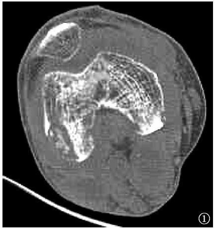
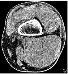
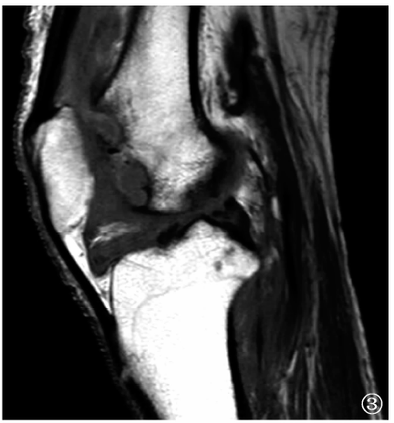
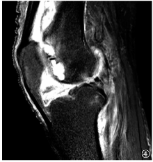
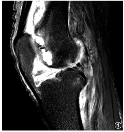
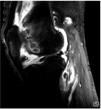
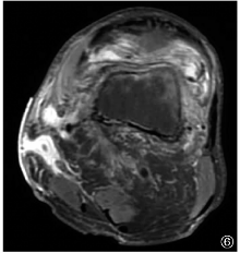
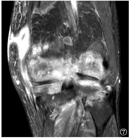
目的 比较CT与MRI检查在成年膝关节结核诊断中的应用价值。 方法 选择2017年1月至2018年6月浙江中医药大学附属中西医结合医院经手术病理证实的36例单侧膝关节结核患者。36例患者年龄18~85岁,平均(52.3±18.9)岁;男26例,女10例。所有患者入院后均行CT与MRI检查,比较两种检查方式的诊断发现率。 结果 CT检查和MRI检查对于膝关节结核骨质破坏、死骨、滑膜增厚、关节积液、钙化的诊断发现率分别为58.3%(21/36)、52.8%(19/36)、22.2%(8/36)、36.1%(13/36)、30.6%(11/36)和91.7%(33/36)、5.6%(2/36)、97.2%(35/36)、80.6%(29/36)、0.0%(0/36),其中CT检查对骨质破坏、滑膜增厚和关节积液的诊断发现率明显低于MRI检查,对死骨和钙化的诊断发现率明显高于MRI检查,差异均有统计学意义(χ 2值分别为8.96、17.21、39.03、14.63、17.24,P值分别为0.003、0.000、0.000、0.000、0.000);CT检查和MRI检查对于关节间隙异常、关节周围冷脓肿及并发窦道形成的诊断发现率分别为97.2%(35/36)、75.0%(27/36)、11.1%(4/36)和94.4%(34/36)、80.6%(29/36)、11.1%(4/36),差异均无统计学意义(χ 2值分别为0.00、0.32、0.00,P值分别为1.000、0.571、1.000)。 结论 CT检查对于发现死骨、钙化占优势;MRI检查对于观察膝关节骨质破坏、滑膜增厚及评估整个关节破坏程度有明显优势。

关键词: 结核,骨关节, 膝关节, 体层摄影术,X线计算机, 磁共振成像, 诊断显像, 对比研究

Abstract:

Objective To compare the application value of CT and MRI in the diagnosis of adult knee joint tuberculosis. Methods Thirty-six patients with unilateral knee joint tuberculosis confirmed by surgical pathology from January 2017 to June 2018 in the Zhejiang Integrated Traditional and Western Medicine Hospital affiliated to Zhejiang Chinese Medical University were selected. The age of 36 patients (26 males and 10 females) ranged from 18 to 85 years, with an average of (52.3±18.9) years old. All patients underwent CT and MRI examinations after admission. The diagnostic discovery rates of the two methods was compared. Results The diagnostic discovery rates of bone destruction, bone death, synovium thickening, joint effusion and calcification were 58.3% (21/36), 52.8% (19/36), 22.2% (8/36), 36.1% (13/36), 30.6% (11/36) by CT, and 91.7% (33/36), 5.6% (2/36), 97.2% (35/36), 80.6% (29/36), 0.0% (0/36) by MRI. The diagnostic discovery rates of bone destruction, synovium thickening, and joint effusion by CT were significantly lower than those by MRI, while the diagnostic discovery rates of bone death, and calcification by CT were obviously higher than those by MRI, all with the statistically significant differences between groups (χ 2 values were 8.96, 17.21, 39.03, 14.63, 17.24, P values were 0.003, 0.000, 0.000, 0.000 and 0.000, respectively). The diagnostic discovery rates of abnormal joint space, cold abscess around the joint, and concurrent sinus formation were 97.2% (35/36), 75.0% (27/36), 11.1% (4/36) by CT, and 94.4% (34/36), 80.6% (29/36), 11.1% (4/36) by MRI, without significant difference between the groups (χ 2 values were 0.00, 0.32, 0.00, P values were 1.000, 0.571, and 1.000, respectively). Conclusion CT examination is dominant in finding dead bone and calcification predominate, while MRI examination has significant advantages in observing knee joint bone destruction, synovial thickening and assessing the degree of joint destruction.

Key words: Tuberculosis,osteoarticular, Knee joint, Tomography,X-ray computed, Magnetic resonance imaging, Diagnostic imaging, Comparative study