Email Alert | RSS 帮助

中国防痨杂志 ›› 2019, Vol. 41 ›› Issue (5): 499-509.doi: 10.3969/j.issn.1000-6621.2019.05.007
赵彬,乔高锋,金锋( ),王成,孙磊,杨秀珍
收稿日期:2019-01-15
出版日期:2019-05-10
发布日期:2019-05-10
Bin ZHAO,Gao-feng QIAO,Feng JIN(),Cheng WANG,Lei SUN,Xiu-zhen. YANG
Received:2019-01-15
Online:2019-05-10
Published:2019-05-10
摘要:
目的 总结分析结核性脓胸并发胸膜血管肉瘤的临床特点,提高临床诊疗水平。方法 对2016年11月山东大学附属山东省胸科医院收治的1例结核性脓胸并发胸膜血管肉瘤患者的临床表现、实验室检查结果、治疗及预后进行分析,并复习国内外文献资料。以“pleural angiosarcoma”为检索词检索PubMed数据库;以“胸膜血管肉瘤”为检索词检索万方医学网和中国知网数据库,检索时间为1995年1月至2018年11月;收集患者的一般情况、既往病史、影像学表现、病理免疫组织化学检测结果、治疗方法、预后及病程时间。结果 患者,男,56岁。因“胸闷、咳嗽超过1个月,痰中带血20d”入院,住院期间多次行胸部手术,病理诊断为结核性脓胸并发胸膜血管肉瘤。患者发病17个月后因病死亡。通过文献复习与筛选共获得相关文献31篇,包含32例胸膜血管肉瘤患者,加上本例患者共计33例;其中男23例,女10例,年龄24~87岁,平均(64.12±13.90)岁。33例患者中有结核性脓胸病史者9例,有脓胸病史者1例;有放化疗病史者2例,单纯放疗病史者1例;有粉尘接触史者1例。病灶位于右侧胸腔14例,左侧胸腔12例,双侧胸腔7例;发病时单纯胸腔积液者4例,胸膜增厚、胸膜肿物者12例,胸腔积液并发胸膜增厚、胸膜肿物者17例;明确有血胸者16例;发生转移病灶5例;CD31阳性者27例,CD34阳性者17例,Vimentin阳性者15例,Ⅷ因子相关抗原阳性者9例。33例患者均为病理确诊。治疗方法有手术(14例)、化疗(10例)、放疗(6例)、介入动脉栓塞(2例)、微波消融(1例)、胸膜固定(3例)、血管靶向药物治疗(5例)。患者病程为2个月至15年,中位时间为7个月;最终24例患者死亡,预后不详、失访和末次随访时存活者各3例。结论 胸膜血管肉瘤临床表现缺乏特异性,结核性脓胸易导致胸膜血管肉瘤的发生,当结核性脓胸患者并发不明原因的渗血性病变时,应考虑到本病的可能,防止误诊、漏诊。
赵彬,乔高锋,金锋,王成,孙磊,杨秀珍. 结核性脓胸并发胸膜血管肉瘤一例并文献复习与分析[J]. 中国防痨杂志, 2019, 41(5): 499-509. doi: 10.3969/j.issn.1000-6621.2019.05.007
Bin ZHAO,Gao-feng QIAO,Feng JIN,Cheng WANG,Lei SUN,Xiu-zhen. YANG. Tuberculous pyothorax combined with pleural angiosarcoma:a case report and review of the literature[J]. Chinese Journal of Antituberculosis, 2019, 41(5): 499-509. doi: 10.3969/j.issn.1000-6621.2019.05.007

图1~3
2011年9月患者行胸部CT扫描,显示左肺下叶后基底段小结节状影,边缘模糊。右侧脏壁层胸膜不规则增厚,条样钙化,其内见包裹性液体及软组织样密度影,并见引流管影通向胸壁外,右肺下叶部分不张

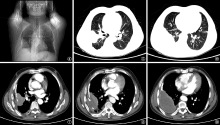
图4~9
2016年11月患者入院时行胸部CT扫描,显示右肺下叶背段存在不规则团块灶,内部密度欠均匀,呈明显强化;右侧脏壁层胸膜不规则增厚,条样钙化,其内见包裹性液体影,右肺下叶部分不张


图10
2016年12月8日患者术中快速病理壁层胸膜标本检查,显示为增生、炎性改变并玻璃样变的纤维组织,其中可见小团不典型肉芽肿性炎,倾向非特异性炎性改变(HE ×100)


图11
2016年12月8日患者术后标本病理检查,显示脏层胸膜病变标本为增生、炎性改变并玻璃样变的纤维组织,另见灶性钙化,其中可见小团可疑肉芽肿(HE ×100)
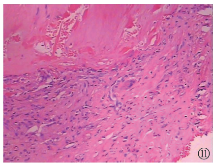

图12~16
2017年1月患者再次入院时行胸部CT扫描,显示手术后右侧脏壁层胸膜呈明显不规则增厚,条样钙化,其内见混杂密度影,右下侧胸壁软组织肿胀
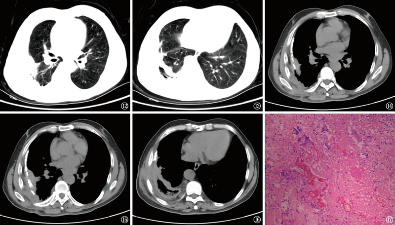
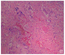
图17
2017年3月15日术后病理检查,显示炎性改变的纤维组织,其内见散在肉芽肿性炎,符合结核感染(HE ×100)
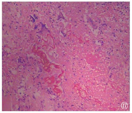

图18~22
2017年5月患者胸部CT扫描,显示胸廓成形术后右下胸壁肿胀,右侧胸膜明显不规则增厚,并可见条样钙化,其内见混杂密度影 图21 2017年8月8日患者手术后脓腔病灶标本病理检查,显示纤维组织增生、炎性改变,部分区域增生活跃,并见坏死(HE ×100) 图22 2017年8月8日患者手术后壁层纤维板标本病理检查,显示增生、炎性改变并玻璃样变的纤维组织(HE ×100)

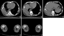
图23~25
2018年1月患者胸部CT扫描,显示右侧下胸壁肿胀,内见低密度区,邻近胸膜出现广泛的不均匀性增厚、钙化,内部密度不均,见不规则低密度区,右侧胸腔内见水样密度影并呈包裹样 图26,27 2018年1月底患者大腿部CT扫描,显示左侧股骨偏下段见溶骨性骨质破坏,并可见死骨形成,其前方见软组织密度影,边界尚清晰,其内密度不均匀
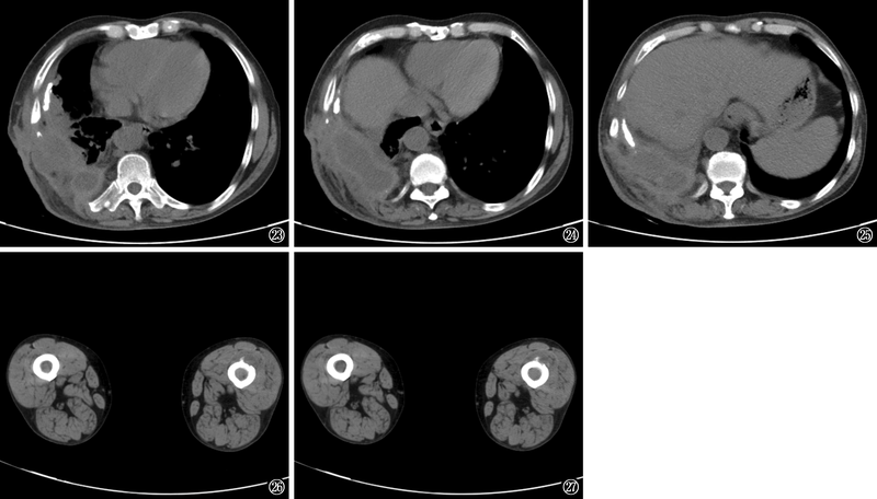
表1
1995年1月至2018年11月文献报告和本文报告的33例胸膜血管肉瘤患者基本情况
| 第一作者 | 例 数 | 年龄 (岁) | 性 别 | 既往史 | 临床表现 | 影像学特征 | 诊断 方法 | 免疫组化 | 病 程 | 治疗方法 | 预后 | |||||||||||
|---|---|---|---|---|---|---|---|---|---|---|---|---|---|---|---|---|---|---|---|---|---|---|
| 1.王涛[ | 1 | 75 | 男 | 无 | 胸痛伴发热3个月;胸腔积液呈黄褐色,稠脓 | 右侧胸腔包裹性积液,胸膜增厚,局部钙化灶 | 开胸 手术 | 阳性:CD31、CD34、细胞角质蛋白CK;阴性:Calrentin、人骨髓内皮细胞标记物HBME-1、EMA、CK7、CK20;Ki-67 约1% | 4 个 月 | 开胸手术 | 术后1个月死亡 | |||||||||||
| 2.高绍荣[ | 1 | 64 | 女 | 无 | 胸闷、咳嗽,胸背疼痛;暗红色血性胸腔积液 | 右侧胸腔大量积液,胸膜增厚,多发结节 | 穿刺 活检 | 阳性:CD31、CD34、Vimentin、CKpan;阴性:HBME-1、Calretinin;Ki-67 约40%~50% | 8.5 个 月 | 吉西他滨/多西他赛+顺铂注药+贝伐单抗/恩度 | 7.5个月后死亡 | |||||||||||
| 3.张敬文[ | 1 | 63 | 女 | 无 | 胸痛2周;黄色胸腔积液 | 右侧胸腔积液,胸膜多发结节 | 穿刺 活检 | 阳性:CD31、CD34、Vimentin、CKpan;阴性:HBME-1、Calretinin | 不 详 | 吉西他滨+顺铂注药+贝伐单抗 | 预后不详 | |||||||||||
| 4.徐佳[ | 1 | 52 | 女 | 无 | 胸闷、发热5个月;血性胸腔积液 | 右侧胸腔大量积液,胸膜多发结节 | 开胸 手术 | 阳性:CD31、CD34;阴性:S-100蛋白、HMB-45 | 6 个 月 | 开胸手术 | 术后2周死亡 | |||||||||||
| 5.常占平[ | 1 | 66 | 男 | 6年前发现结核性胸膜炎 | 反复咯血、左胸肿物逐渐增大4年,1年前左胸肿物增大,开胸手术后反复发热,伤口不愈合 | 左侧胸腔巨大肿物 | 穿刺 活检 | 阳性:CD34、CD31、Vimentin、Ⅷ、CK;阴性:EMA、CEA、TTF-1、Calretinin | 58 个 月 | 未治疗 | 术后10个月(确诊后1个月)死亡 | |||||||||||
| 6. Filippiadis[ | 1 | 74 | 男 | 7年前胃肿瘤手术+伊马替尼 | 顽固血胸、贫血,微波消融后出血好转 | 右侧胸腔下部脊柱旁结节样肿物 | 开胸 手术 | 不详 | 5 个 月 | 开胸手术+介入栓塞+放疗+微波消融 | 确诊4个月后死亡 | |||||||||||
| 7. Cabibi[ | 1 | 50 | 男 | 9年前甲状腺手术+碘131放疗 | 呼吸困难 | 左侧胸腔胸膜增厚、多发结节 | 胸腔镜 活检 | 阳性:CD31、CK7、D2-40、c-MYC、CKpan;阴性:CD34、Ⅷ、WTI、CK5/6、Calretinin、EMA、HBME-1、CEA、p63、EpCAM、Bcl-2、TTFI | 不 详 | 不详 | 预后不详 | |||||||||||
| 8. Miller[ | 1 | 75 | 男 | 4年前左上肺鳞癌放疗+化疗 | 呼吸困难 | 左侧胸腔包裹性积液,胸膜结节;正电子发射体层摄影(PET)-CT示胸膜表面放射性浓聚 | 胸腔镜 活检 | 阳性:CD31、CD34 | 4 个 月 | 吉西他滨+多西他赛 | 4个月后死亡 | |||||||||||
| 9. Yamaguchi[ | 1 | 72 | 女 | 5年前左乳腺癌手术+放疗+化疗 | 左胸胀满感,活检后腔镜窦道转移 | 左侧胸腔积液,PET-CT显示阴性;活检后有肿块沿窦道凸入胸腔 | 胸腔镜 活检 | 阳性:CD31 | 5 个 月 | 胸膜粘连治疗+帕唑帕尼+放疗 | 4个月后死亡 | |||||||||||
| 10. Matsuda[ | 1 | 87 | 男 | 采石场工作15年 | 昏迷、贫血、血胸、呼吸困难 | 右侧胸膜2个结节,双侧胸腔积液,双侧胸膜轻度增厚 | 尸检 | 阳性:CD31、Vimentin、CD34、CK;阴性:Calretinin、D2-40、desmin、EMA、CK5/6、WT-1、 | 1.5 个 月 | 未治疗 | 1.5个月后死亡 | |||||||||||
| 第一作者 | 例 数 | 年龄 (岁) | 性 别 | 既往史 | 临床表现 | 影像学特征 | 诊断 方法 | 免疫组化 | 病 程 | 治疗方法 | 预后 | |||||||||||
| 11. Zhang[ | 1 | 76 | 男 | 无 | 咳嗽、咳痰4个月,加重1周;1个月内体质量降低4kg | 左侧胸腔巨大肿物,局部钙化,胸膜广泛增厚、钙化;PET-CT示肿物外周有放射浓聚,中心区正常 | 开胸 手术 | 阳性:CD31、CD34、Vimentin、CK;阴性:D2-40、Glut-1、Calretinin、CK5/6、Syn、WT-1;局部Ki-67约90% | 超 过 11 个 月 | 开胸手术 | 随访7个月 | |||||||||||
| 12. Bruixola[ | 1 | 68 | 男 | 幼年时患肺结核、结核性脓胸 | 咯血、贫血、胸痛,术后胸痛复发加重、呼吸困难,抗结核治疗无效 | 右侧胸腔肿物,周边钙化,术后原位肿物复发并肝脏、脊柱、腰大肌、髂骨、臀部肿物,PET-CT示所有病灶均有放射浓聚 | 开胸手 术+复 发后穿 刺活检 | 不详 | 2 年 | 开胸手术(切缘阴性)+复发后放疗+多西他赛+异环磷酰胺 | 发现2年后死亡 | |||||||||||
| 13. Onur[ | 1 | 79 | 女 | 无 | 渐进性呼吸困难1个月,胸背痛、咳嗽、乏力,血胸 | 左侧胸腔积液,胸膜增厚、胸膜结节 | 穿刺 活检 | 阳性:CD34、CD31、CKpan;阴性:CEA、Calretinin、Mesothelin、CK5/6、Vimentin、TTF-1 | 至 少 7 个 月 | 未治疗 | 6个月后失访 | |||||||||||
| 14. Quesada[ | 1 | 58 | 女 | 无 | 呼吸困难、低热10d,血胸 | 双侧积液(右侧为多),胸膜小结节 | 胸腔镜 活检 | 阳性:CD31、Vimentin、D2-40、Nestin、VEGF-A、SMA;阴性:CD34、Calretinin、CK5/6、CK7、CK20、Bcl-20、HMB45、CKpan、Ⅷ、Melan-A;Ki-67 30% | 5 个 月 | 胸膜化学药物固定,紫杉醇4周期,索拉菲尼 | 5个月后死亡 | |||||||||||
| 15. Abu-Zaid[ | 1 | 63 | 男 | 无 | 右胸痛3个月,伴呼吸困难、咯血,血胸 | 双侧胸腔积液,右肋膈隐窝膈肌表面肿物 | 穿刺 活检 | 阳性:Vimentin、CD31、CD68、Fli-1 | 3 个 月 | 未治疗 | 确诊1周后心源性猝死 | |||||||||||
| 16. Chen[ | 1 | 69 | 男 | 无 | 间断右胸痛4个月,体质量减轻8kg,血胸 | 右侧胸腔包裹性积液,胸膜结节 | 开胸 手术 | 阳性:CD31、Ⅷ、CK;阴性:Calretinin | 5 个 月 | 开胸手术 | 1个月后死亡 | |||||||||||
| 17. Lorentziadis[ | 1 | 77 | 男 | 无 | 呼吸困难20d,贫血,血胸 | 双侧胸腔积液,右侧为著;手术后右侧胸膜增厚 | 开胸 手术 | 阳性:CD31、CD34、Ⅷ;阴性:CK7、CK5/6、EMA、CKpan、Calretinin | 1 个 月 | 开胸手术 | 1周后呼吸衰竭死亡 | |||||||||||
| 18. Kao[ | 1 | 49 | 男 | 无 | 间断右胸痛1个月,伴渐进性呼吸困难;血胸;胸腔镜术后胸膜肿物形成 | 右侧胸腔积液、胸膜增厚;胸腔镜术后右侧胸膜肿块形成 | 开胸 手术 | 阳性:CD31、FLI-1、CK7;阴性:CD34、Ⅷ、Calretinin、CK5/6、HBME-1、WT-1 | 至 少 1 年 | 胸腔镜+开胸切除手术+放疗+5周期化疗(美司钠、异环磷酰胺、阿霉素、达卡巴嗪)+2周期化疗(顺铂+阿霉素)+3周期化疗(阿霉素+依托泊苷+沙利度安) | 诊断后存活9个月失访 | |||||||||||
| 19. Otsubo[ | 1 | 79 | 男 | 19岁时因肺结核行有机玻璃球胸膜外置入术;76岁时右侧腋下脓肿形成 | 贫血、乏力、肉眼血尿,尸检示右上胸腔渗血性肿块,肾上腺、骨、胃、膀胱转移 | 右侧胸腔上部巨大肿块,内见多枚规整圆形空泡,双侧肾上腺结节 | 尸检 | 阳性:CD31、Vimentin | 2 个 月 | 不详 | 1个月后死亡 | |||||||||||
| 20. Baisi[ | 1 | 75 | 男 | 无 | 左胸痛3个月,术后贫血,手术见脏层、壁层胸膜、膈肌多处血性囊肿 | 左侧胸腔积液,胸膜增厚,胸膜肿物;PET-CT示壁层胸膜肿物、膈肌肿物均有放射性浓聚 | 胸腔镜 活检 | 阳性:CD34、CD31、Ⅷ、Vimentin;阴性:S-100、EMA、CEA、CD15、CD99、TTF1 | 13 个 月 | 滑石粉胸膜固定 | 10个月后死亡 | |||||||||||
| 21. Dainese[ | 1 | 62 | 男 | 无 | 呼吸困难数天,血胸 | 双侧胸腔积液,以左侧为著 | 胸腔镜 活检 | 阳性:Vimentin、WT1、CD31、Ⅷ、CD34、CKpan、CK7、CK8/18;阴性:Calretinin、MART-1、CK5、CK20、CEA、HHV8 | 不 详 | 未治疗 | 3d后死亡 | |||||||||||
| 22. Kurtz[ | 1 | 61 | 男 | 无 | 血胸;胸腔镜1个月后左脸渗血性肿物,2个月后背部肿物,出现乏力、呼吸困难、牙龈出血;尸检示双肺、皮肤、黏膜多处转移 | 双侧胸腔积液(由右侧进展至双侧) | 左脸肿 物活检 +尸检 | 阳性:CD31、Vimentin;阴性:CK、desmin、leukocyte、Carcinoembryonic | 3 个 月 | 未治疗 | 3个月后死亡 | |||||||||||
| 23. Chen[ | 1 | 39 | 男 | 无 | 右胸痛,呼吸困难,咯血,短暂意识不清,血胸 | 右侧胸腔包裹性积液,胸膜广泛增厚,胸膜肿物 | 开胸 手术 | 阳性:CD31、CD34;阴性:CKpan、Calretinin、CK5/6、CD15、CEA、TTF-1 | 8 个 月 | 开胸手术 | 8个月后死亡 | |||||||||||
| 24. Kimura[ | 1 | 70 | 女 | 57岁时 诊断结核性脓胸并行胸廓成形术 | 间断头痛、暂时性意识不清;术后脑内病变复发并脑内转移 | 左脑肿物,右侧胸腔肿物,MR扫描显示脑病变为出血性 | 颅 脑 手术+ 尸 检 | 阳性:CD34、CD31、Vimentin、CK;Ki-67:脑病灶65.3%,胸膜病灶42.6% | 2 个 月 | 颅脑手术,胸腔病灶未治疗 | 2个月后死亡 | |||||||||||
| 25. Del Frate[ | 1 | 74 | 男 | 无 | 间断右胸痛1.5年,加重2个月 | 右侧胸腔积液,胸膜增厚,胸膜结节,侵及胸壁及肺、上腔静脉,右下肺叶斑片影;PET-CT示纵隔面及膈肌面放射浓聚 | 穿刺 活检 | 阳性:CD31、角化蛋白;阴性:CEA、TTF-1、Calretinin | 2 年 | 未治疗 | 5个月后失访 | |||||||||||
| 26. Roh[ | 1 | 34 | 女 | 4个月前诊断结核病,抗结核药物治疗无效 | 呼吸困难,胸痛数月 | 左侧胸膜增厚,胸膜结节 | 开胸 手术 | 阳性:CD31、CD34、Vimentin、Ⅷ;阴性:CK、EMA、S-100、SMA | 1 年 | 开胸手术+阿霉素、异环磷酰胺化疗2周期 | 随访5个月仍有疼痛 | |||||||||||
| 27. Hattori[ | 1 | 63 | 男 | 20岁时 患结核性脓胸,28岁时左侧胸痛,慢性出血性脓胸 | 血胸,入院10d后突发咯血,呼吸困难,前胸壁血肿形成 | 左侧胸腔包裹性积液,胸膜增厚、钙化,壁层胸膜肿物形成,侵及胸壁 | 尸检 | 阳性:CD31、Ⅷ、CAM5.2、CD34、Vimentin、AE1/AE3;阴性:S-100 | 不 详 | 介入支气管动脉栓塞 | 院内出血死亡 | |||||||||||
| 28. Alexiou[ | 1 | 57 | 女 | 无 | 胸闷、发热6周,血胸,术中见胸膜血性囊肿 | 右侧胸腔包裹性积液,左侧胸膜结节 | 开胸 手术 | 阳性:CD31、Vimentin | 1 年 | 开胸手术+异环磷酰胺化疗 | 10个月后因糖尿病、败血症死亡 | |||||||||||
| 29. Alexiou[ | 1 | 24 | 女 | 无 | 感冒后查体发现;左肺啰音 | 左侧胸腔脊柱旁肿物 | 开胸 手术 | 阳性:Ⅷ | 15 年 | 开胸手术 | 术后随访15年未见复发 | |||||||||||
| 30. Pramesh[ | 1 | 55 | 男 | 诊断结核病,抗结核治疗5个月无效 | 咳嗽、右胸痛1年 | 右侧胸腔巨大肿物,边缘钙化 | 穿刺 活检 | 阳性:CD31、CD34、Vimentin;阴性:HBME-1、Calretinin、CK、EMA | 至 少 1 年 | 开胸手术 | 预后不详 | |||||||||||
| 第一作者 | 例 数 | 年龄 (岁) | 性 别 | 既往史 | 临床表现 | 影像学特征 | 诊断 方法 | 免疫组化 | 病 程 | 治疗方法 | 预后 | |||||||||||
| 31. Hagiwara[ | 1 | 74 | 男 | 50年前患结核病行胸廓成形术 | 意识不清 | 颅脑血肿;左上胸腔肿物,轻度强化;PET-CT扫描发现左胸肿物放射浓聚 | 颅脑 手术 | 阳性:CD31;阴性:CD34 | 2 年 | 开颅手术+颅脑、胸部放疗+化疗 | 2年后死亡 | |||||||||||
| 32. Saitou[ | 1 | 76 | 男 | 尸检示慢性脓胸 | 干咳,左侧胸痛,呼吸困难3个月 | 左侧胸膜肿物,胸膜钙化 | 尸检 | 阳性:Ⅷ | 5 个 月 | 未治疗 | 2个月后死亡 | |||||||||||
| 33.本例 | 1 | 56 | 男 | 25年前患肺结核、结核性胸腔积液,脓胸形成 | 咳嗽、胸闷、咯血1个月,血胸,贫血 | 右侧胸腔包裹性积液、胸膜增厚;术后右侧胸腔多处包裹性积液 | 开胸 手术 | 阳性:CD31;阴性:CD34、CK5/6、MC、WT-1 | 17 个 月 | 开胸手术+放疗+化疗 | 16个月后死亡 | |||||||||||
| [1] | Naka N, Ohsawa M, Tomita Y , et al. Angiosarcoma in Japan. A review of 99 cases. Cancer, 1995,75(4):989-996. |
| [2] |
Aozasa K, Naka N, Tomita Y , et al. Angiosarcoma developing from chronic pyothorax. Mod Pathol, 1994,7(9):906-911.
URL pmid: 7892158 |
| [3] |
王涛, 林鹏, 胡晓 , 等. 胸壁上皮样血管肉瘤误诊为包裹性脓胸一例. 中华外科杂志, 2014,52(3):239-240.
doi: 10.3760/cma.j.issn.0529-5815.2014.03.028 URL |
| [4] |
高绍荣, 宋成村, 徐红燕 , 等. 血管靶向药联合化疗治疗胸膜血管肉瘤1例. 中外医学研究, 2015,13(23):162-164.
doi: 10.14033/j.cnki.cfmr.2015.23.090 URL |
| [5] |
张敬文, 徐宁 . 胸膜血管肉瘤1例报告. 山东医药, 2013,53(46):105-106.
doi: 10.3969/j.issn.1002-266X.2013.46.043 URL |
| [6] |
徐佳, 徐浩 . 原发性胸膜血管肉瘤1例并文献复习. 全科医学临床与教育, 2014,12(5):587-588.
doi: 10.3969/j.issn.1672-3686.2014.05.040 URL |
| [7] | 常占平 . 结核性脓胸伴发上皮样血管肉瘤临床病理观察. 中国现代医药杂志, 2012,14(8):92-93. |
| [8] | Filippiadis DK, Kapetanakis EI, Spiliopoulos S , et al. Bleeding remission with microwave ablation in a transfusion-dependent patient with hemorrhaging angiosarcoma of the pleura. J Vasc Interv Radiol, 2018,29(9):1298-1300. |
| [9] |
Cabibi D, Pipitone G, Porcasi R , et al. Pleural epithelioid angiosarcoma with lymphatic differentiation arisen after radiometabolic therapy for thyroid carcinoma: immunohistochemical findings and review of the literature. Diagn Pathol, 2017,12(1):60.
doi: 10.1186/s13000-017-0652-1 URL pmid: 28810922 |
| [10] |
Miller R, Mudambi L, Vial MR , et al. Radiation-induced angiosarcoma as a cause of pleural effusion. Am J Respir Crit Care Med, 2017,196(4):e10-e11.
doi: 10.1164/rccm.201702-0442IM URL pmid: 28510475 |
| [11] |
Yamaguchi K, Yoshino K, Imafuku K , et al. Case of primary pleural angiosarcoma with malignant seeding along the pleural tap tract. J Dermatol, 2017,44(4):e75-e76.
doi: 10.1111/1346-8138.13620 URL pmid: 27667743 |
| [12] |
Matsuda K, Yamaryo T, Akazawa Y , et al. Primary pleural angiosarcoma associated with pneumoconiosis: An autopsy case. Pathol Int, 2015,65(11):603-607.
doi: 10.1111/pin.12347 URL pmid: 26314557 |
| [13] |
Zhang S, Zheng Y, Liu W , et al. Primary epithelioid angiosarcoma of the pleura: a case report and review of literature. Int J Clin Exp Pathol, 2015,8(2):2153-2158.
URL pmid: 25973118 |
| [14] |
Bruixola G, Díaz-Beveridge R, Jiménez E , et al. Pleuropulmonary angiosarcoma involving the liver, the jejunum and the spine, developed from chronic tuberculosis pyothorax: Multidisciplinary approach and review of literature. Lung Cancer, 2014,86(1):105-111.
doi: 10.1016/j.lungcan.2014.07.012 URL pmid: 25097031 |
| [15] |
Onur ST, Günlüoglu Z, Dalar L , et al. Pleural angiosarcoma: a rare cause of spontaneous haemothorax. J Pak Med Assoc, 2013,63(2):265-267.
doi: 10.1007/s00109-012-0952-6 URL pmid: 23894910 |
| [16] |
Quesada A, Quesada J, Khalil K , et al. Morphoproteomic study of primary pleural angiosarcoma of lymphangioendothelial lineage: a case report. Ann Clin Lab Sci, 2013,43(3):317-322.
URL pmid: 23884228 |
| [17] |
Abu-Zaid A, Mohammed S . Primary pleural angiosarcoma in a 63-year-old gentleman. Case Rep Pulmonol, 2013,2013:974567.
doi: 10.1155/2013/974567 URL pmid: 3697234 |
| [18] |
Chen CY, Wu YC, Chou TY , et al. Pleural angiosarcoma mimicking pleural haematoma. Interact Cardiovasc Thorac Surg, 2013,17(5):886-888.
doi: 10.1093/icvts/ivt269 URL pmid: 3805188 |
| [19] |
Lorentziadis M, Sourlas A . Primary de novo angiosarcoma of the pleura. Ann Thorac Surg, 2012,93(3):996-998.
doi: 10.1016/j.athoracsur.2011.07.023 URL pmid: 22365000 |
| [20] |
Kao YC, Chow JM, Wang KM , et al. Primary pleural angiosarcoma as a mimicker of mesothelioma: a case report **VS ** . Diagn Pathol, 2011,6:130.
doi: 10.1186/1746-1596-6-130 URL pmid: 22208720 |
| [21] |
Otsubo S, Tanaka S, Kamiryo Y , et al. A case of primary pleural angiosarcoma metastasing to the bladder. Nihon Hinyokika Gakkai Zasshi, 2011,102(5):686-690.
doi: 10.5980/jpnjurol.102.686 URL pmid: 22191277 |
| [22] |
Baisi A, Raveglia F, De Simone M , et al. Primary multifocal angiosarcoma of the pleura. Interact Cardiovasc Thorac Surg, 2011,12(6):1069-1070.
doi: 10.1510/icvts.2011.267708 URL pmid: 21429871 |
| [23] |
Dainese E, Pozzi B, Milani M , et al. Primary pleural epithe-lioid angiosarcoma. A case report and review of the literature. Pathol Res Pract, 2010,206(6):415-419.
doi: 10.1016/j.prp.2009.11.008 URL pmid: 20089367 |
| [24] |
Kurtz JE, Serra S, Duclos B , et al. Diffuse primary angiosarcoma of the pleura: a case report and review of the literature. Sarcoma, 2004,8(4):103-106.
doi: 10.1080/1357-7140400003596 URL |
| [25] | Chen L, Shih HJ, Seguerra E Jr , et al. Pathologic quiz case: a 39-year-old man with diffuse pleural thickening and massive hemothorax. Epithelioid angiosarcoma of pleura. Arch Pathol Lab Med, 2004,128(11):1299-1300. |
| [26] |
Kimura M, Ito H, Furuta T , et al. Pyothorax-associated angiosarcoma of the pleura with metastasis to the brain. Pathol Int, 2003,53(8):547-551.
doi: 10.1046/j.1440-1827.2003.01510.x URL pmid: 12895234 |
| [27] |
Del Frate C, Mortele K, Zanardi R , et al. Pseudomesotheliomatous angiosarcoma of the chest wall and pleura. J Thorac Imaging, 2003,18(3):200-203.
doi: 10.1097/00005382-200307000-00011 URL pmid: 12867819 |
| [28] |
Roh MS, Seo JY, Hong SH . Epithelioid angiosarcoma of the pleura: a case report. J Korean Med Sci, 2001,16(6):792-795.
doi: 10.3346/jkms.2001.16.6.792 URL pmid: 3054810 |
| [29] | Hattori H . Epithelioid angiosarcoma arising in the tuberculous pyothorax: report of an autopsy case. Arch Pathol Lab Med, 2001,125(11):1477-1479. |
| [30] |
Alexiou C, Clelland CA, Robinson D , et al. Primary angiosarcomas of the chest wall and pleura. Eur J Cardiothorac Surg, 1998,14(5):523-526.
doi: 10.1016/S1010-7940(98)00211-5 URL pmid: 9860212 |
| [31] | Pramesh CS, Madur BP, Raina S , et al. Angiosarcoma of the pleura. Ann Thorac Cardiovasc Surg, 2004,10(3):187-190. |
| [32] | Hagiwara S, Miyazaki T, Ishikawa N , et al. Pyothorax-associated angiosarcoma metastasized to the brain with multiple and progressively expanding hematomas: Case report and litera-ture review. Asian J Neurosurg, 2018,13(3):803-809. |
| [33] |
Saitou M, Niitsuma K . A case of pyothorax-associated pleural angiosarcoma diagnosed by autopsy. Kekkaku, 2009,84(7):531-534.
URL pmid: 19670800 |
| [34] | 金锋, 张运曾 . 加强结核相关性肿瘤的研究. 中国防痨杂志, 2019,41(3):245-247. |
| [35] |
张明霞, 周建科, 梁俊红 . 胆固醇氧化产物毒害性作用的研究进展. 中国动脉硬化杂志, 2002,10(5):451-454.
doi: 10.3969/j.issn.1007-3949.2002.05.026 URL |
| [1] | 梁瑞云, 方伟军, 任会丽, 黎惠如, 张晖. 非结核分枝杆菌肺病并发与未并发糖尿病患者的CT征象研究[J]. 中国防痨杂志, 2020, 42(9): 962-967. |
| [2] | 段李明, 丁超, 刘玉钢, 韦林, 谷振宁. 电视辅助胸腔镜手术在慢性结核性脓胸治疗中的应用价值[J]. 中国防痨杂志, 2020, 42(8): 850-853. |
| [3] | 夏露, 卢水华, 李涛, 刘旭晖, 刘平, 席秀红. 19例先天性结核病患儿临床特征分析并文献复习[J]. 中国防痨杂志, 2020, 42(8): 854-857. |
| [4] | 姜丽, 张晓强, 刘伶俐, 王晗, 刘峰, 李智越, 沈生荣. 结核病患者营养素缺乏临床特征研究进展[J]. 中国防痨杂志, 2020, 42(7): 741-746. |
| [5] | 蒋玲, 曾尉峰, 唐娜, 罗廷茹, 蒙昌平, 彭羽静, 钱春芳. 15例妊娠并发结核病孕产妇的临床分析[J]. 中国防痨杂志, 2020, 42(6): 634-637. |
| [6] | 鲍锐, 刘晓阳, 任鹏, 张峰, 梁海燕, 王茹, 付玲, 甘地守. 脊柱结核并发HIV感染患者手术治疗特点分析[J]. 中国防痨杂志, 2020, 42(6): 645-648. |
| [7] | 李邦银, 蒲育, 何敏, 何磊, 环明苍, 蔡玉郭, 刘林, 蒋曦. HIV检测阳性脊柱结核患者的临床特征及强化围手术期管理的效果分析[J]. 中国防痨杂志, 2020, 42(5): 449-453. |
| [8] | 何敏, 蒲育, 蔡玉郭, 李邦银, 何磊, 环明苍. 23例脊柱结核并发艾滋病患者的手术疗效观察[J]. 中国防痨杂志, 2020, 42(5): 454-458. |
| [9] | 梁瑞云, 李城城, 罗杰棋, 方伟军, 任会丽, 黎惠如. 脊柱非结核分枝杆菌病的CT表现特征分析[J]. 中国防痨杂志, 2020, 42(5): 459-464. |
| [10] | 林建, 林淑芳, 戴志松, 赵永, 魏淑贞, 周银发. 福建省非结核分枝杆菌菌种分布及其流行病学特征初步研究[J]. 中国防痨杂志, 2020, 42(5): 518-522. |
| [11] | 张兴,王凤鸣,吕旭峰,华天齐,张学军,蒋靖怡,丁陈丽,朱伟,夏国栋,吉俊敏,赵飞. HIV感染/AIDS者结核感染的影响因素分析[J]. 中国防痨杂志, 2020, 42(4): 360-365. |
| [12] | 李芳,吕平欣,贺伟,吕岩,李成海,周新华. 胸部CT扫描显示簇状微结节样病灶对肺结核的诊断价值[J]. 中国防痨杂志, 2020, 42(3): 210-214. |
| [13] | 洪盟,过丽芳,郭佳,关晓姣,王仁贵. 回盲部结核、癌与淋巴瘤的CT扫描特征分析[J]. 中国防痨杂志, 2020, 42(3): 215-221. |
| [14] | 洪盟,过丽芳,张建梅,王梦君,王仁贵. CT扫描对腹膜弥漫性病变的鉴别诊断价值[J]. 中国防痨杂志, 2020, 42(3): 227-232. |
| [15] | 姜爽爽,郑海伦,曹丽娅,罗萍. 颈部淋巴结结核的二维超声声像图表现及病理对照分析[J]. 中国防痨杂志, 2020, 42(3): 245-248. |
| 阅读次数 | ||||||
|
全文 |
|
|||||
|
摘要 |
|
|||||
 京公网安备11010202007215号
ip访问总数: ip当日访问总数: 当前在线人数:
京公网安备11010202007215号
ip访问总数: ip当日访问总数: 当前在线人数:
 本作品遵循Creative Commons Attribution 3.0 License授权许可