Email Alert | RSS    帮助

中国防痨杂志 ›› 2018, Vol. 40 ›› Issue (7): 696-701.doi: 10.3969/j.issn.1000-6621.2018.07.006

• 论著 • 上一篇    下一篇

CT引导下经皮肺穿刺活检术诊断菌阴肺结核的应用研究

周震,吕岩,吕平欣(),周新华,李成海,王东坡   

  1. 101149 首都医科大学附属北京胸科医院放射科
  • 收稿日期:2018-04-19 出版日期:2018-07-10 发布日期:2018-09-07

Application of CT guided percutaneous pulmonary biopsy in the diagnosis of smear and culture negative pulmonary tuberculosis

Zhen ZHOU,Yan LYU,Ping-xin LYU(),Xin-hua ZHOU,Cheng-hai LI,Dong-po. WANG   

  1. Department of Radiology of Beijing Chest Hospital, Capital Medical University, Beijing 101149,China
  • Received:2018-04-19 Online:2018-07-10 Published:2018-09-07

摘要:

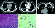
目的 分析CT引导下经皮肺穿刺活检术诊断菌阴肺结核时取材部位的病灶形态、密度与诊断阳性率的关系,以提高菌阴肺结核的诊断准确率。方法 搜集首都医科大学附属北京胸科医院2017年1—12月收治的103例经穿刺活检病理证实或临床试验性治疗确诊的菌阴肺结核患者,回顾性分析经CT引导下穿刺活检时各类病灶CT征象与诊断阳性率之间的关系。统计学处理采用IBM SPSS 24.0 软件,计数资料的比较采用χ 2检验,以P<0.05为差异有统计学意义。 结果 (1)病变形态:磨玻璃样影穿刺活检阳性率33.33%(1/3)。结节、实变、空洞、团块病灶穿刺活检阳性率分别为91.67%(33/36)、94.74%(18/19)、100.00%(21/21)、75.00%(18/24),该4种形态病灶穿刺活检阳性率差异有统计学意义(χ 2=8.918,P=0.030);两两比较,结节组与实变组、空洞组、团块组比较(χ 2=0.174,P=0.677;χ 2=1.847,P=0.174;χ 2=3.137,P=0.077),实变组与空洞组、团块组比较(χ 2=1.134,P=0.287;χ 2=3.031,P=0.082),空洞组与团块组比较(χ 2=8.058,P=0.014)。差异无统计学意义。(2)病变密度:纵隔窗不可测量组3例,病灶穿刺活检阳性率33.33%(1/3)。0~20HU无强化组、>20HU无强化或强化不明显组、>20HU且强化明显组的穿刺活检阳性率分别为96.88%(31/32)、94.34%(50/53)、60.00%(9/15),差异有统计学意义(χ 2=17.790,P=0.000)。该3种密度病灶穿刺阳性率两两比较, 0~20HU无强化与>20HU且强化明显组比较,差异有统计学意义(χ 2=10.956,P=0.001)。>20HU无强化或强化不明显组与>20HU且强化明显组比较,差异有统计学意义(χ 2=12.005,P=0.001)。0~20HU无强化与>20HU无强化或强化不明显组比较差异无统计学意义(χ 2=0.286,P=0.593)。(3)病理检测:HE染色阳性33例、抗酸染色阳性87例、TB-DNA检测阳性91例, HE染色阳性率32.04%(33/103)、抗酸染色阳性率84.47%(87/103)、TB-DNA检测阳性率88.35%(91/103)比较,差异有统计学意义(χ 2=94.084,P=0.001)。 结论 CT引导下经皮肺穿刺活检术对菌阴肺结核诊断具有重要价值,正确选择取材病灶的形态、密度进行穿刺能够提高穿刺活检的阳性率。

关键词: 结核, 肺, 体层摄影术, X线计算机, 活组织检查, 针吸, 诊断, 鉴别

Abstract:

Objective To analyze the relationship of morphology, density and diagnostic positive rate of lesion at the sampling site using CT guided percutaneous pulmonary biopsy, in order to improve its diagnostic accuracy.Methods The data of the relationship between various features of CT imaging and diagnostic positive rates using CT guided percutaneous pulmonary biopsy were analyzed retrospectively in 103 patients with smear and culture negative diagnosis confirmed by pathology or diagnostic antituberculosis treatment admitted in Beijing Chest Hospital, Capital Medical University from January to December 2017. The statistics were processed by IBM SPSS 24.0 software, and the enumeration data were analyzed by χ 2 test and the differences were considered significantly when P<0.05. Results According to lesion morphology of CT, the positive rate of lesion biopsy was 33.33% (1/3) in ground glass opacity lesions and were 91.67% (33/36), 94.74% (18/19), 100.00% (21/21) and 75.00% (18/24) in nodule, consolidation, cavity, mass lesion with significant difference statistically in these four lesions (χ 2=8.918,P=0.030). A comparison was made between nodular group and solid group (χ 2=0.174, P=0.677), cavity group (χ 2=1.847,P=0.174), and mass group (χ 2=3.137,P=0.077), when the comparison was made between consolidation group and cavity group (χ 2=1.134,P=0.287), mass group (χ 2=3.031,P=0.082), and the comparison between the cavity group and the mass group (χ 2=8.058,P=0.014), the differences were no significant statistically due to the modified P' more than 0.008. According to lesion density of CT, the positive rate of lesion biopsy was 33.33% (1/3) in mediastinal window unmeasurable group and were 96.88% (31/32), 94.34% (50/53) and 60.00% (9/15) in non-enhancement group with 0-20 HU, non-enhancement or enhancement not obvious group with more than 20 HU, and obvious enhancement group with more than 20 HU with significant differences statistically (χ 2=17.790,P=0.000). A comparison was made between non-enhancement group with 0-20 HU and obvious enhancement group with more than 20 HU with significant difference statistically (χ 2=10.956,P=0.001), and between non-enhancement group or enhancement not obvious group with more than 20 HU and obvious group with more than 20 HU with also significant difference statistically (χ 2=12.005,P=0.001). However, a comparison was made between non-enhancement group with 0-20 HU and non-enhancement or enhancement not obvious group with more than 20 HU without significant difference statistically (χ 2=0.286,P=0.593).According to pathological detection, the diagnosis were confirmed in 33 cases (32.04%, 33/103) using HE staining,in 87 cases (84.47%, 87/103) using acid-fast stain and in 91 cases (88.35%, 91/103) using TB-DNA. The diagnostic accuracy rates were compared between different two groups with significant differences statistically (χ 2=94.084,P=0.001). Conclusion CT-guided percutaneous pulmonary biopsy is of great value in the diagnosis of smear and culture negative pulmonary tuberculosis. The correct sampling according to morphology, density of lesions using this technique can improve the positive rate of puncture biopsy.

Key words: Tuberculosis, pulmonary, Tomography, X-ray computed, Biopsy, needle, Diagnosis, differential