Email Alert | RSS    帮助

中国防痨杂志 ›› 2020, Vol. 42 ›› Issue (11): 1171-1176.doi: 10.3969/j.issn.1000-6621.2020.11.006

• 论著 • 上一篇    下一篇

肺结核中医证候与CT空洞征象特征的相关性研究

杨红梅, 陈亮, 裴宁(), 钟秀君, 邹成韵, 江燕, 王海英()   

  1. 201203 上海中医药大学(杨红梅、邹成韵);上海德济医院放射科(陈亮、江燕);上海市公共卫生临床中心结核科(裴宁);上海中医药大学附属岳阳中西医结合医院呼吸科(钟秀君),医学检验科(王海英)
  • 收稿日期:2020-08-30 出版日期:2020-11-10 发布日期:2020-11-13
  • 通信作者: 裴宁,王海英 E-mail:peining1125@163.com;wanghaiying@shyueyanghospital.com
  • 基金资助:
    上海市卫生健康委员会科技教育处面上项目(KY110.01.222)

Correlation between traditional Chinese medicine syndromes of pulmonary tuberculosis and CT cavity characteristics

YANG Hong-mei1, CHEN Liang, PEI Ning(), ZHONG Xiu-jun, ZOU Cheng-yun, JIANG Yan, WANG Hai-ying()   

  1. Shanghai University of Traditional Chinese Medicine,Shanghai,201203,China
  • Received:2020-08-30 Online:2020-11-10 Published:2020-11-13
  • Contact: PEI Ning,WANG Hai-ying E-mail:peining1125@163.com;wanghaiying@shyueyanghospital.com

摘要:

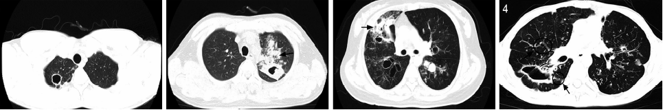
目的 探索肺结核不同中医证候与CT空洞征象特征间的关系,为肺结核中医辨证分型提供客观依据。方法 纳入2019年1—8月上海市公共卫生临床中心结核科确诊的初治肺结核患者173例,采集患者中医病史、临床表现及舌脉象等信息进行中医证候分型,筛选胸部CT检查提示有结核性空洞者 71例,根据患者胸部CT征象的特征,分析中医证候与肺结核空洞CT征象特征的相关性,单因素分析采用χ2检验或Fisher精确检验,多因素分析采用多分类logistic回归分析,均以P<0.05为差异有统计学意义。结果 173例初治肺结核患者,肺阴亏虚证89例(51.45%),阴虚火旺证38例(21.96%),气阴两虚证37例(21.39%),阴阳两虚证9例(5.20%)。其中胸部CT检查显示有空洞者71例,包括肺阴亏虚证31例(43.66%),阴虚火旺证22例(30.99%),气阴两虚证15例(21.13%),阴阳两虚证3例(4.22%)。单因素分析发现气阴两虚证患者空洞周围渗出发生率(66.67%,10/15)最高,显著高于肺阴亏虚证患者(32.26%,10/31)(χ2=4.870,P=0.027); 肺阴亏虚证、阴虚火旺证、气阴两虚证患者空洞个数分别为2.00(1.00,5.00)、4.50(1.00,8.25)、3.50(1.75,8.50)(Z=2.952,P=0.229);空洞体积分别为1884.00(435.50,5569.50)、7969.50(2958.25,29710.00)、3250.00(1162.00,8492.00)mm3 (Z=10.534,P=0.005);空洞面积分别为420.50(191.75,753.00)、 888.00(487.00,2283.00)、572.00(190.50,1264.50)mm2 (Z=6.822,P=0.033);空洞壁厚度分别为3.00(1.25,4.00)、4.00(3.13,5.88)、3.50(2.50,5.00)mm (Z=10.436,P=0.005)。阴虚火旺证患者空洞体积、空洞面积、空洞壁厚度显著高于肺阴亏虚证患者(Z值分别为-17.017、-13.792和-16.695;P值分别为0.004、0.027、0.004)。比较空洞形态,肺阴亏虚证薄壁空洞发生率(74.19%,23/31)显著高于阴虚火旺证(31.82%,7/22),差异有统计学意义(χ2=9.407,P=0.004)。多因素分析显示,空洞周围渗出是影响中医肺结核证候分型的独立相关因素(OR=0.238;95%CI:0.076~0.741;P=0.013)。结论 肺结核中医各证候间CT空洞征象的特征表现不同。肺结核肺阴亏虚证阶段,空洞形态以薄壁空洞为主;进展到阴虚火旺型,空洞范围出现增大趋势,空洞壁逐渐增厚,空洞形态向厚壁空洞转变;气阴两虚证空洞周围更容易出现渗出。

关键词: 结核,肺, 中医证候, 体层摄影术,X线计算机, 空洞, 疾病特征, 数据说明,统计, 因素分析,统计学

Abstract:

Objective To explore the correlation between different TCM syndromes of pulmonary tuberculosis and chest CT cavity characteristics, to provide objective evidence for pulmonary tuberculosis TCM syndrome differentiation. Methods A total of 173 new tuberculosis patients diagnosed in the department of tuberculosis, Shanghai Public Health Clinical Center from January 2019 to August 2019 were included. TCM medical history, clinical manifestations, tongue pulse and other information of the patients were collected for TCM syndrome identifying. Senventy-one patients were proved to be having tuberculous cavities by chest CT. Correlation between TCM syndrome types and the characteristics of tuberculosis cavities was analyzed. Chi-square test or Fisher’s exact test were used for univariate analysis. Multi-nomial logistic regression analysis was used for multivariate analysis, P<0.05 was considered statistically significant. Results Among the 173 patients, 89 cases (51.45%) were diagnosed by TCM as having pulmonary Yin deficiency (PYD) syndrome, 38 cases (21.96%) having hyperactivity of fire due to Yin deficiency (HFYD) syndrome, 37 cases (21.39%) having deficiency of Qi and Yin (DQY) syndrome, and 9 cases (5.20%) having deficiency of Yin and Yang (DYY) syndrome. Senventy-one patients with tuberculous cavities included 31 cases (43.66%) with PYD, 22 cases (30.99%) with HFYD, 15 cases (21.13%) with DQY, and 3 cases (4.22%) with DYY; Univariate analysis showed that patients with DQY had the highest incidence of exudation around the cavity, significantly higher than that of patients with PYD (χ2=4.870 P=0.027); For patients with syndrome of PYD,HFYD and DQY: median numbers of cavities were 2.00 (1.00,5.00),4.50 (1.00,8.25), 3.50 (1.75,8.50),(Z=2.952,P=0.229);Median volume of cavities were 1884.00 (435.50,5569.50), 7969.50 (2958.25,29710.00) and 3250.00 (1162.00,8492.00)mm 3,(Z=10.534,P=0.005); Median areas of cavities were 420.50 (191.75,753.00),888.00 (487.00,2283.00),572.00 (190.50,1264.50)mm2, (Z=6.822, P=0.033); Median thickness of cavity walls were 3.00 (1.25, 4.00), 4.00 (3.13, 5.88) and 3.50 (2.50, 5.00)mm respectively, (Z=10.436, P=0.005).The volume, area and wall thickness of cavities in patients with HFYD were significantly higher than those with FYD (Z values were -17.017,-13.792 and -16.695, respectively; P values are 0.004, 0.027 and 0.004, respectively). Comparing shapes of cavities in different patients, the incidence of thin-walled cavities in PYD patients (74.19%, 23/31) was significantly higher than that in HFYD patients (31.82%, 7/22) for which difference was statistically significant (χ2=9.407, P=0.004).Multivariate analysis showed that exudation around cavity was an independent factor affecting the TCM classification of pulmonary tuberculosis syndromes (OR=0.238, 95%CI:0.076-0.741; P=0.013). Conclusion There are differences in characteristics of CT cavities between different TCM syndromes of pulmonary tuberculosis. In the stage of PYD of pulmonary tuberculosis, cavities were mainly thin-walled cavities, when they progressed to HFYD, cavity areas appeared to expand, and cavity walls gradually thickened. Exudation is more likely to occur around the cavity in DQY patients.

Key words: Tuberculosis,pulmonary, Chinese medicine syndromes, Tomography,X-ray computed, Cavity, Disease attributes, Data interpretation,statistical, Factor analysis,statistical