Email Alert | RSS    帮助

中国防痨杂志 ›› 2020, Vol. 42 ›› Issue (11): 1142-1152.doi: 10.3969/j.issn.1000-6621.2020.11.002

• 论著 • 上一篇    下一篇

对胸腔积液患者利用临床及内科胸腔镜下特征构建诺谟图诊断结核性胸膜炎的价值

柳芳超, 杨新婷, 姜慧, 段鸿飞, 梁清涛, 李华, 杨扬, 郭超, 张芸, 邵玲玲, 陈效友()   

  1. 101149 首都医科大学附属北京胸科医院 北京市结核病胸部肿瘤研究所(陈效友已调至:100015 首都医科大学附属北京地坛医院)
  • 出版日期:2020-11-10 发布日期:2020-11-13
  • 通信作者: 陈效友 E-mail:chenxy1998@hotmail.com
  • 基金资助:
    北京市医院管理局“登峰”计划专项经费资助项目(DFL20151501);北京市医院管理局“登峰”计划专项经费资助项目(DFL20181601);北京市科学技术委员会重点项目(D181100000418003);北京市科学技术委员会重点项目(Z191100006619078);北京市卫生和计划生育委员会北京市卫生与健康科技成果和适宜技术推广项目(2018-TG-43)

Evaluation of the diagnosis value of nomogram based on the clinical and internal thoracoscopic features in pleural effusion patients complicated with tuberculous pleurisy

LIU Fang-chao, YANG Xin-ting, JIANG Hui, DUAN Hong-fei, LIANG Qing-tao, LI Hua, YANG Yang, GUO Chao, ZHANG Yun, SHAO Ling-ling, CHEN Xiao-you()   

  1. Beijing Chest Hospital, Capital Medical University, Beijing Tuberculosis and Thoracic Tumor Research Institute, Beijing 101149, China (*Now in: Beijing Ditan Hospital, Capital Medical University, Beijing 100015, China)
  • Online:2020-11-10 Published:2020-11-13
  • Contact: CHEN Xiao-you E-mail:chenxy1998@hotmail.com

摘要:

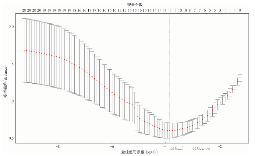
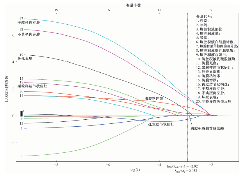
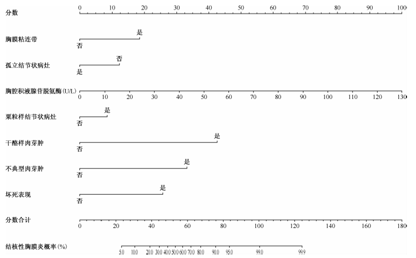
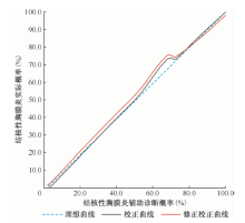
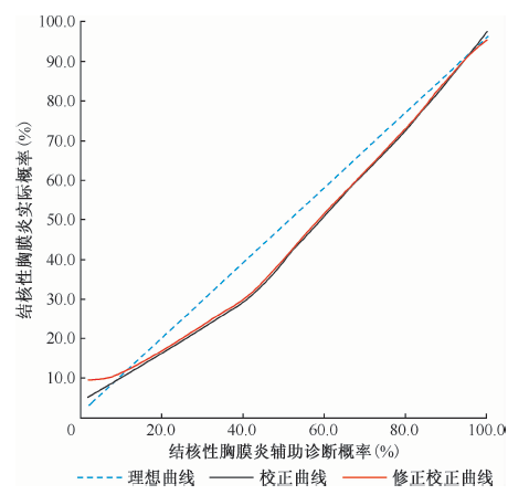
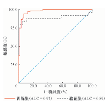
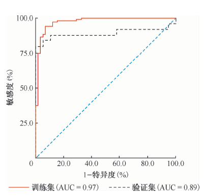
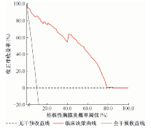
目的 利用临床及内科胸腔镜下特征对胸腔积液患者构建诺谟图(nomogram),探讨其诊断结核性胸膜炎的价值。 方法 采用前瞻性研究的方法,收集2015—2018年首都医科大学附属北京胸科医院(99例)、首都医科大学附属北京朝阳医院(67例)、北京积水潭医院(16例)及北京医院(24例)收治的206例胸腔积液患者。其中确诊为结核性胸膜炎的患者129例,作为结核性胸膜炎组;确诊为其他原因胸腔积液的患者77例,作为其他原因胸腔积液组。收集两组患者的一般资料、胸腔积液实验室检查结果、胸腔镜下形态学特征和病理形态学特征信息,按照4∶1的比例通过SAS随机分组过程,将206例患者完全随机分为训练集(162例)和验证集(44例)。利用套索(least absolute shrinkage and selection operator,LASSO)回归筛选结核性胸膜炎的诊断因素,构建辅助诊断诺谟图,分别验证模型的区分度、校准度及收益,综合评价诺谟图的辅助诊断能力。 结果 训练集162例胸腔积液患者中,共104例(64.2%)确诊为结核性胸膜炎。经LASSO回归,共7个高诊断价值因素被筛选出,分别为病理形态学上的干酪样肉芽肿、坏死表现、不典型肉芽肿,胸腔镜观察下的粟粒样结节状病灶、胸膜粘连带、孤立结节状病灶,以及胸腔积液检查指标中的腺苷脱氨酶水平。训练集中ROC曲线下面积(AUC)为0.97,敏感度为92.31%,特异度为93.42%,在验证集中模型AUC为0.89,敏感度为80.44%,特异度为100.00%。 结论 诺谟图能够在胸腔积液患者中有效诊断结核性胸膜炎,有一定的辅助诊断价值。

关键词: 结核,胸膜, 病理状态,体征和症状, 胸腔镜, 诺谟图, 诊断

Abstract:

Objective Constructing nomogram based on clinical and internal thoracoscopic features of pleural effusion patients, to explore its value of diagnosis of tuberculous pleurisy. Methods A total of 206 patients treated in 4 hospitals in Beijing from 2015 to 2018 were collected in this prospective study, and of them, 129 patients were tuberculous pleurisy patients (tuberculous pleurisy group), 77 patients were pleural effusion patients (pleural effusion group), including the patients of tuberculous pleurisy and patients with pleural effusion. All the patients were from Beijing Chest Hospital affiliated to Capital Medical University (n=99), Beijing Chaoyang Hospital affiliated to Capital Medical University (n=67), Beijing Jishuitan Hospital (n=16) and Beijing Hospital (n=24). The general information, results of pleural effusion, internal thoracoscopic and pathologic characteristics were collected. The 206 patients were randomly divided into the training set (n=162) and the validation set (n=44). The predictive factors of tuberculous pleurisy were screened by least absolute shrinkage and selection operator (LASSO), regression, and the predictive nomogram was constructed to verify its discrimination, calibration and benefits, comprehensively evaluating the diagnostic and predictive ability of the nomogram. Results Among the 162 pleural effusion patients, 104 (64.2%) were diagnosed with tuberculous pleurisy. By LASSO regression, 7 predictors of high diagnosis value were selected, which were caseous granulomas, necrotic manifestations, atypical granulomas, miliary nodules, pleural adhesions, isolated nodules with thoracoscopy and adenosine dehydrogenase level (an indicator in pleural effusion examination). The AUC, sensitivity and specificity in the training set were 0.97, 92.31% and 93.42%, respectively, while in the model of the verification set, those were 0.89, 80.44% and 100.00%, respectively. Conclusion The nomogram could effectively predict the diagnosis of tuberculous pleurisy in patients with pleural effusion, it had applied diagnosis value to some extent.

Key words: Tuberculosis,pleural, Pathological conditions,signs and symptoms, Thoracoscopes, Nomogram, Diagnosis